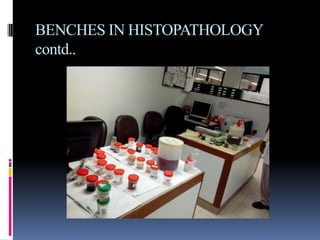
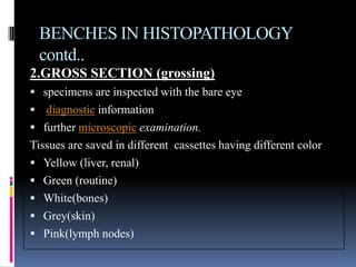
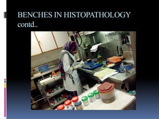
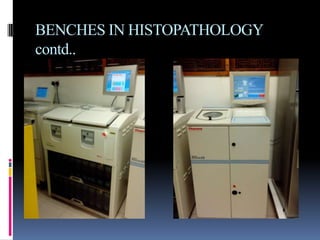
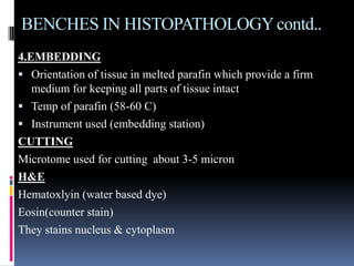
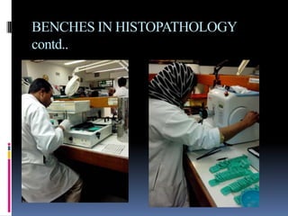
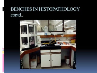
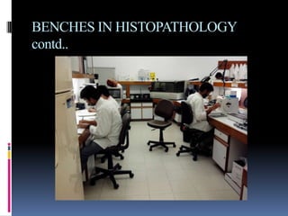
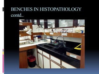
The document provides an overview of the department of histopathology and its various benches. It describes histopathology as the microscopic examination of tissue to study disease manifestations. The key benches mentioned are processing, gross sectioning, tissue processing, embedding, cutting, staining including H&E, immunohistochemistry, special stains, cytology, cytogenetics, and semen analysis. The roles of each bench are briefly outlined.
2. INTRODUCTION TO DEPARTMENT OF
HISTOPATHOLOGY
Histopathology is the department of clinical lab which
deals with the study of diff types of tissues
The department is based on following benches:
Processing
Gross
Tissue processing
Embedding,cutting,H&E
Immunohistochemistry
Special stains
Cytology
Semen DR
Cytogenetics
3. WHAT IS HISTOPATHOLOGY?
Greek word
Histo- tissue
Pathos- disease suffering
refers to the microscopic examination of tissue in order
to study the manifestations of disease.
histopathology refers to the examination of a biopsy or
surgical specimen by a pathologist,
after the specimen has been processed and histological
sections have been placed onto glass slides
6. BENCHES IN HISTOPATHOLOGY
contd..
2.GROSS SECTION (grossing)
specimens are inspected with the bare eye
diagnostic information
further microscopic examination.
Tissues are saved in different cassettes having different color
Yellow (liver, renal)
Green (routine)
White(bones)
Grey(skin)
Pink(lymph nodes)
8. BENCHES IN HISTOPATHOLOGY
contd..
3.TISSUE PROCESSING
fixing tissue into paraffin
dehydration and clearing
tissue is infiltrated with the embedding agent, (paraffin)
Tissue processing is always automated for the large volumes of
tissues.
Automation consists of an instrument that moves the tissues
around through the various agents on a preset time scale.
10. BENCHES IN HISTOPATHOLOGYcontd..
4.EMBEDDING
Orientation of tissue in melted parafin which provide a firm
medium for keeping all parts of tissue intact
Temp of parafin (58-60 C)
Instrument used (embedding station)
CUTTING
Microtome used for cutting about 3-5 micron
H&E
Hematoxlyin (water based dye)
Eosin(counter stain)
They stains nucleus & cytoplasm
13. BENCHES IN HISTOPATHOLOGY
contd..
5.IMMUNOHISTOCHEMISTRY
Ag-Ab specific reaction
Clinical diagnosis and distinguishing diagnosis of tumor
histogenesis
Can be used to locate particular cells and proteins
Can be used to identify cellular events – e.g.apoptosis
15. BENCHES IN HISTOPATHOLOGY
contd..
6.SPECIAL STAINS
Required when diagnosis is not identified with H&E
staining
Some of the stains used are
Connective tissue ( rapid trichome)
Nucleic acid (methylene green pyronin)
Carbohydrates(PAS & PASD)
17. BENCHES IN HISTOPATHOLOGY
contd..
7.CYTOGENETICS
Study of chromosomes and its abnormalities
chromosome number
structure
function
behavior in relation to gene inheritance,
organization and expression
Types of disorders:
Deletions
Inversions
Translocation
Addition(insertion)
20. BENCHES IN HISTOPATHOLOGY
contd..
8.CYTOLOGY
Study of cells to know the infection,pre malignant and
malignanent changes
2 types of samples
Gyane
(pap smears)
Non Gyane
(bodyfluids,CSF,urine,pericardial,pleural.asitic.synovial)
23. BENCHES IN HISTOPATHOLOGY
contd..
9.SEMEN DR
Fresh sample of seminal fluid is examined for :
Liquefaction
Motility
Number
Morphology
Elements of analysis
Quantity
Color
Liquefaction
pH