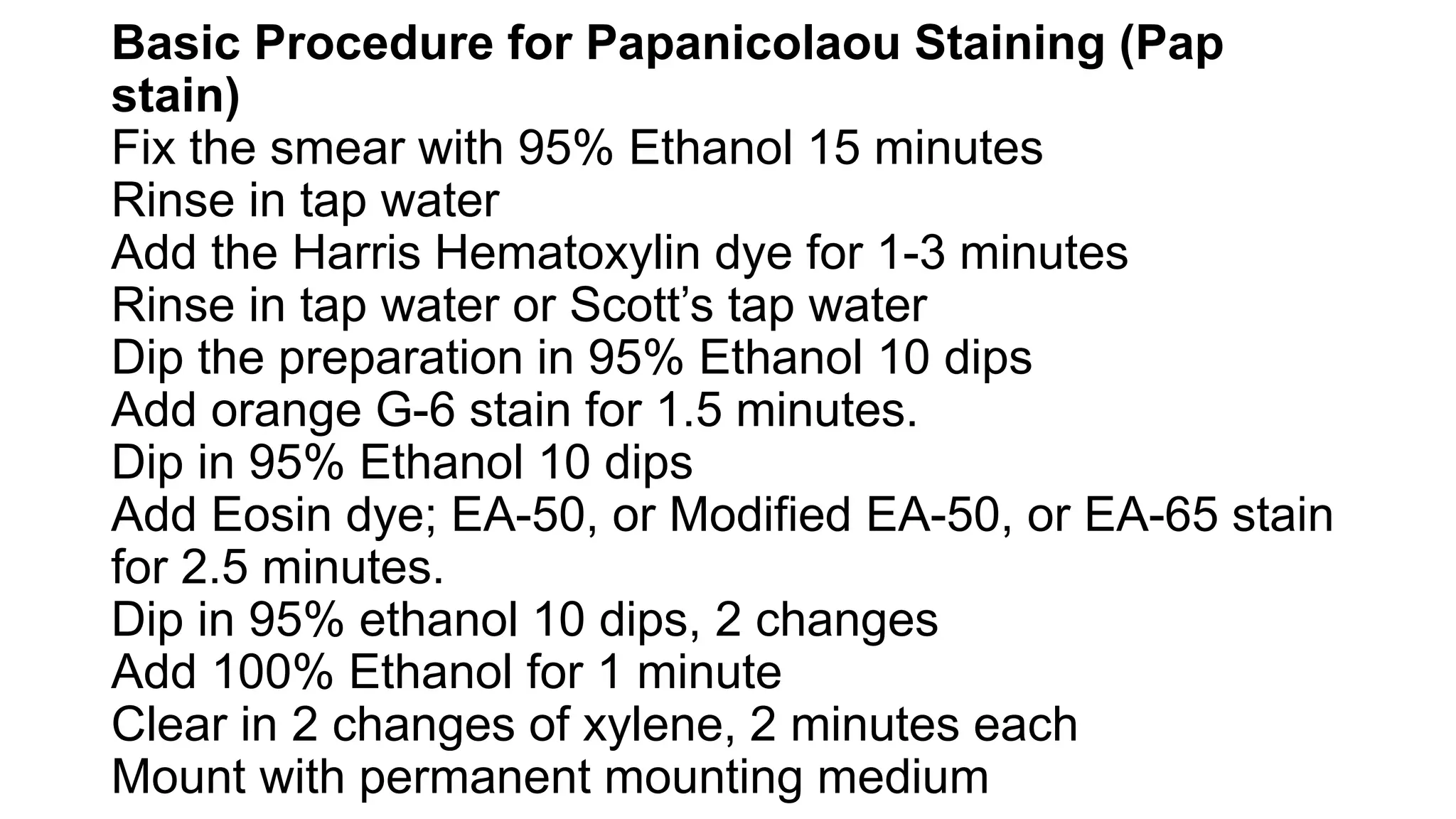
2. What is Papanicolaou Staining (Pap stain)?
Papanicolaou stain is also known as the pap stain and the procedure of the stain is
known as a pap smear.
It is a polychromatic stain that uses multiple dyes to differentially stain various
components of the cells.
It is a histological and cytopathological staining technique used to differentiate cells in a
smear preparation.
It is the most common screening method for cervical cancer.
Several specimens can be used to prepare the pap smear depending on the screening
infection, including sputum, urine, cerebrospinal fluid, abdominal fluid, tumor biopsies,
synovial fluid, fine needle aspirates, pleural fluids.
The technique was developed by George Papanicolaou in 1942.
3. Pap stain was first developed by Dr GN Papanicoloau in 1947 and since then it
has been used successfully to screen cervical cancer. In fact, it has reduced the
incidence of cervical cancer by 70% especially in developed countries having
well planned screening programs. However, the stain has undergone various
modifications from regressive conventional method to progressive rapid Pap
staining where the time taken for staining was reduced. Further the stain was
modified as ultrafast Papanocoloau stain and modified ultrafast Pap stain were
air dried cervical smears were used and staining time was reduced. Later the
other modifications were Enviro- Pap stain which was environmentally friendly
with results similar to conventional method; REAP stain which was rapid and
economical and Cytocolor developed by Merck where isopropyl alcohol is
replaced by Propanol. Each method has advantages and disadvantages.
Hence laboratories should develop their own protocol and standardize the
staining technique.
4. Objectives of Papanicolaou Staining (Pap
stain)
To define the cell nuclear to aid in the
identification of nuclear abnormalities of
cancer cells.
To stain the cytoplasm and make it
transparent for visualization
To differentiate and identify certain cell types
such as acidophils and basophils.
5. Principle of Papanicolaou Staining (Pap
stain)
The stain uses both basic and acidic dyes
such that the basic dye stains acidic
components of the cell while the acidic dyes
stain the basic components of the cells. This
is based on the ionic charges of the
components of the cell with the principle of
attraction and repulsion of the ions and the
dyes. Five dyes are used in three solutions as
the main reagents used in the stain.
6. Hematoxylin: This is a natural dye that stains
the cell nuclear blue. The dye attaches to the
sulfate groups of DNA because it has a high
affinity for nuclear chromatins. The most
common hematoxylin dyes used are Harris’
hematoxylin, Gills’ H is the commonest
cytologically although Gills’ hematoxylin and
Hematoxylin S.
7. Orange Green 6: It is an acidic
counterstain that stains the cytoplasm of
mature keratinized cells. The
components of the target stain orange in
varying intensities of the dye.
8. Eosin Azure: It is the second counterstain, a
combination of eosin Y, light green SF, and
Bismarck brown. Eosin Y stains the
cytoplasm of mature squamous cells,
nucleoli, Red blood cells, and cilia pink. The
eosin dyes commonly used are EA 31 and EA
50, while EA 65. Light green SF stains the
cytoplasm of active cells such as columnar
cells, parabasal squamous cells, and
intermediate squamous cells, blue. Bismarck
brown Y stains nothing and sometimes it is
often omitted.
9. Composition of the Reagents of
Papanicolaou Staining (Pap stain)
Harris’ hematoxylin
Hematoxylin = 2.5g
Ethanol = 25ml
Potassium alum = 50g
Distilled water (50°C) = 500ml
Mercuric oxide = 1-3g
Glacial acetic acid = 20ml
10. Orange G 6
Orange G (10% aqueous) = 25ml
Alcohol = 475ml
Phosphotungstic acid = 0-8g
EA 50
0.04 M light green SF = 5ml
0.3M eosin Y = 10ml
Phosphotungstic acid = 1g
Alcohol = 365ml
Methanol = 125ml
Glacial acetic acid = 10ml
Other reagents include 95% ethanol, 100%
ethanol, tap water, Scott’s tap water, xylene
11. Basic Procedure for Papanicolaou Staining (Pap
stain)
Fix the smear with 95% Ethanol 15 minutes
Rinse in tap water
Add the Harris Hematoxylin dye for 1-3 minutes
Rinse in tap water or Scott’s tap water
Dip the preparation in 95% Ethanol 10 dips
Add orange G-6 stain for 1.5 minutes.
Dip in 95% Ethanol 10 dips
Add Eosin dye; EA-50, or Modified EA-50, or EA-65 stain
for 2.5 minutes.
Dip in 95% ethanol 10 dips, 2 changes
Add 100% Ethanol for 1 minute
Clear in 2 changes of xylene, 2 minutes each
Mount with permanent mounting medium
12. Results and Interpretation of Papanicolaou Staining (Pap
stain)
Staining dyes will stain different components of the cell with
different colors and intensities as follows:
Nuclei: Blue
Acidophilic cells: Red
Basophilic cells: Blue Green
Erythrocytes: Orange-red to dark pink
Keratin: Orange-red
Superficial cells: Pink
Intermediate and Parabasal Cells: Blue-Green
Eosinophil: Orange Red
Metaplastic cells: May contain both blue/green and pink
Candida: Red
Trichomonas: Grey-green