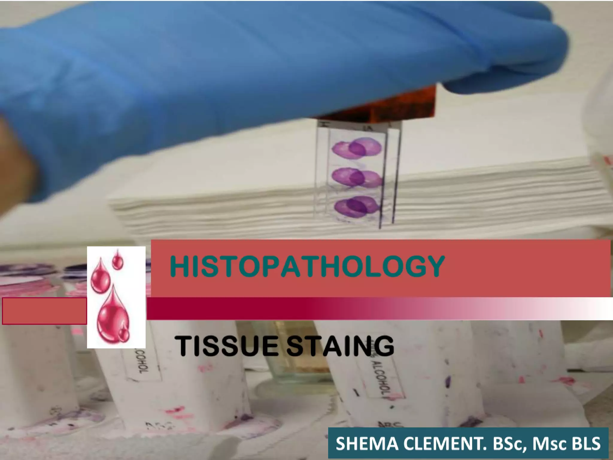
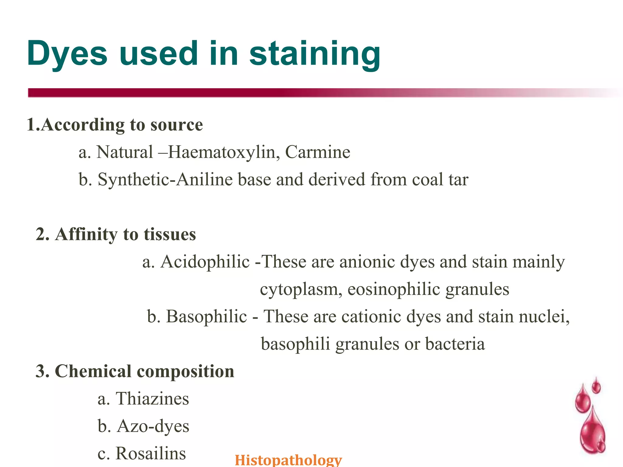
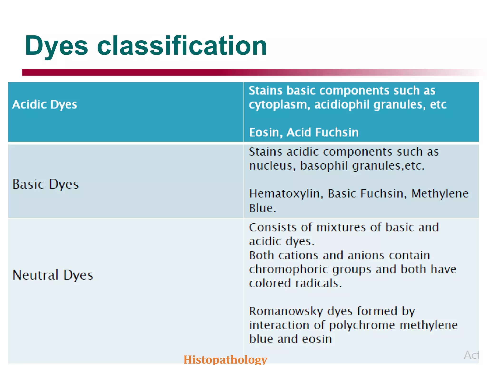
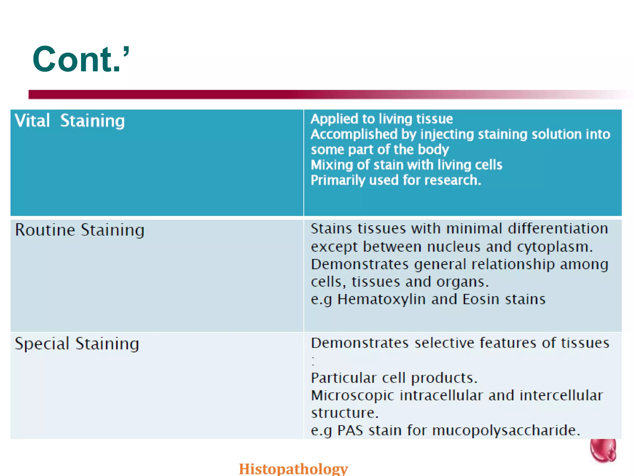
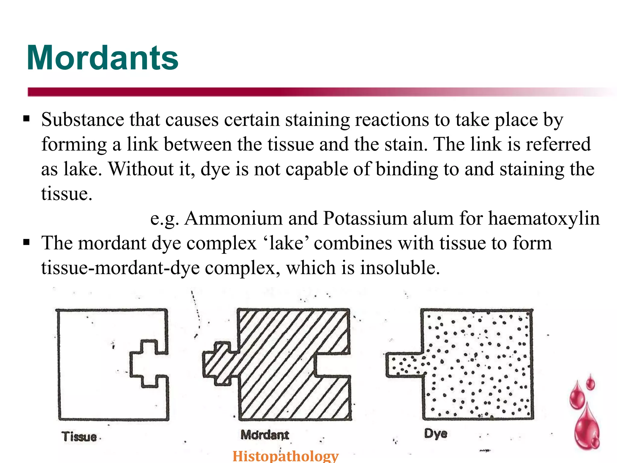
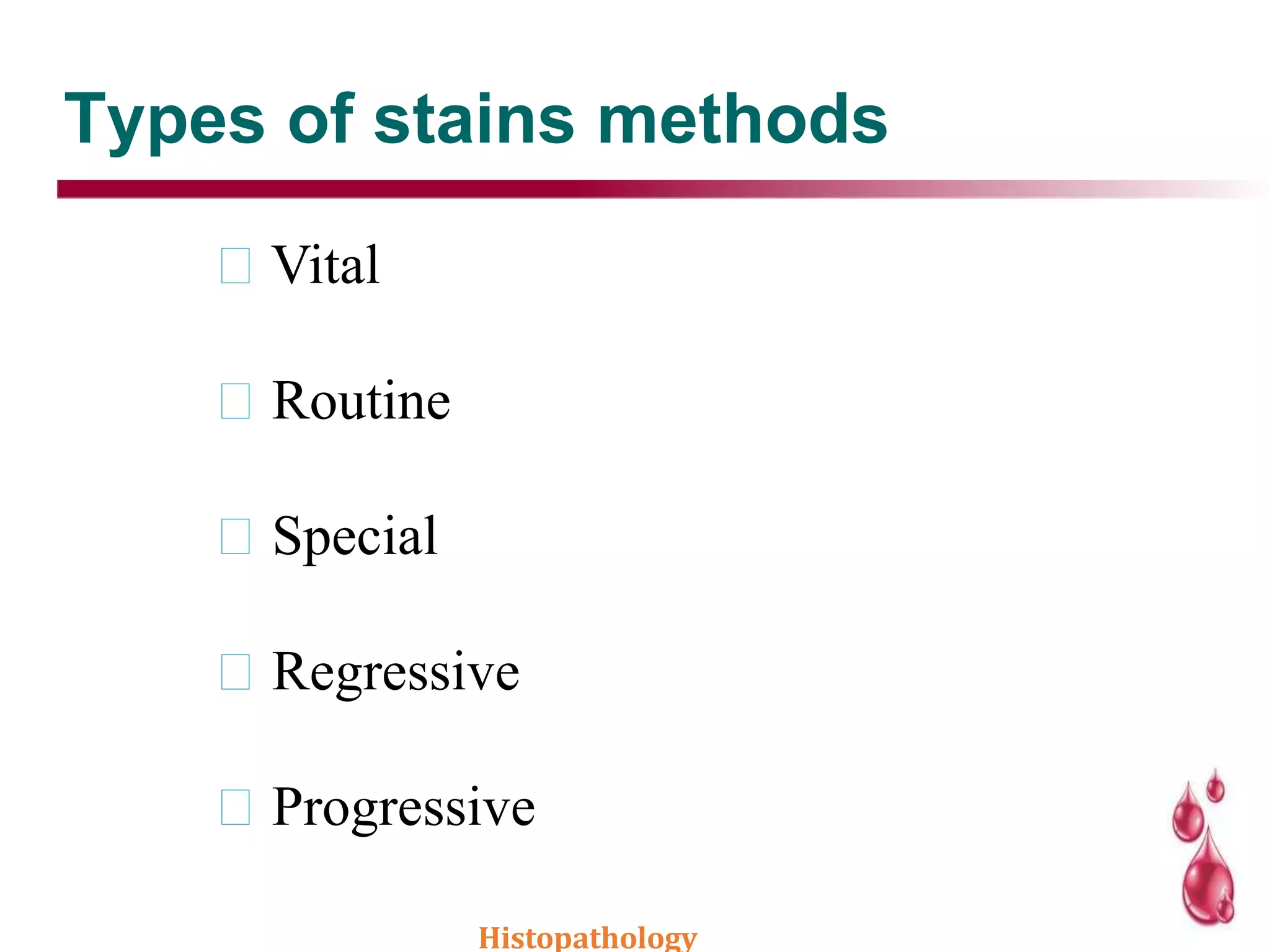
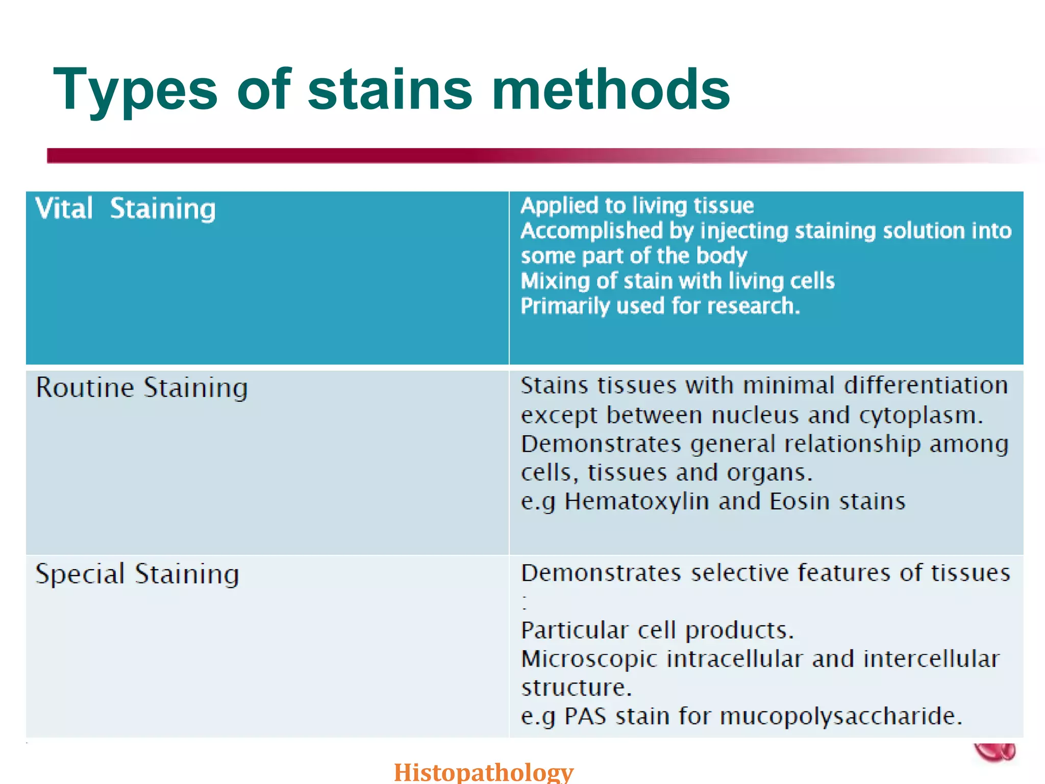
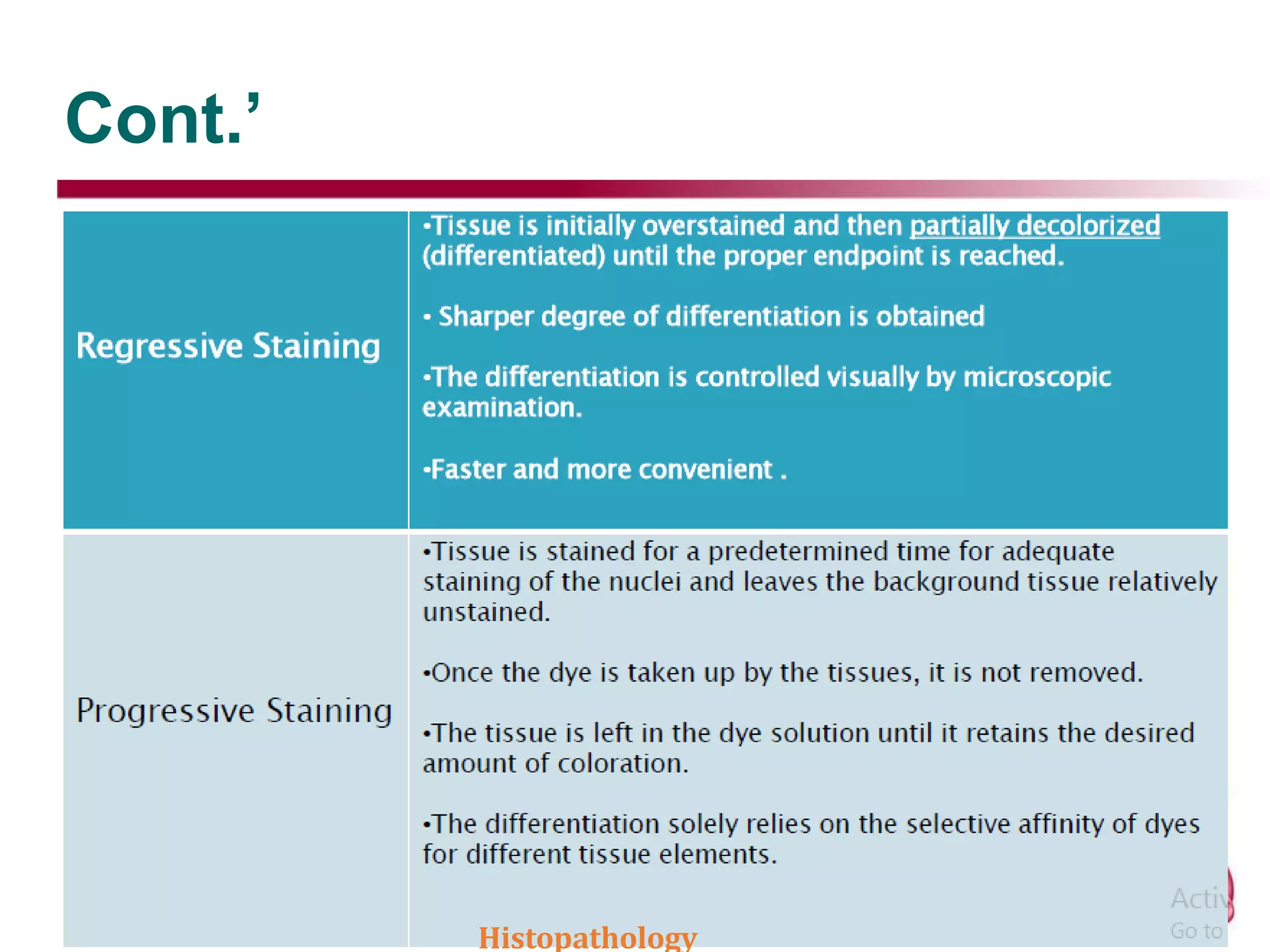
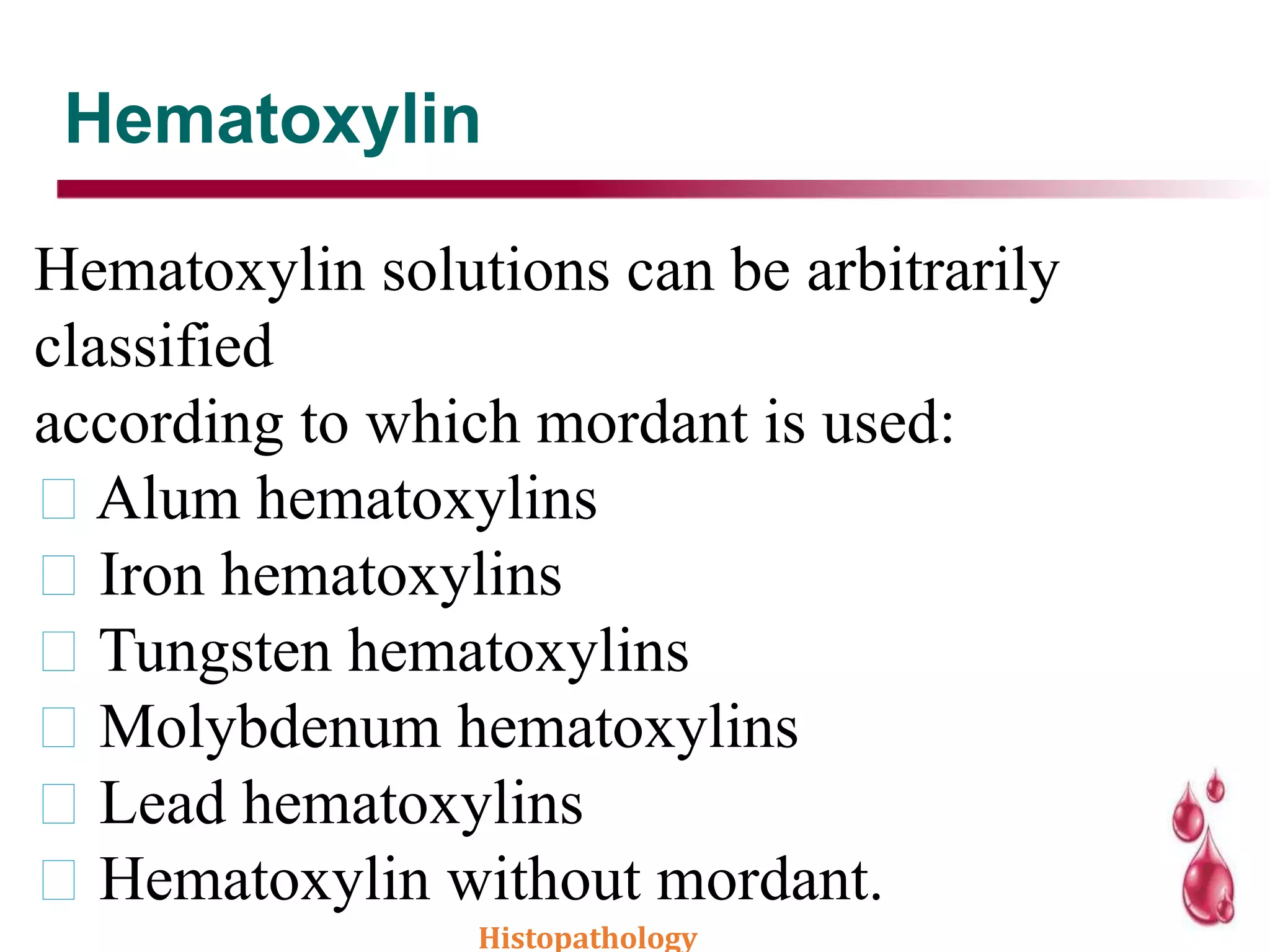
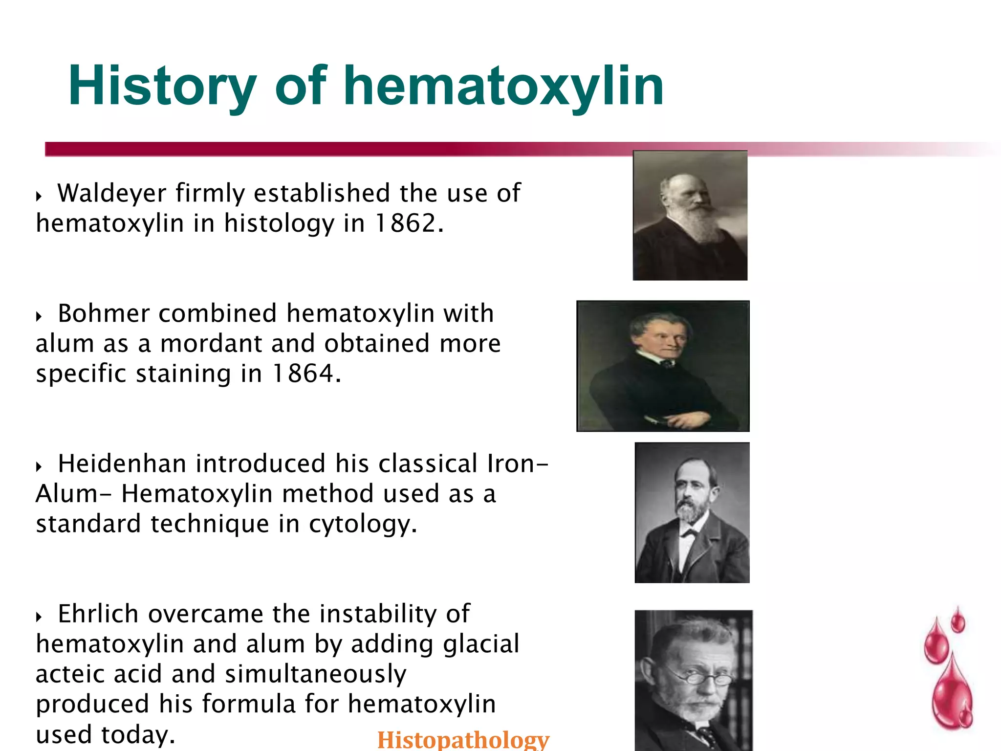
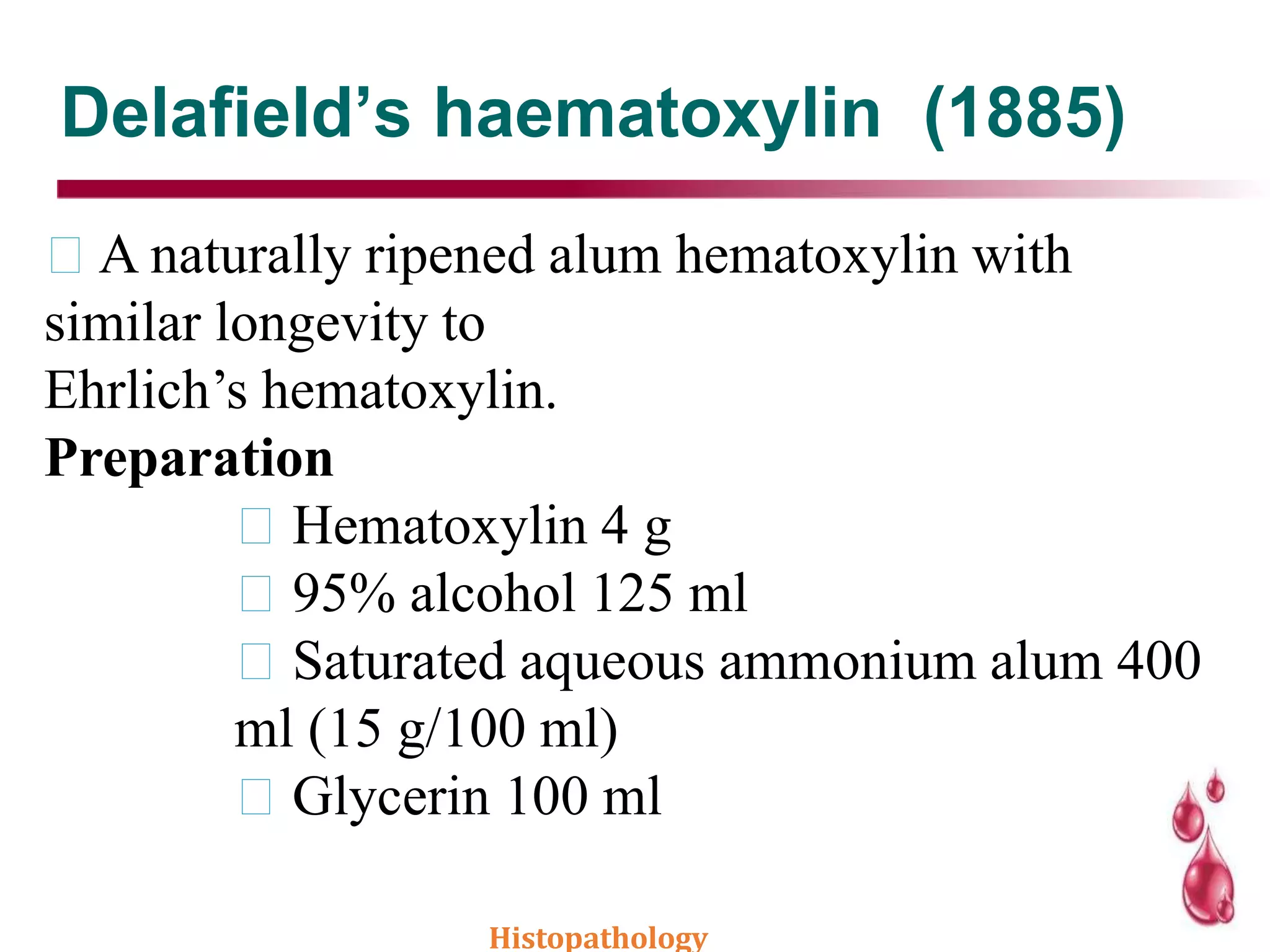
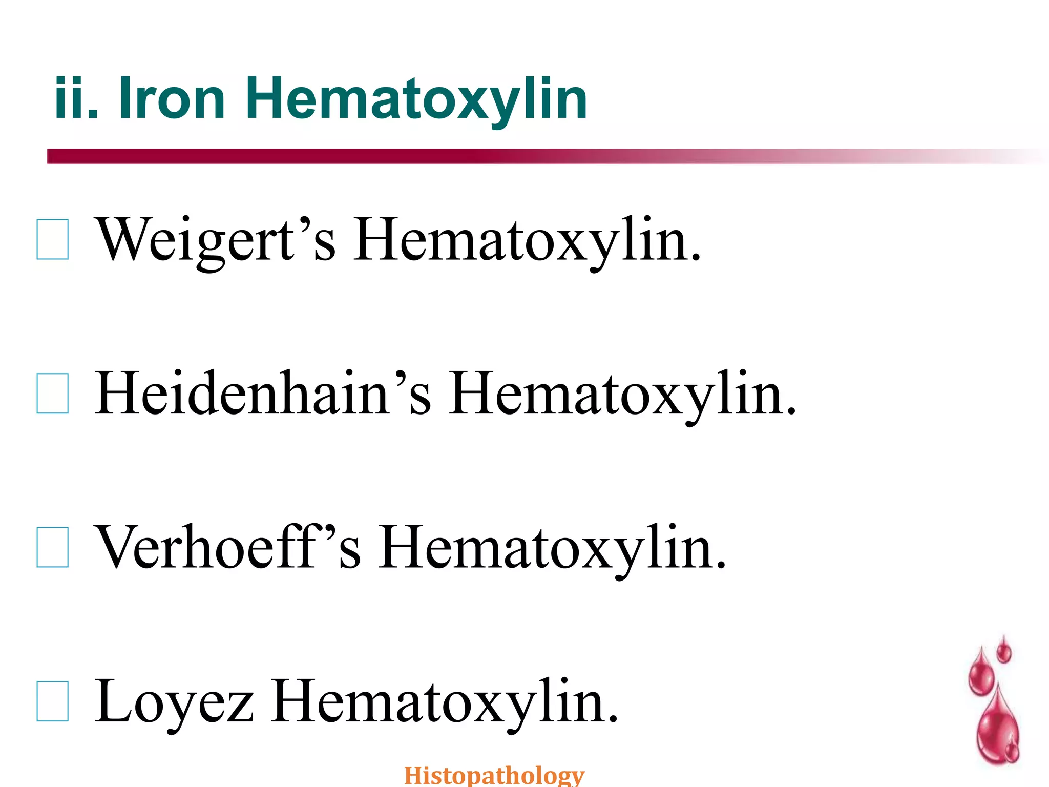
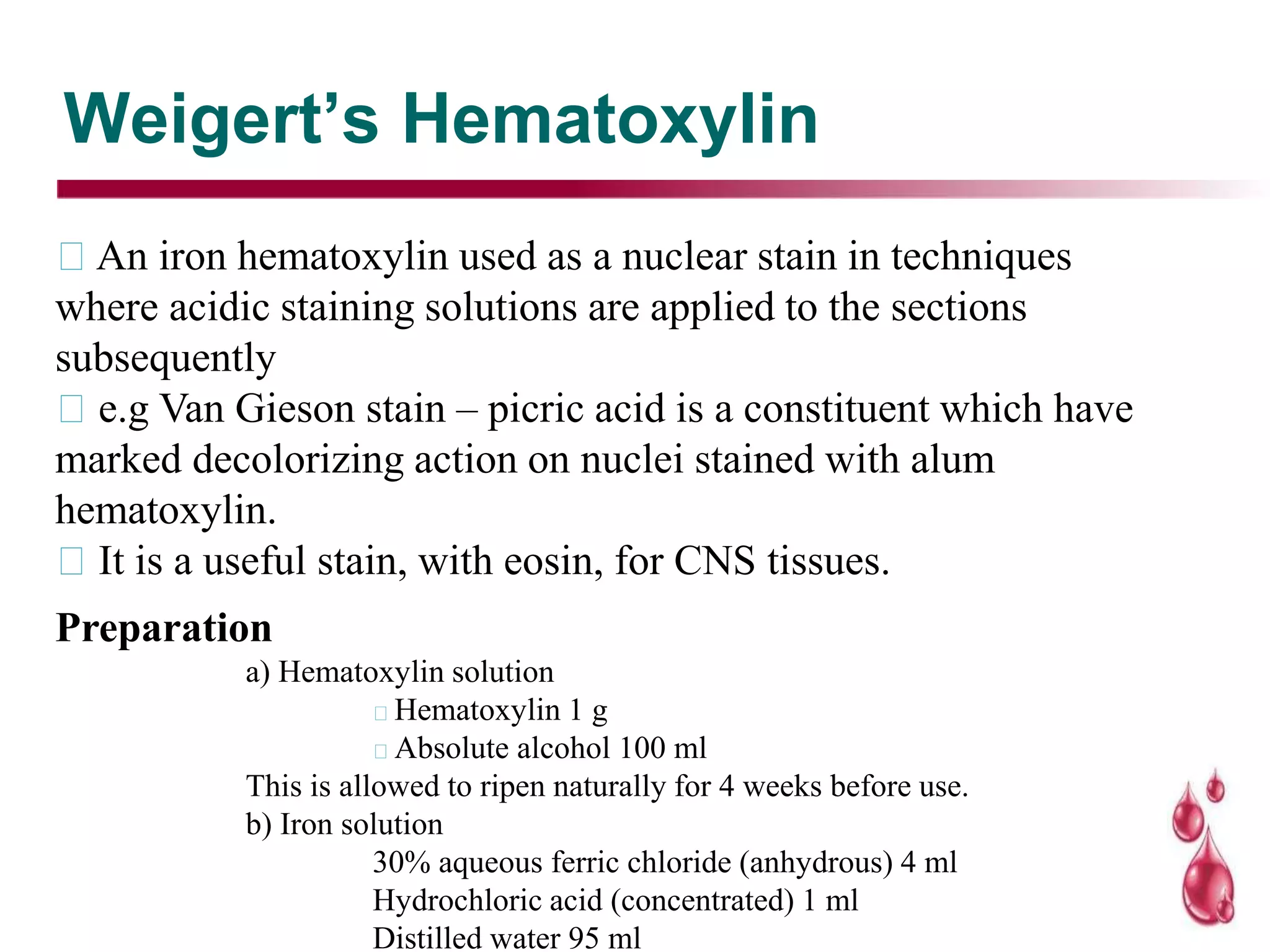
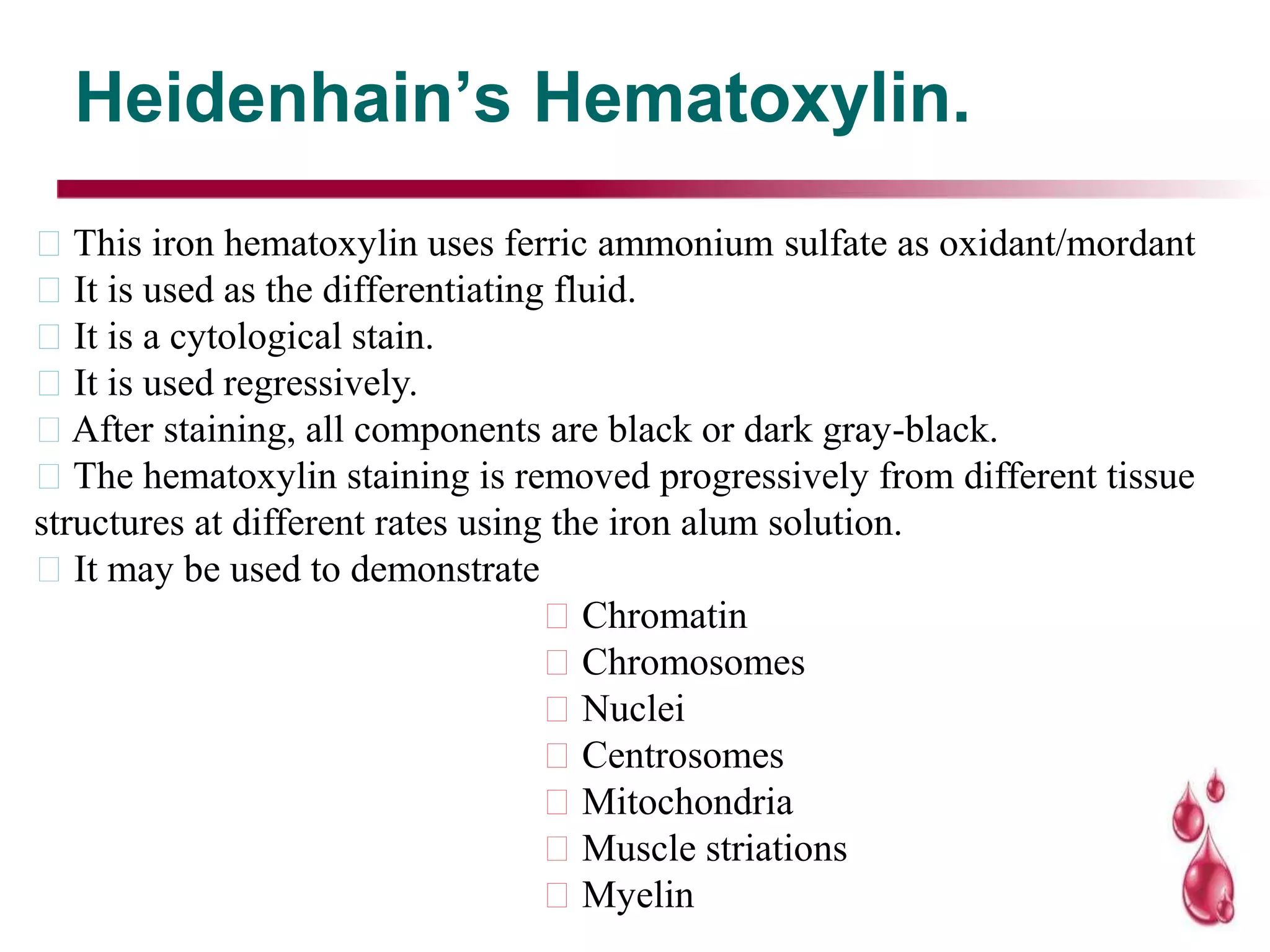
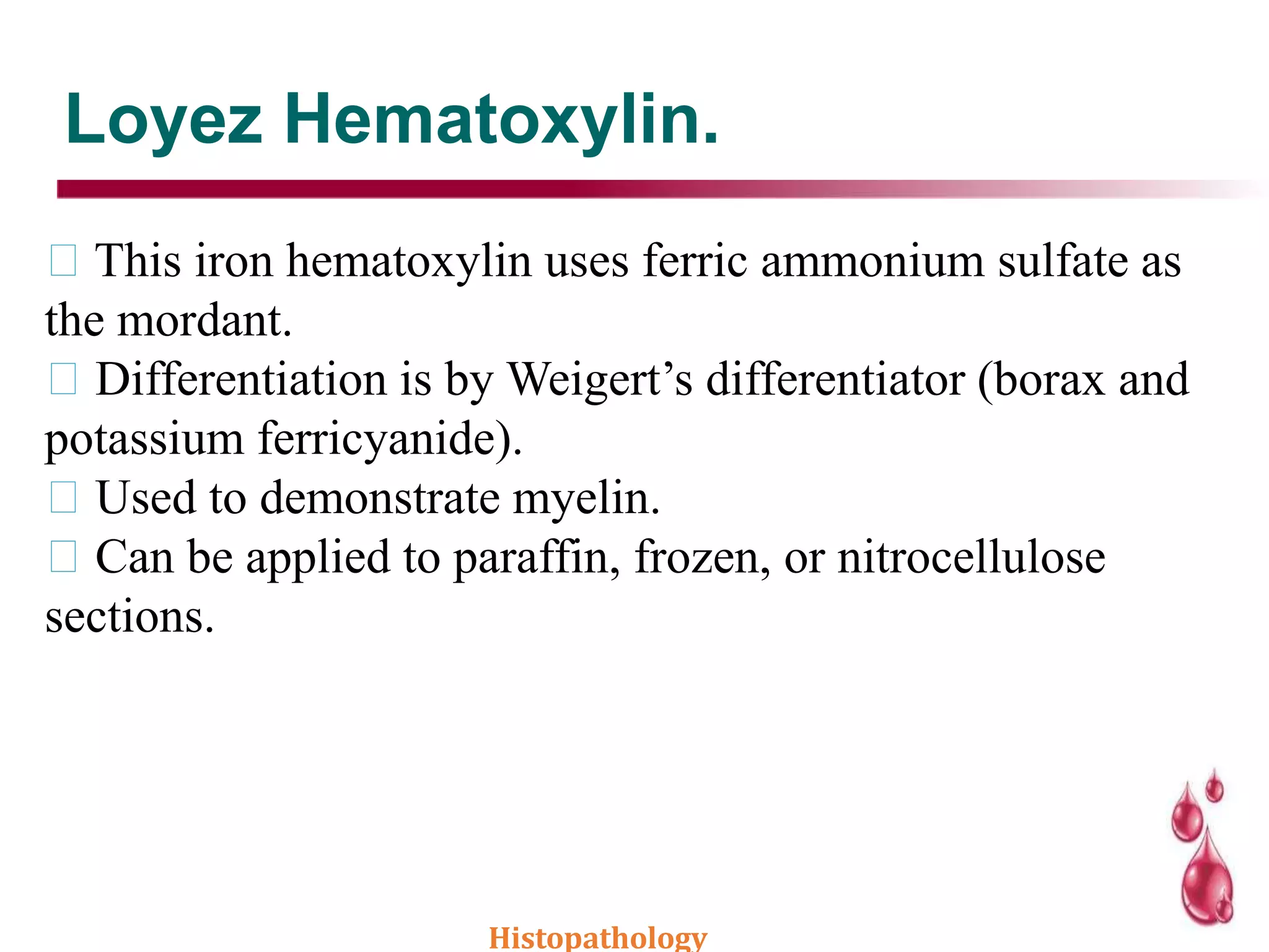
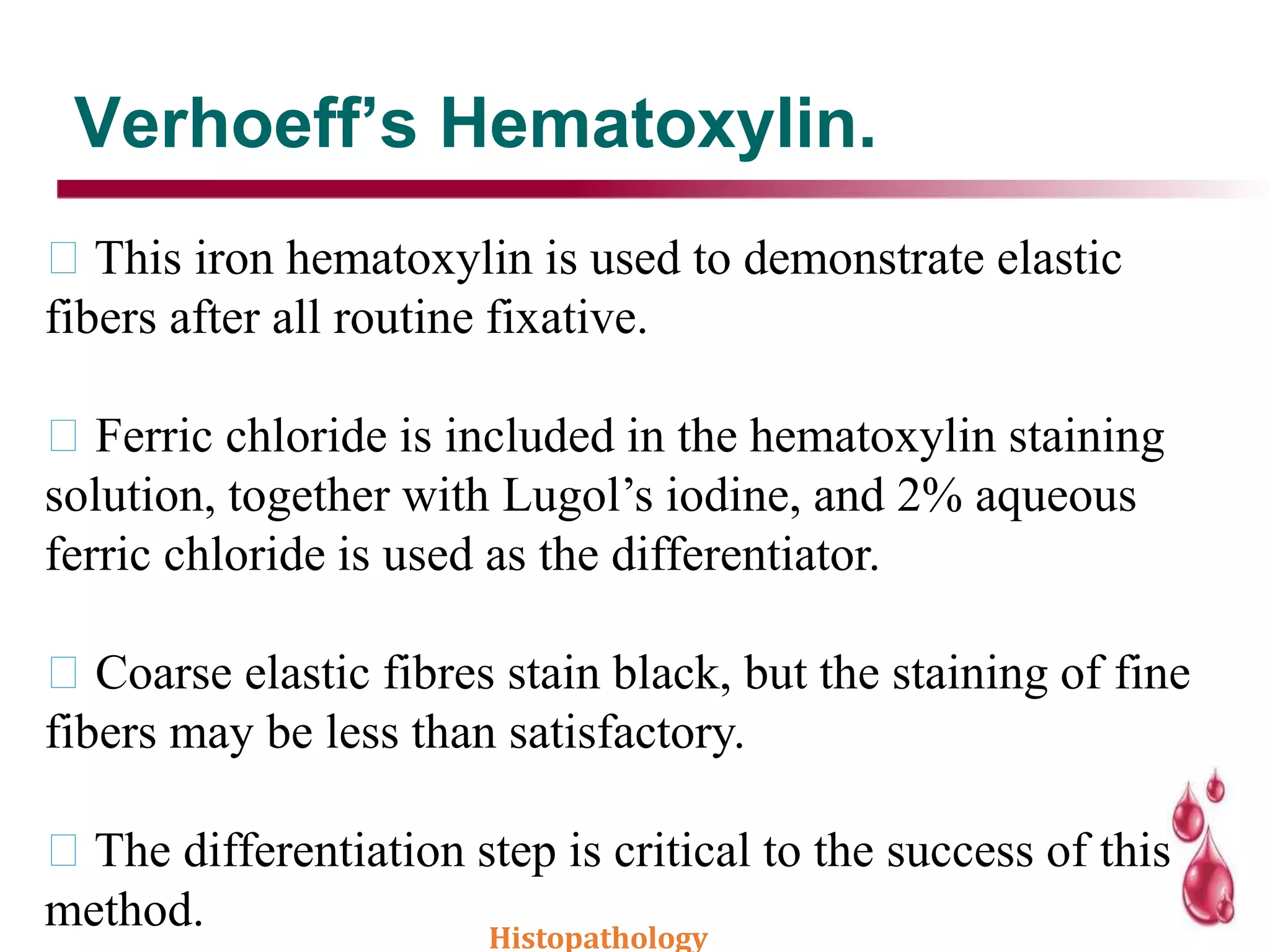
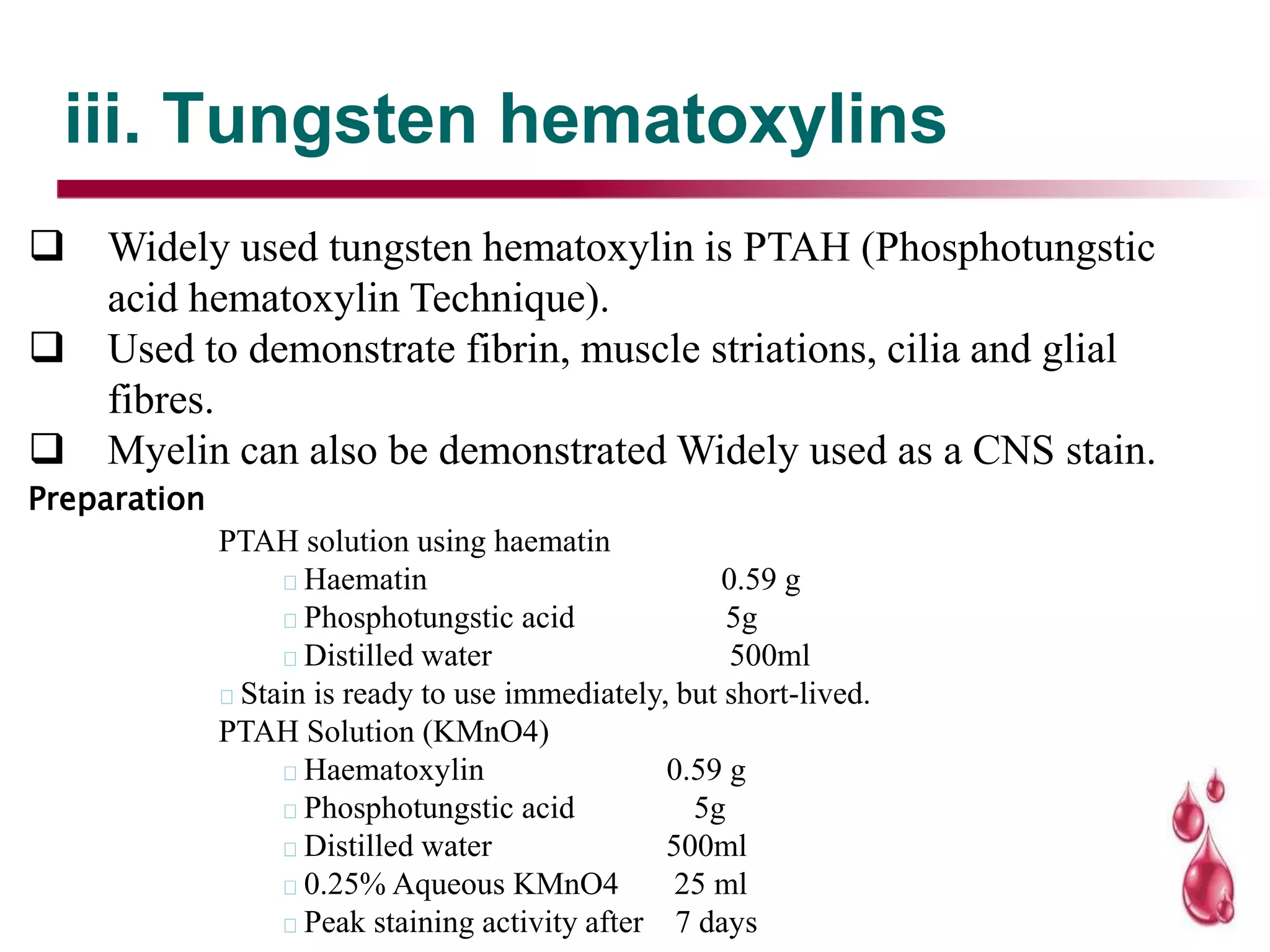
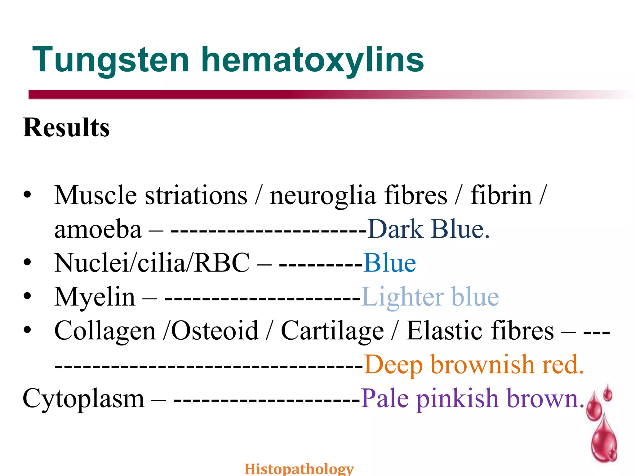
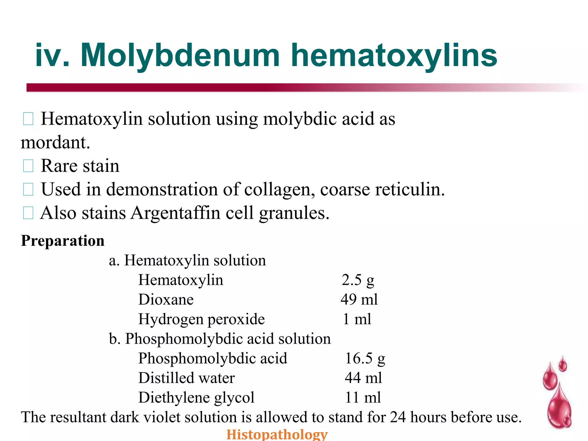
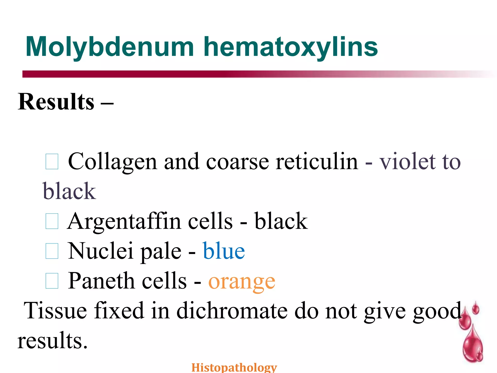
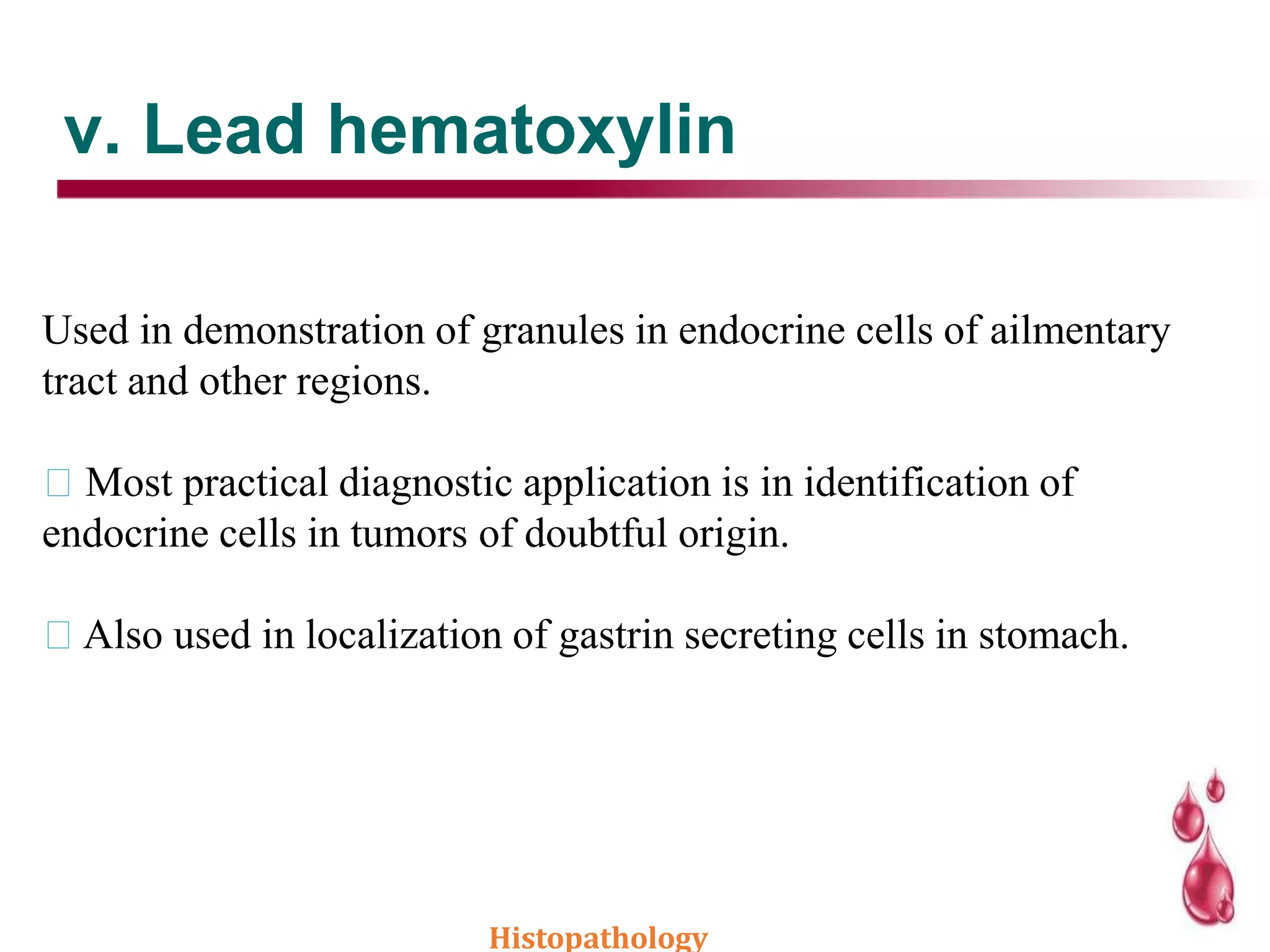
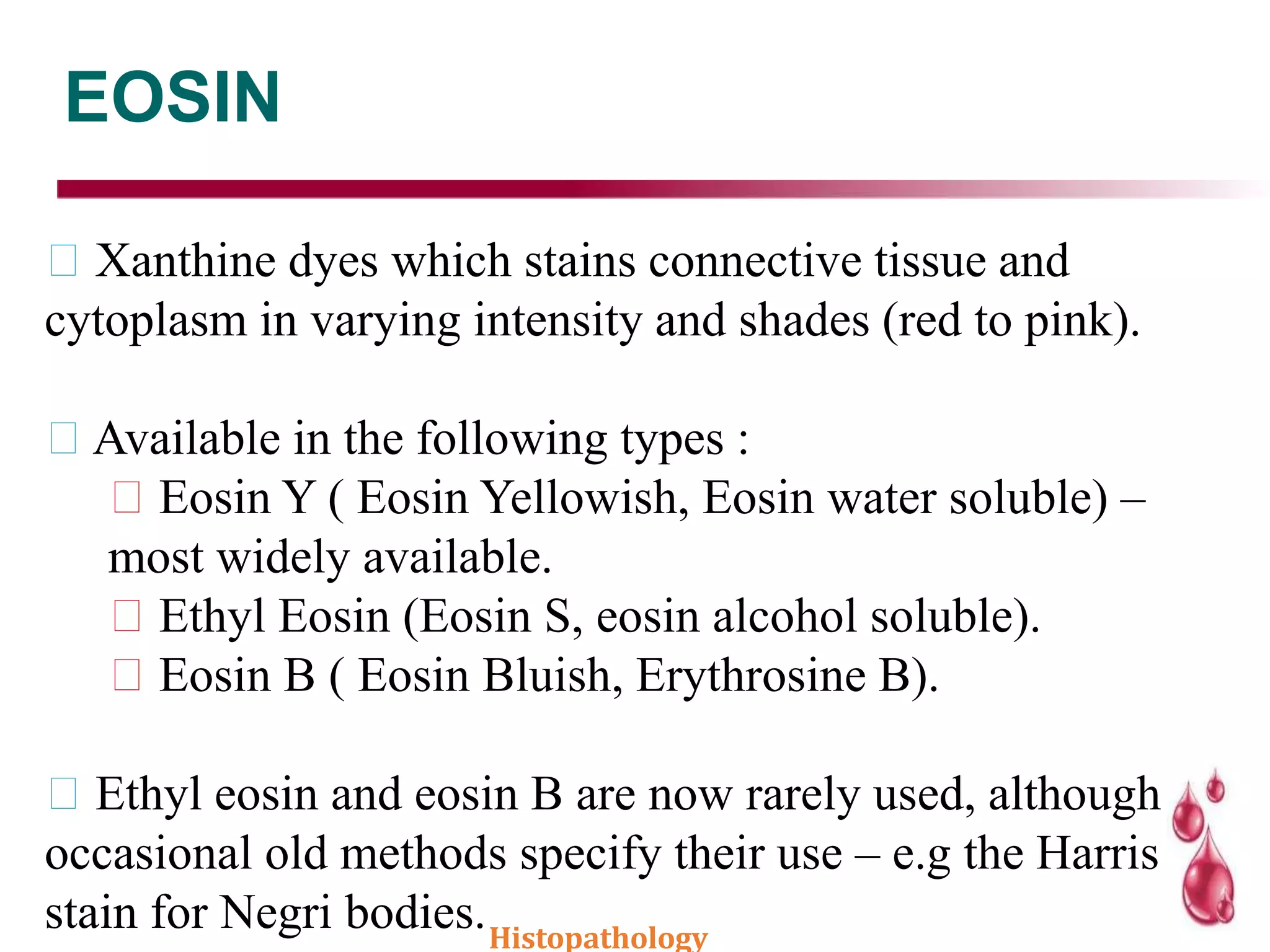
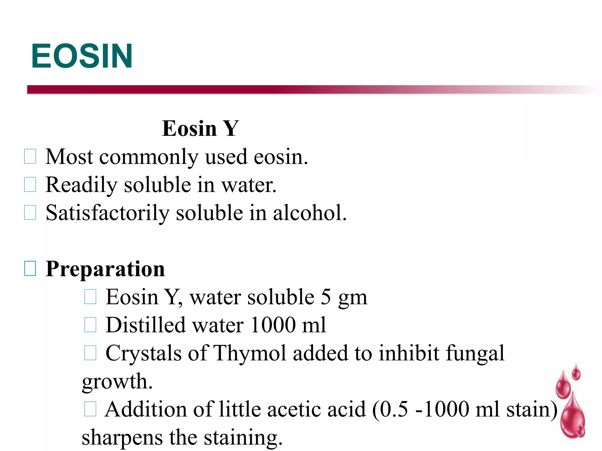
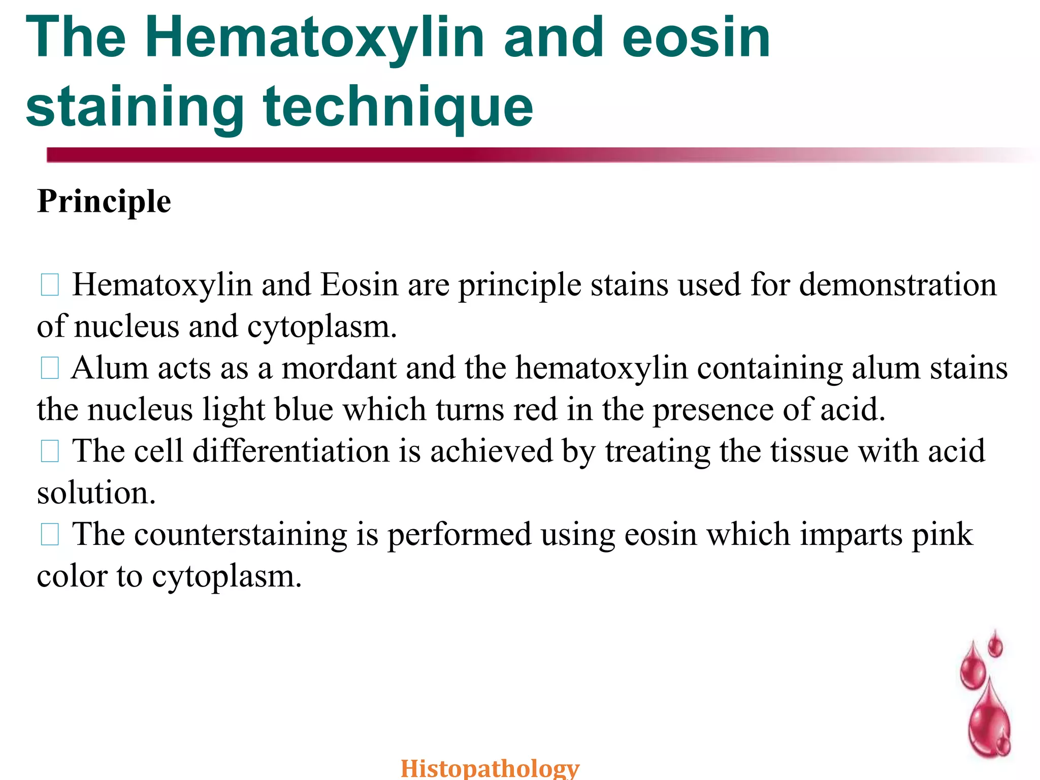
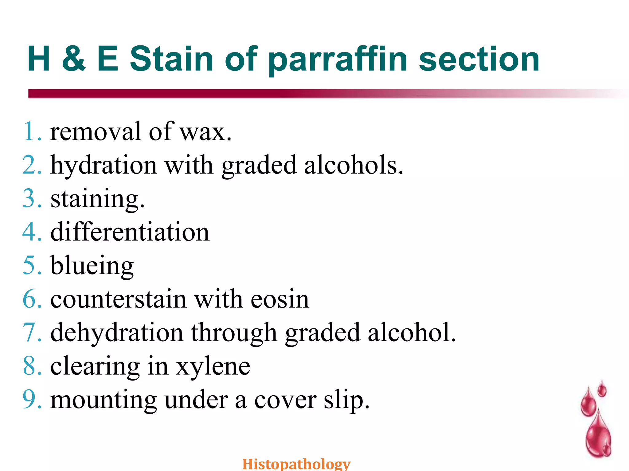
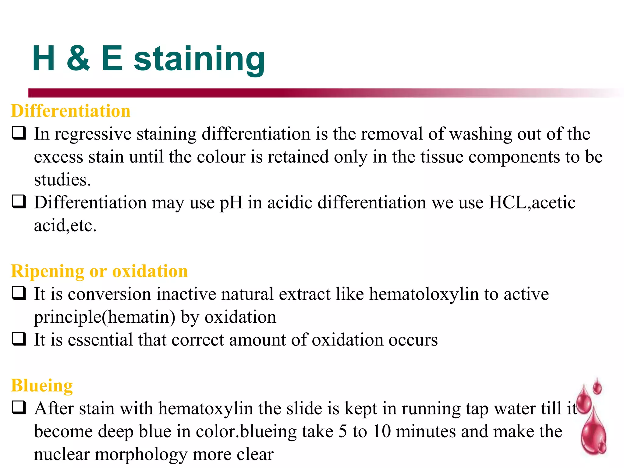
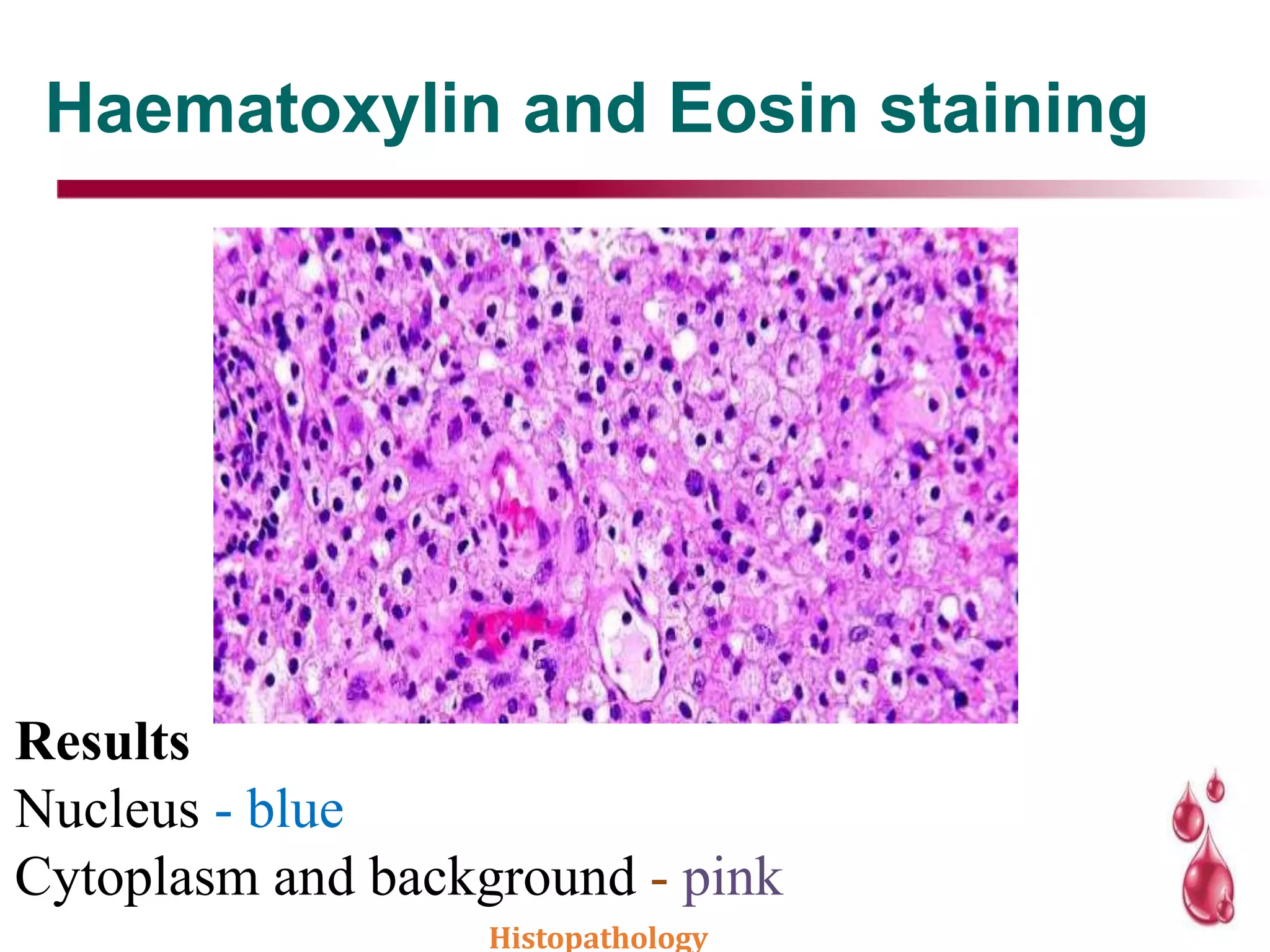
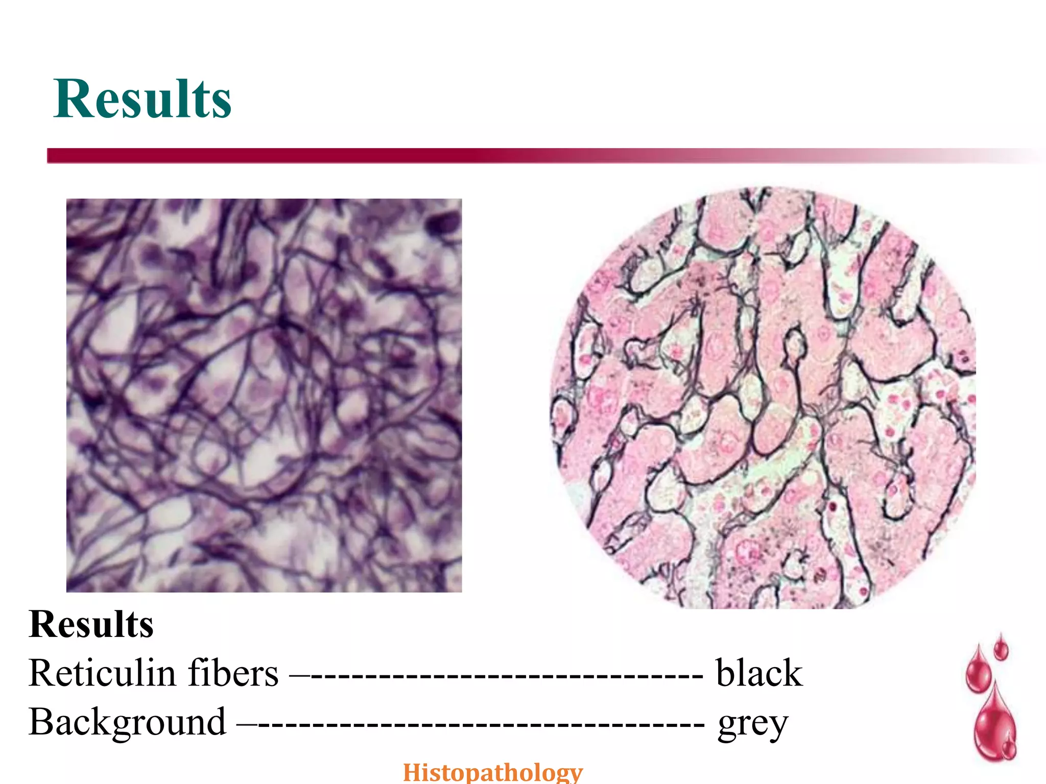
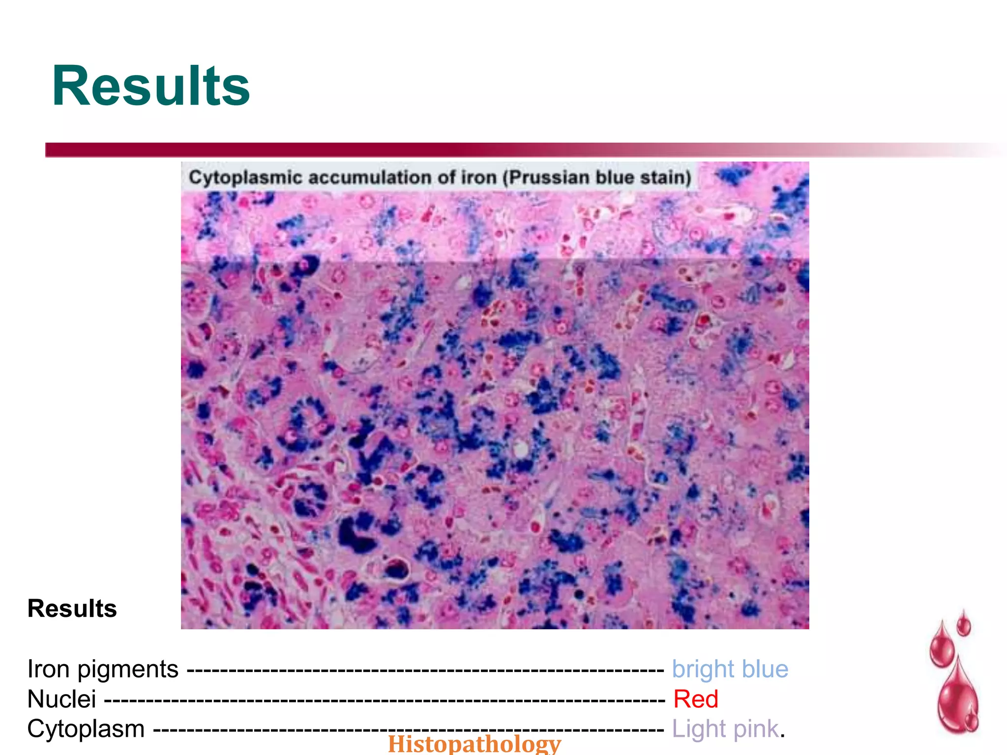
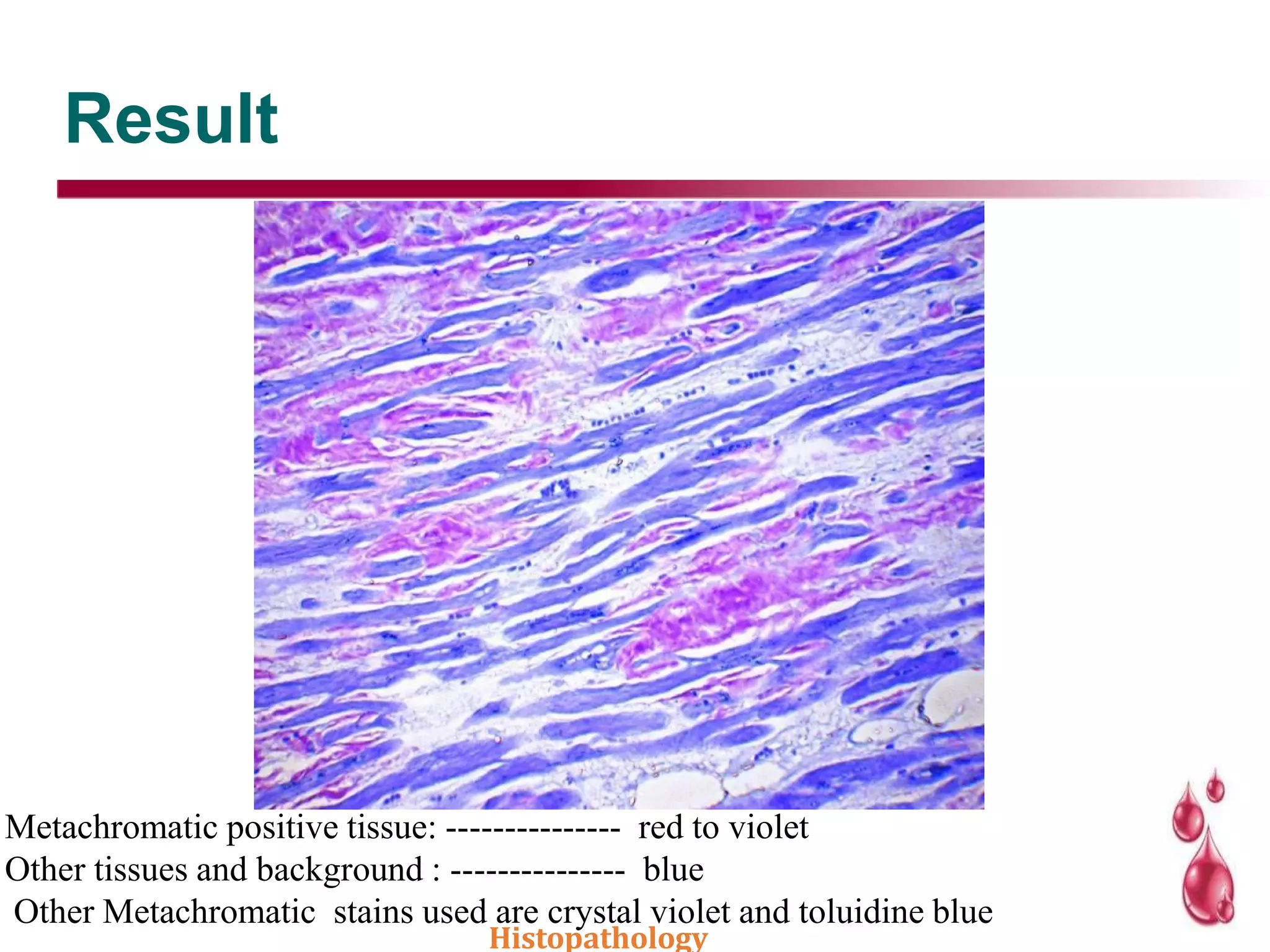
SlideShare a Scribd company logo
1 of 86
2
Histopathology
Definition
3
 Staining is used to highlight important features of the tissue as well as to
enhance the tissue contrast.
 Due to their transparent nature, the cellular and intracellular structure of
tissue samples can not be microscopically examined until they are colored
by dyes.
 Dyes used to stain tissue samples in the histology laboratory for
microscopic evaluation are called biological dyes or biological stains.
Biological dyes can be grouped into the following two categories:
 Natural: Dyes that are derived from natural resources. The most
important natural dye in the histopathology laboratory is hematoxylin.
 Artificial: Dyes that are derived through chemical reactions. Artificial
dyes greatly outnumber natural dyes.
STAINING
 The sections, as they are prepared, are colourless
and different components cannot be appreciated.
 Staining them by different coloured dyes, having
affinities of specific components of tissues, makes
identification and study of their morphology
possible.
 If unstained slide is observed under microscope
we can make out only nucleus and cell boundary.
 Staining is a technique used to highlight
important features of the tissue as well as to
enhance the tissue contrast.
Dyes used in staining
1.According to source
a. Natural –Haematoxylin, Carmine
b. Synthetic-Aniline base and derived from coal tar
2. Affinity to tissues
a. Acidophilic -These are anionic dyes and stain mainly
cytoplasm, eosinophilic granules
b. Basophilic - These are cationic dyes and stain nuclei,
basophili granules or bacteria
3. Chemical composition
a. Thiazines
b. Azo-dyes
c. Rosailins Histopathology
Dyes classification
Histopathology
Cont.’
Histopathology
Counter stains
 A counter stain is the application to the original stain,
usually nuclear, of one or more dyes that by contrast will
bring out difference between the various cells and
tissues.
 A heavy counterstain is to be avoided lest it mask the
nuclear stain. It can be done either by using dilute stain
or cutting down the staining time.
 Some counterstains which are acidic may lighten or
remove the nuclear stains.
Histopathology
Mordants
 Substance that causes certain staining reactions to take place by
forming a link between the tissue and the stain. The link is referred
as lake. Without it, dye is not capable of binding to and staining the
tissue.
e.g. Ammonium and Potassium alum for haematoxylin
 The mordant dye complex ‘lake’ combines with tissue to form
tissue-mordant-dye complex, which is insoluble.
Histopathology
Mordant-dyes application
Mordant is applied first, followed by the
dye.
e.g Heidenhain’s iron hematoxylin
Mordant and dye are mixed together and
then applied.
Commonly done in histo-technology
e.g Alum hematoxylin solutions
Dye applied first, followed by the mordant.
Hardly done in histo-technology.
Histopathology
Accentuators:
Accentuators:
Substances which increase the staining power of dye.
 They increase the intensity & selectivity of stain.
 These are substances that causes an increase in the selectively or in the staining
power of dye. Thus they lead to more intense staining.
e.g KOH in Lofflers methylene blue phenol in carbol fuschin &
carbol thionin.
Accelerators
Accentuators used in metallic impregnation technique for the
nervous system.
e.g chloral hydrate
Trapping agents
Agents which holds dye combination with tissue or bacteria .
e.g tannic acid/iodine
Histopathology
Types of stains reactions
Absorption or direct staining – tissue penetrated
by dye solution.
Indirect staining – using intermediate treatment
with mordant
Physical staining – simple solubility of dye in
element of tissue.
Chemical staining – formation of new substance
e.g. PAS
Adsorption phenomenon – accumulation on the
surface of the compound.
Histopathology
Types of stains methods
Vital
Routine
Special
Regressive
Progressive
Histopathology
Types of stains methods
Histopathology
Cont.’
Histopathology
Hematoxylin
Hematoxylin solutions can be arbitrarily
classified
according to which mordant is used:
Alum hematoxylins
Iron hematoxylins
Tungsten hematoxylins
Molybdenum hematoxylins
Lead hematoxylins
Hematoxylin without mordant.
Histopathology
History of hematoxylin
 Waldeyer firmly established the use of
hematoxylin in histology in 1862.
 Bohmer combined hematoxylin with
alum as a mordant and obtained more
specific staining in 1864.
 Heidenhan introduced his classical Iron-
Alum- Hematoxylin method used as a
standard technique in cytology.
 Ehrlich overcame the instability of
hematoxylin and alum by adding glacial
acteic acid and simultaneously
produced his formula for hematoxylin
used today. Histopathology
i. Alum Hematoxylin
Used routinely in the hematoxylin and eosin stain and produce
good nuclear staining.
The mordant is aluminum in the form of ‘potash alum -
aluminum potassium sulfate or ‘ammonium alum -aluminum
ammonium sulfate
Alum Hematoxylin can be used progressively or regressively.
Haematoxylin and eosin staining :is the most common staining
technique in the histopathology. This uses a combination of two
dyes, haematoxylin and eosin used for demonstration of nucleus
and cytoplasmic inclusions in clinical specimens.
Histopathology
Classification of Alum Hematoxylin
Ehrilch’s haematoxylin
Mayer’s haematoxylin
Harris’ haematoxylin
Gill’s haematoxylin
Cole’s haematoxylin
Delafield’s haematoxylin
Carazzi’s haematoxylin
Histopathology
Ehrilch’s haematoxylin ( 1886)
Naturally ripened strong alum hematoxylin.
Stains nuclei intensely and crisply - stained sections fade
much more slowly .
Stains mucin in salivary glands, cartilage and cement lines of
bones
Suitable for tissues subjected to acid decalcification.
Suitable for tissues that have been stored for a long period in
formalin fixatives which have gradually become acidic over the
storage period.
Suitable for Bouin’s fixed tissue.
not ideal for frozen sections.
Histopathology
Mayer’s haematoxylin (1903)
A widely used hematoxylin stain.
Chemically ripened with sodium iodate.
More vigorous in action than Ehrlich’s hematoxylin.
Used as both progressive and regressive stain.
Used as a nuclear counterstain in the demonstration of
glycogen (PAS,mucicarmine) in various enzyme histological
techniques.
Stain applied for a short period - 5-10 mins until nuclei
are stained and then blued without any differentiation which
might destroy/decolor the stained cytoplasmic components.
Histopathology
Harris’ haematoxylin (1900)
This alum hematoxylin was traditionally chemically ripened
with mercuric oxide (sodium or potassium iodate is frequently
used as a substitute for oxidation).
It gives particular clear nuclear staining.
It is used as a regressive stain in routine histology practice.
It is used as a progressive stain in diagnostic exfoliative
cytology.
When using it as a progressive stain, an acetic acid-alcohol
rinse provides a more controllable method in removing excess
stain from tissue components and the glass slide.
Histopathology
Gill’s haematoxylin (1974)
Available in 3 concentrations –
◦ Gill’s I (normal)
◦ Gill’s II (double)
◦ Gill’s III (triple) most concentrated.
More frequently used than Mayer’s hematoxylin for
routine H&E staining.
More stable than Harris’s hematoxylin, as auto-
oxidation is inhibited to the extent.
Histopathology
Cole’s haematoxylin (1943)
Alum hematoxylin, artificially ripened with an alcoholic
iodine solution.
Has good keeping qualities and is suitable for use
especially in sequence with celestine blue unlike Ehrlich’s
hematoxylin
Preparation
Hematoxylin 1.5 g
Saturated aqueous potassium alum 700 ml
1% iodine in 95% alcohol 50 ml
Distilled water 250 ml
Histopathology
Carazzi’s haematoxylin (1911)
Alum hematoxylin which is chemically ripened using potassium
iodate
Used as a progressive nuclear counterstain.
Largely confined to use with frozen sections because it gives
excellent and clear nuclear staining with a very short staining time.
Preparation –
Hematoxylin 5 g
Glycerol 100 ml
Potassium alum 25 g
Distilled water 400 ml
Potassium iodate 0.1 g
Histopathology
A naturally ripened alum hematoxylin with
similar longevity to
Ehrlich’s hematoxylin.
Preparation
Hematoxylin 4 g
95% alcohol 125 ml
Saturated aqueous ammonium alum 400
ml (15 g/100 ml)
Glycerin 100 ml
Delafield’s haematoxylin (1885)
Histopathology
ii. Iron Hematoxylin
Weigert’s Hematoxylin.
Heidenhain’s Hematoxylin.
Verhoeff’s Hematoxylin.
Loyez Hematoxylin.
Histopathology
Weigert’s Hematoxylin
An iron hematoxylin used as a nuclear stain in techniques
where acidic staining solutions are applied to the sections
subsequently
e.g Van Gieson stain – picric acid is a constituent which have
marked decolorizing action on nuclei stained with alum
hematoxylin.
It is a useful stain, with eosin, for CNS tissues.
Preparation
a) Hematoxylin solution
Hematoxylin 1 g
Absolute alcohol 100 ml
This is allowed to ripen naturally for 4 weeks before use.
b) Iron solution
30% aqueous ferric chloride (anhydrous) 4 ml
Hydrochloric acid (concentrated) 1 ml
Distilled water 95 ml
Heidenhain’s Hematoxylin.
This iron hematoxylin uses ferric ammonium sulfate as oxidant/mordant
It is used as the differentiating fluid.
It is a cytological stain.
It is used regressively.
After staining, all components are black or dark gray-black.
The hematoxylin staining is removed progressively from different tissue
structures at different rates using the iron alum solution.
It may be used to demonstrate
Chromatin
Chromosomes
Nuclei
Centrosomes
Mitochondria
Muscle striations
Myelin
Loyez Hematoxylin.
This iron hematoxylin uses ferric ammonium sulfate as
the mordant.
Differentiation is by Weigert’s differentiator (borax and
potassium ferricyanide).
Used to demonstrate myelin.
Can be applied to paraffin, frozen, or nitrocellulose
sections.
Histopathology
This iron hematoxylin is used to demonstrate elastic
fibers after all routine fixative.
Ferric chloride is included in the hematoxylin staining
solution, together with Lugol’s iodine, and 2% aqueous
ferric chloride is used as the differentiator.
Coarse elastic fibres stain black, but the staining of fine
fibers may be less than satisfactory.
The differentiation step is critical to the success of this
method.
Verhoeff’s Hematoxylin.
Histopathology
iii. Tungsten hematoxylins
 Widely used tungsten hematoxylin is PTAH (Phosphotungstic
acid hematoxylin Technique).
 Used to demonstrate fibrin, muscle striations, cilia and glial
fibres.
 Myelin can also be demonstrated Widely used as a CNS stain.
Preparation
PTAH solution using haematin
Haematin 0.59 g
Phosphotungstic acid 5g
Distilled water 500ml
Stain is ready to use immediately, but short-lived.
PTAH Solution (KMnO4)
Haematoxylin 0.59 g
Phosphotungstic acid 5g
Distilled water 500ml
0.25% Aqueous KMnO4 25 ml
Peak staining activity after 7 days
Tungsten hematoxylins
Results
• Muscle striations / neuroglia fibres / fibrin /
amoeba – ---------------------Dark Blue.
• Nuclei/cilia/RBC – ---------Blue
• Myelin – ---------------------Lighter blue
• Collagen /Osteoid / Cartilage / Elastic fibres – ---
---------------------------------Deep brownish red.
Cytoplasm – --------------------Pale pinkish brown.
Histopathology
iv. Molybdenum hematoxylins
Hematoxylin solution using molybdic acid as
mordant.
Rare stain
Used in demonstration of collagen, coarse reticulin.
Also stains Argentaffin cell granules.
Preparation
a. Hematoxylin solution
Hematoxylin 2.5 g
Dioxane 49 ml
Hydrogen peroxide 1 ml
b. Phosphomolybdic acid solution
Phosphomolybdic acid 16.5 g
Distilled water 44 ml
Diethylene glycol 11 ml
The resultant dark violet solution is allowed to stand for 24 hours before use.
Histopathology
Molybdenum hematoxylins
Results –
Collagen and coarse reticulin - violet to
black
Argentaffin cells - black
Nuclei pale - blue
Paneth cells - orange
Tissue fixed in dichromate do not give good
results.
Histopathology
v. Lead hematoxylin
Used in demonstration of granules in endocrine cells of ailmentary
tract and other regions.
Most practical diagnostic application is in identification of
endocrine cells in tumors of doubtful origin.
Also used in localization of gastrin secreting cells in stomach.
Histopathology
EOSIN
Xanthine dyes which stains connective tissue and
cytoplasm in varying intensity and shades (red to pink).
Available in the following types :
Eosin Y ( Eosin Yellowish, Eosin water soluble) –
most widely available.
Ethyl Eosin (Eosin S, eosin alcohol soluble).
Eosin B ( Eosin Bluish, Erythrosine B).
Ethyl eosin and eosin B are now rarely used, although
occasional old methods specify their use – e.g the Harris
stain for Negri bodies.Histopathology
EOSIN
Eosin Y
Most commonly used eosin.
Readily soluble in water.
Satisfactorily soluble in alcohol.
Preparation
Eosin Y, water soluble 5 gm
Distilled water 1000 ml
Crystals of Thymol added to inhibit fungal
growth.
Addition of little acetic acid (0.5 -1000 ml stain)
sharpens the staining.
The Hematoxylin and eosin
staining technique
Principle
Hematoxylin and Eosin are principle stains used for demonstration
of nucleus and cytoplasm.
Alum acts as a mordant and the hematoxylin containing alum stains
the nucleus light blue which turns red in the presence of acid.
The cell differentiation is achieved by treating the tissue with acid
solution.
The counterstaining is performed using eosin which imparts pink
color to cytoplasm.
Histopathology
H & E Stain of parraffin section
1. removal of wax.
2. hydration with graded alcohols.
3. staining.
4. differentiation
5. blueing
6. counterstain with eosin
7. dehydration through graded alcohol.
8. clearing in xylene
9. mounting under a cover slip.
Histopathology
H & E staining
Differentiation
 In regressive staining differentiation is the removal of washing out of the
excess stain until the colour is retained only in the tissue components to be
studies.
 Differentiation may use pH in acidic differentiation we use HCL,acetic
acid,etc.
Ripening or oxidation
 It is conversion inactive natural extract like hematoloxylin to active
principle(hematin) by oxidation
 It is essential that correct amount of oxidation occurs
Blueing
 After stain with hematoxylin the slide is kept in running tap water till it
become deep blue in color.blueing take 5 to 10 minutes and make the
nuclear morphology more clear
Haematoxylin and Eosin
staining
Procedure
♦ Deparaffinize in hot air oven.
♦ Hydrate the section.
i) 3 dips in xylene (2 Min. each)
ii) 3 dips in acetone / alcohol (2 Min. each)
iii) In running tap water for 5 Minutes.
♦ Mayer's haemotoxylin for 15 minutes.
♦ Wash in running tap water for 20 minutes
♦ Counter stain with eosin for 2 minutes
♦ Dehydrate the section in 95% and absolute alcohol/ acetone
2 changes (2minutes each).
♦ Clear in xylene 3 changes (2 minutes each)
♦ Mount in DPX
Histopathology
Haematoxylin and Eosin staining
Results
Nucleus - blue
Cytoplasm and background - pink
Histopathology
4
4
SPECIAL STAININGS
Histopathology
1.Van gieson method
This stain is used for staining of collagen fibres
Principle :
 It is based on the differential staining of collagen and other tissues
(e.g muscle) depending upon the porosity of tissue and the size of
the dye molecule.
 In the routine staining method collagen, elastic fibres and smooth
muscle appear pink or reddish in colour.
 In the Van Gieson stain, collagen and most reticulin stain selectively
with acid aniline dyes (acid fuchsin).
 Picric acid acts as counter stain for muscle and cytoplasm and form
complex with the dyes. This complex has special affinity for
collagen.
Histopathology
Cont.’
Reagents
1. Solution A
(a) Haematoxylin 1.0 gm
(b) Alcohol 95% 100 ml
2. Solution B
(a) 29% (w/v) ferric chloride 4 ml
(b) Conc. Hydrochloric acid 1.0 ml
(c) Distilled water 95.0 ml
3. Weight's iron heamatoxylin solution :mix equal quantities of solution
A and solution B. Colour of this reagent should appear violet black.
4. Van Gieson’s solution
(a) Saturated aqueous picric acid – 10 ml
(b) 1% (m/v) acid fuchsin – 1.5ml
It should be freshly prepared
Histopathology
Cont.’
Procedure
(1) Deparaffinize with xylene
(2) Hydration take sections to water
(3) Stain with Weigert’s haematoxylin for 20-40
minutes
(4) Wash in distilled water
(5) Van Gieson stain 1-3 min
(6) Rinse well in distilled water
(7) Dehydrate in absolute alcohol (2 changes)
(8) Clear in xylene (2 changes)
(9) Mount in DPX
Histopathology
Results
Results
1. Collagen --------------------------------Red
2. Muscle and
Cornified epithelium -----------------Yellow
3. Nuclei -----------------------------------Blue to Black
Clinical significance
 Several pathological changes are associated with
cellular changes in connective tissues.
 Histological diagnosis of collagen diseases is
based on the study of the section of various
connective tissues.
Histopathology
2.Gomori’s method for reticulum
This is used for demonstration of reticulin
Principle :
 In the connective tissue, reticulin appears as a fibrillary extracellular framework.
 Reticular fibres have low natural affinity for silver salts and require pretreatment
with heavy metal solutions like fernic ammonium sulphate to enhance the
selectivity of impregnation Silver in alkaline solution is in a state readily able to
precipitate as metallic silver.
 Upon treatment with a reducing agent, silver taken up by the tissue in unreduced
form is, converted to metallic silver which is deposited at the sensitised site.
Histopathology
Cont.’
Reagents
1. Ammonical silver solution
To 10 ml of 10% silver nitrate solution add 2.5 ml of 10% aqueous
solution of potassium hydroxide, add 28% ammonium hydroxide drop
by drop while shaking the container continuously until the precipitate
in completely dissolved. Add again 4 drops of silver nitrate solution for
every 10 ml of silver nitrate used. Make the solution with distilled
water to twice its volume use acid clean glassware
2. 0.5% potassium permanganate –
Potassium permanganate –0.5 gm
Distilled water 100 ml
Histopathology
Cont.’
3. 2% potassium metabisulphite
Potassium metabisulphate 2.0 gm
Distilled water 100 ml
4. 2% ferric ammonium sulfate solution
Ferric ammonium sulphate 2 gm
Distilled water 100 ml
5. 20% formation solution
Formaldehyde 20 ml
Distilled water 80 ml
6. 0.2% gold chloride solution
Gold chloride solution 1% - 10 ml
Distilled water - 40 ml
7. 2% sodium thiosulphate solution
sodium thiosulphate 2 gm
Distilled water 100 ml
Histopathology
Cont.’
Procedure
1. Deparaffinze and hydrate to distilled water
2. Oxidize in potassium permagnate solution for 1 minute
3. Rinse well in tap water – 2 min
4. Differentiate with potassium metabisulphite solution for 1 minute.
5. Wash in tap water for 2 min
6. Sensitize in Ferric ammonium sulphate solution for 1 min.
7. Wash in tap water for 2 minutes follow with two changes of distilled water 30 seconds 8.
Impregnate in the silver solution for 1 minute
9. Rinse in distilled water for 20 seconds
10. Reduce in formalin solution for 3 minutes
11. Wash in tap water for 3 minutes
12. Tone in gold chloride solution for 10 minutes
13. Rinse in distilled water
14. Reduce in potassium metabisulfite solution for 1 minute
15. Fix in sodium thiosulfate solution for 1 minute.
16. Wash in tap water for 2 minutes.
17. Dehydrate in 95% alcohol, absolute alcohol and clear in xylene 2 changes
18. Mount in DPX.
Histopathology
Results
Results
Reticulin fibers –----------------------------- black
Background –--------------------------------- grey
Histopathology
Clinical significance
 This type of staining is used for tumors of
uncertain origin.
 The pattern of deposition of reticulin in a tumor
is characteristic to identify the origin of tumor.
 Mild degree of fibrosis in organs can also be
recognized by reticulin stain especially in liver to
assess cirrhosis.
Histopathology
3.McManus for glycogen (pas)
 This stain is used for demonstration of glycogen and muco
polysaccharides.
 PAS positive substances are glycogen, amyloid, colloid,
neutral mucin, basement membranes and hyaline cast
Principle :
 Tissue structures like liver & heart, striated muscles are
studied by Periodic acid Shiff stain.
 Periodic acid reacts with aldehyde group of the carbohydrates
and afterwards reaction with the schiff’s reagent produces a
red or purple red colour.
Histopathology
Reagents
Reagents
1. 0.5% w/v periodic acid solution
2. Schiff’s reagent
(a) Dissolve 1.0 gm of basic fuchsin in ≈100 ml of boiling
distilled
water cool to about 60 degrees and filter.
(b) Add 20 ml of 0.1 N hydrochloric acid, cool further and add
1.0 gm of sodium metabisulphite and mix well.
(c) Keep in the dark for 24-48 hours. When the solution
becomes
straw coloured, add 300 mg of activated charcoal, shake
vigorously, filter and store.
3. 1 N Hydrochloric acid
4. 0.1 gm of light green in 100 ml of 0.1% (v/v) acetic acid.
5. Harris haematoxylin stain Histopathology
Procedure
Procedure
1. Deparaffinize and hydrate to distilled water.
2. Oxidize in periodic acid solution for 5 minutes
3. Rinse in distilled water
4. Schiff’s regent solution for 15 minutes.
5. Wash in running water for 10 minutes for pink colour to develop.
6. Harris haematoxylin for 6 minutes or light green counter stain for a few
seconds.
7. Wash in running water.
8. Differentiate in 1% acid alcohol solution 3-10 quick dips.
9. Wash in running water.
10. Dip in ammonia water to blue the sections
11. Wash in running water for 10 minutes
12. Dehydrate in 95% alcohol, absolute alcohol, clear in xylene two changes
each.
13. Mount in DPX. Histopathology
Results
Results
With hematoxylin counterstain
1. Nuclei --------------------------------------blue
2. Glycogen, mucin, hyaluronic acid,
reticulin, colloid droplets, amyloid
infiltration, thrombi. –------------------- purple red
3. Fungi – -------------------------------------Red
4. Background –------------------------------ pale green (with light green counter staining).
Histopathology
4.Mayer’s mucicarmine method
To demonstrate mucin in a tumor or epithelium
Principle :
 The rationale for specificity of mucicarmine for mucin is not fully
understood by the probable mechanism is that the aluminium salts in
the solution form a chelate compound with carmine, thus producing
a net positive charge on the molecule and consequent binding to the
tissue polyanions.
 The compound has a large molecular size and allows that dye
complex to penetrate and bind to acidic substrates of low density
like mucins other acidic substances like nucleic acid are of high
density hence exclude the mucicarmine.
Histopathology
Reagents
Reagents
(1) Weigert’s Iron haematoxylin solution
(a) Solution A
Hematoxylin crystals 1.0 gm
Alcohol 95% 100 ml
(b) Solutions B
Ferric chloride 29% aqueous 4 ml
Hydrochloric acid conc. 1 ml
Distilled water 95 ML
Working solution – Add equal parts of solution A and solution B
(2) Mucicarmine solution
Carmine 1 gm
Aluminium chloride anhydrous 0.5 gm
Distilled water 2 ml
(3) 0.25% Metanil yellow solution
Melanil Yellow - 0.25 gm
Distilled water - 100 ml
Glacial acetic acid - 0.25 ml
Histopathology
Procedure
1. Deparaffinize and hydrate to distilled water.
2. Working solution of Weigert’s Haematoxylin for 7
minutes.
3. Wash in running water for 10 minutes
4. Diluted mucicarmine solution for 60 minutes
5. Rinse quickly in distilled water.
6. Metanil yellow solution for 1 minute.
7. Rinse quickly in distilled water
8. Dehydrate in 95% alcohol, absolute alcohol and clear in
xylene 2 changes each.
9. Mount with DPX.
Histopathology
Results
Results
Mucin --------------------------------------------------------------- Deep rose to red.
Capsule of cryptcoccus----------------------------------------- deep rose to red.
Nuclei --------------------------------------------------------------- Black
Other tissue elements ------------------------------------------ yellow
5.FITE’s method for acid fast
organisms
to demonstrate weakly acid fast bacilli like mycobacterium Lepre and
Nocardia.
Principle:
 The mycobacteria are the bacteria which are relatively resistant to
staining because of the lipid capsule which surrounds them, when
stained by a strong stain (eg carbol fuchsin) they resist
decolourization by acid.
 In this method a red dye carbol fuchsin is forced into the bacteria
and other structures with heat and is then removed from other
structures with acid or alcohol ; tubercle bacillus because of lipid
capsule however resists decolourization.
Procedure
Procedure
1. Deparaffinize through 2 changes of xylene peanut oil solution for 12
minutes each.
2. Drain wipe off excess oil & blot to opacity.
3. Carbol fuchsin solution for 30 minute
4. Wash in tap water for 3 minutes & blot dry.
5. Differentiate slides with sulfuric acid solution till sections are faint
pink for 1-2 minutes.
6. Wash in running water for 3 minutes
7. Counterstain lightly with working methylene blue solution (few
seconds).
8. Rinse off excess methylene blue in tap water.
9. Blot and let it stand for few minutes to air dry thoroughly.
10. Dip slides in xylene before mounting.
11. Mount with DPX
Results
Results
M. Leprae ------------------------------------------------------- red
Nocardia filaments -------------------------------------------- blue grey
Histopathology
6.Gomori's method for iron
Staining of section for haemosiderin (a tissue
pigment)
Principle:
 Haemosiderin is a brown granular pigment
occurring at the site of previous haemorrhage.
 It is a product of the breakdown of haemoglobin.
 It reacts with potassium ferrocyanide in acid
medium and yields a Prussian blue colour.
Reagents
1. Solution A - 20% hydrocholroc acid solution (stock)
Hydrochloric acid (conc.) 20ml
Distilled water 80ml
2. Solution B - 10% potassium ferrocyanide solution (stock)
Potassium ferrocyanide 10 gm
Distilled water 100ml
3. Acidified potassium ferrocyanide solution.
Prepare fresh by mixing equal part of solution A and solution B and
leave for 20 minutes
4. Nuclear fast red stain.
Dissolve 5.0 gm of aluminium sulphate is hot distilled water and add
0.1 gm of nuclear fast red mix well and filter.
Add a crystal of thymol as a preservative.
Histopathology
Procedure
Procedure
1. Deparaffinize and hydrate to distilled water
2. Put the slides in acidified potassium ferrocyanide
solution for 30 minutes
3. Rinse in distilled water
4. Counterstain in nuclear fast red solution for 5 minutes.
5. Rinse in distilled water
6. Dehydrate in 95% alcohol, absolute alcohol, and clear in
xylene 2 changes each
7. Mount with DPX
Histopathology
Results
Results
Iron pigments --------------------------------------------------------- bright blue
Nuclei ------------------------------------------------------------------- Red
Cytoplasm ------------------------------------------------------------- Light pink.
Histopathology
7.Von kossa's method for
calcium
Staining of the sections for calcium
Principle –
 The calcium salts in the form of phosphates, carbonates
or oxalates occur as components of oxalates occur as
components of laminated concentration in various
organs such as the kidneys, urinary bladder, lymph
nodes.
 Von Kossa's technique in a metal substituted for calcium
by metallic salt formation with the anion of the calcium
salt.
Histopathology
Reagent
Reagent :
1. 5% W/v silver nitrate solution (stored in amber coloured
bottle)
2. 5% W/v sodium thosulphate solution
3. Nuclear fast red stain. Dissolve 0.1 gm nuclear fast red
no 100 ml of
5% solution of aluminium sulphate with the aid of heat.
Cool filter add grain of thymol as a preservative.
Histopathology
Procedure
Procedure: (Use control slides and chemically clean glassware)
1. Deparaffinize and hydrate to distilled water
2. Silver nitrate solution expose to bright sunlight or under the light
of 100 watt bulb for 60 minutes
3. Rinse in Distilled water
4. 5% sodium thiosulfate solution for 2 minutes
5. Rinse well in distilled water
6. Counter stain with nuclear fast led solution for 5 minutes
7. Rinse in distilled water
8. Dehydrate in 95% alcohol, absolute alcohol and clear in xylene 2
changes each
9. Mount with DPX
Histopathology
Results
Results
Calcium salt ------------------------------------------------------ Black
Nuclei -------------------------------------------------------------- Red
Cytoplasm -------------------------------------------------------- Light pink
Histopathology
8.Hall's method for bilirubin
To stain the bilirubin or bile pigment in tissue section
Principle
- Bilirubin is oxidized to biliverdin and stains olive green to
emerald green depending on the concentration of bilirubin in
solution.
Histopathology
Reagents
Reagents
1. Fouchet's Reagent
Trichloroacetic acid --------------------------------------------------- 25.0gm
Distilled water ------------------------------------------------------------100ml
Mix and add 10 % ferric chloride
2. 10% Ferric chloride solution
Ferric chloride --------------------------------------------------------- 10gm
Distilled water --------------------------------------------------------- 100ml
3. Van Gieson's solution
(a) Saturated aqueous picric acid ---------------------------------10ml
(b) 1% w/v acid fuchsin -----------------------------------------------1.5ml
Histopathology
Procedure
Procedure
1. Deparaffinize and hydrate to distilled water
2. Fouchet's reagent for 5 minutes
3. Wash in running water, then in distilled water
4. Van Gieson's solution for 5 minutes
5. Dehydrate in 95% alcohol, absolute alcohol and
clear in xylene 2 changes each
6. Mount with DPX
Histopathology
Results
Results
Biliverdin ------------------------------------------------green
Collagen -------------------------------------------------red
Muscle ---------------------------------------------------YellowHistopathology
9. Sudan black/ oil red O stain
Both these stains are used for demonstration of fat
Principle:
 Both SB and oil red O staining are based on physical
combination of the stain with fat. It involves
differential solubility of stain in fat because these
stains are more soluble in fat than the solvent in which
these are prepared.
 The stain leaves the solvent and goes into the fatHistopathology
Result
Result:
Oil red O
fat -------------------------------------------------------------- bright red
nuclei ---------------------------------------------------------- blue
Sudan black
fat ------------------------------------------------------------- black
nuclei --------------------------------------------------------- red
Histopathology
10.Methyl violet stain
 This is a metachromatic stain i.e. the tissues are stained in a colour
which is different from the colour of the stain itself.
 It is used for demonstration of amyloid in tissue. Other
metachromatic positive substances are mucin and hyaline
Principle:
 This depends upon the type of dye(stain) used and character of the
tissue which unites with dye
 Tissues containing S04,PO4,or COOH groups react with basis dyes
and cause their polymerisation which in turn leads to production of
colour different from the original dye
Histopathology
Result
Metachromatic positive tissue: --------------- red to violet
Other tissues and background : --------------- blue
Other Metachromatic stains used are crystal violet and toluidine blue
Histopathology
Knowledge checking
1) Which are the special stains used for
demonstration of fat in tissue sections?
2) Van Gieson stain is used for which purpose and
what is a positive colour?
3) What is meant by silver impregnation?
4) What are the applications of PAS stain?
5) What are the main diagnostic stains used for
demonstration of amyloid?
6) What is the principle of prussian blue stain?
Histopathology
Question?
Histopathology
References
1. Bancroft, J.D. and Stevens, A.: theory and practice of histological
techniques ed.3, Churchill livingstone inc. 1990. Edinburgh.
London, Melbourne and New York.
2. Lillie, R.D.: Histopathologic technique and practice histo-
chemistry ed. 3, New York, 1965 McGraw Hill Book co.
3. Manual of histologic and special staining techniques ed. 2, New
York, 1960, The Blakiston Division McGraw Hill Book Co.
Histopathology
BOOKS

More Related Content

What's hot

Hematoxylin and Eosin Staining (H& E Staining)
Hematoxylin and Eosin Staining (H& E Staining)Hematoxylin and Eosin Staining (H& E Staining)
Hematoxylin and Eosin Staining (H& E Staining)SUNIL KUMAR PEDDANA
 
Theory of staining
Theory of stainingTheory of staining
Theory of stainingishita1994
 
Special stains by sowmya
Special stains by sowmyaSpecial stains by sowmya
Special stains by sowmyaSowmya Srinivas
 
General 1 fixatives cytology
General  1 fixatives cytologyGeneral  1 fixatives cytology
General 1 fixatives cytologyNem Shrestha
 
Histotechnique
HistotechniqueHistotechnique
HistotechniqueDrSyed Asif
 
Histopathology staining method
Histopathology staining methodHistopathology staining method
Histopathology staining methodD. JASMINE PRIYA
 
Staining by hematoxylin and eosin
Staining by hematoxylin and eosinStaining by hematoxylin and eosin
Staining by hematoxylin and eosinSowmya Srinivas
 
Fixation & fixatives in histopathology, dr naveen reddy
Fixation & fixatives in histopathology, dr naveen reddyFixation & fixatives in histopathology, dr naveen reddy
Fixation & fixatives in histopathology, dr naveen reddyNaveen Parvathareddy
 
Demonstration of protein and nucleic acids
Demonstration of protein and nucleic acidsDemonstration of protein and nucleic acids
Demonstration of protein and nucleic acidsMAHMOUD IBRAHIM
 
Perl's stain
Perl's stainPerl's stain
Perl's stainmanoj pant
 
Histopatholgy staining by suchit kumar
Histopatholgy staining by suchit kumarHistopatholgy staining by suchit kumar
Histopatholgy staining by suchit kumarsuchitkumar24
 
Frozen section and cryostat
Frozen section and cryostatFrozen section and cryostat
Frozen section and cryostatMAHMOUD IBRAHIM
 
Fixatives in Histopathology
Fixatives in HistopathologyFixatives in Histopathology
Fixatives in HistopathologyIshwar9
 

What's hot (20)

Theories of Staining
Theories of StainingTheories of Staining
Theories of Staining
 
Hematoxylin and Eosin Staining (H& E Staining)
Hematoxylin and Eosin Staining (H& E Staining)Hematoxylin and Eosin Staining (H& E Staining)
Hematoxylin and Eosin Staining (H& E Staining)
 
Theory of staining
Theory of stainingTheory of staining
Theory of staining
 
Special stains by sowmya
Special stains by sowmyaSpecial stains by sowmya
Special stains by sowmya
 
Decalcificaion
DecalcificaionDecalcificaion
Decalcificaion
 
Connective Tissue
Connective TissueConnective Tissue
Connective Tissue
 
H & E staining part 1
H & E staining part 1H & E staining part 1
H & E staining part 1
 
Cytochemistry i
Cytochemistry iCytochemistry i
Cytochemistry i
 
General 1 fixatives cytology
General  1 fixatives cytologyGeneral  1 fixatives cytology
General 1 fixatives cytology
 
Histotechnique
HistotechniqueHistotechnique
Histotechnique
 
Histology
HistologyHistology
Histology
 
Histopathology staining method
Histopathology staining methodHistopathology staining method
Histopathology staining method
 
Staining by hematoxylin and eosin
Staining by hematoxylin and eosinStaining by hematoxylin and eosin
Staining by hematoxylin and eosin
 
Fixation & fixatives in histopathology, dr naveen reddy
Fixation & fixatives in histopathology, dr naveen reddyFixation & fixatives in histopathology, dr naveen reddy
Fixation & fixatives in histopathology, dr naveen reddy
 
Demonstration of protein and nucleic acids
Demonstration of protein and nucleic acidsDemonstration of protein and nucleic acids
Demonstration of protein and nucleic acids
 
Perl's stain
Perl's stainPerl's stain
Perl's stain
 
Histopatholgy staining by suchit kumar
Histopatholgy staining by suchit kumarHistopatholgy staining by suchit kumar
Histopatholgy staining by suchit kumar
 
Frozen section and cryostat
Frozen section and cryostatFrozen section and cryostat
Frozen section and cryostat
 
Fixatives in Histopathology
Fixatives in HistopathologyFixatives in Histopathology
Fixatives in Histopathology
 
Lecture 2. tissue processing
Lecture 2. tissue processingLecture 2. tissue processing
Lecture 2. tissue processing
 

Similar to Histopathology Staining Techniques Explained

routine staining of tissues.pptx
routine staining of tissues.pptxroutine staining of tissues.pptx
routine staining of tissues.pptxAbdulRashidAdams
 
Hematoxylin and Eosin Staining
Hematoxylin and Eosin StainingHematoxylin and Eosin Staining
Hematoxylin and Eosin StainingDr. Varughese George
 
Dental Anatomy and Dental Histology Project on Hematoxylin & Eosin Stain - 1S...
Dental Anatomy and Dental Histology Project on Hematoxylin & Eosin Stain - 1S...Dental Anatomy and Dental Histology Project on Hematoxylin & Eosin Stain - 1S...
Dental Anatomy and Dental Histology Project on Hematoxylin & Eosin Stain - 1S...deepupadhyaya
 
Hematoxylin and Eosin
Hematoxylin and EosinHematoxylin and Eosin
Hematoxylin and EosinMAHMOUD IBRAHIM
 
H & e staining- part 1 & 2
H & e staining- part 1 & 2H & e staining- part 1 & 2
H & e staining- part 1 & 2Sonal Aggarwal
 
Hematoxylin & eosin stain
Hematoxylin & eosin stainHematoxylin & eosin stain
Hematoxylin & eosin stainUmair Raza
 
hematoxylin and eosin stains
hematoxylin and eosin stainshematoxylin and eosin stains
hematoxylin and eosin stainsUpama Sishan
 
4_5839287714297417902.ppt
4_5839287714297417902.ppt4_5839287714297417902.ppt
4_5839287714297417902.pptOfkalaTariku
 
Hematoxylin and eosin staining
Hematoxylin and eosin stainingHematoxylin and eosin staining
Hematoxylin and eosin stainingAarthiprabha KR
 
GENERAL PRINCIPLES OF STAINING AND H & E STAIN.pptx
GENERAL PRINCIPLES OF STAINING AND H & E STAIN.pptxGENERAL PRINCIPLES OF STAINING AND H & E STAIN.pptx
GENERAL PRINCIPLES OF STAINING AND H & E STAIN.pptxDr.Shubham Patel
 
Routine H & E
Routine H & ERoutine H & E
Routine H & EJoelle Weaver
 
H and E staingin.ppt
H and E staingin.pptH and E staingin.ppt
H and E staingin.pptPravin Amabade
 
Principles of Staining
Principles of StainingPrinciples of Staining
Principles of StainingCharlotte McLean
 
Hematoxylin & eosinstaining
Hematoxylin & eosinstainingHematoxylin & eosinstaining
Hematoxylin & eosinstainingLayal Fahad
 
Glossary of staining methods, reagents, immunostaining, terminology and eponyms
Glossary of staining methods, reagents, immunostaining, terminology and eponymsGlossary of staining methods, reagents, immunostaining, terminology and eponyms
Glossary of staining methods, reagents, immunostaining, terminology and eponymsPravin Amabade
 
Notes for STAINING AND ANALYSIS of HISTOLOGICAL PREPARATIONS
Notes for STAINING AND ANALYSIS of HISTOLOGICAL PREPARATIONSNotes for STAINING AND ANALYSIS of HISTOLOGICAL PREPARATIONS
Notes for STAINING AND ANALYSIS of HISTOLOGICAL PREPARATIONSimprovemed
 
Staining Technique in microbiology
Staining Technique in microbiologyStaining Technique in microbiology
Staining Technique in microbiologyPriyaDixit46
 

Similar to Histopathology Staining Techniques Explained (20)

routine staining of tissues.pptx
routine staining of tissues.pptxroutine staining of tissues.pptx
routine staining of tissues.pptx
 
Hematoxylin and Eosin Staining
Hematoxylin and Eosin StainingHematoxylin and Eosin Staining
Hematoxylin and Eosin Staining
 
H & e staining part 1
H & e staining part 1H & e staining part 1
H & e staining part 1
 
Dental Anatomy and Dental Histology Project on Hematoxylin & Eosin Stain - 1S...
Dental Anatomy and Dental Histology Project on Hematoxylin & Eosin Stain - 1S...Dental Anatomy and Dental Histology Project on Hematoxylin & Eosin Stain - 1S...
Dental Anatomy and Dental Histology Project on Hematoxylin & Eosin Stain - 1S...
 
Hematoxylin and Eosin
Hematoxylin and EosinHematoxylin and Eosin
Hematoxylin and Eosin
 
H & e staining- part 1 & 2
H & e staining- part 1 & 2H & e staining- part 1 & 2
H & e staining- part 1 & 2
 
Hematoxylin & eosin stain
Hematoxylin & eosin stainHematoxylin & eosin stain
Hematoxylin & eosin stain
 
hematoxylin and eosin stains
hematoxylin and eosin stainshematoxylin and eosin stains
hematoxylin and eosin stains
 
4_5839287714297417902.ppt
4_5839287714297417902.ppt4_5839287714297417902.ppt
4_5839287714297417902.ppt
 
Hematoxylin and eosin staining
Hematoxylin and eosin stainingHematoxylin and eosin staining
Hematoxylin and eosin staining
 
GENERAL PRINCIPLES OF STAINING AND H & E STAIN.pptx
GENERAL PRINCIPLES OF STAINING AND H & E STAIN.pptxGENERAL PRINCIPLES OF STAINING AND H & E STAIN.pptx
GENERAL PRINCIPLES OF STAINING AND H & E STAIN.pptx
 
Routine H & E
Routine H & ERoutine H & E
Routine H & E
 
Staining seminar 2
Staining seminar 2Staining seminar 2
Staining seminar 2
 
H and E staingin.ppt
H and E staingin.pptH and E staingin.ppt
H and E staingin.ppt
 
Principles of Staining
Principles of StainingPrinciples of Staining
Principles of Staining
 
H & e staining part 2
H & e staining part 2H & e staining part 2
H & e staining part 2
 
Hematoxylin & eosinstaining
Hematoxylin & eosinstainingHematoxylin & eosinstaining
Hematoxylin & eosinstaining
 
Glossary of staining methods, reagents, immunostaining, terminology and eponyms
Glossary of staining methods, reagents, immunostaining, terminology and eponymsGlossary of staining methods, reagents, immunostaining, terminology and eponyms
Glossary of staining methods, reagents, immunostaining, terminology and eponyms
 
Notes for STAINING AND ANALYSIS of HISTOLOGICAL PREPARATIONS
Notes for STAINING AND ANALYSIS of HISTOLOGICAL PREPARATIONSNotes for STAINING AND ANALYSIS of HISTOLOGICAL PREPARATIONS
Notes for STAINING AND ANALYSIS of HISTOLOGICAL PREPARATIONS
 
Staining Technique in microbiology
Staining Technique in microbiologyStaining Technique in microbiology
Staining Technique in microbiology
 

Recently uploaded

Call Girl Number in Panvel Mumbai📲 9833363713 💞 Full Night Enjoy
Call Girl Number in Panvel Mumbai📲 9833363713 💞 Full Night EnjoyCall Girl Number in Panvel Mumbai📲 9833363713 💞 Full Night Enjoy
Call Girl Number in Panvel Mumbai📲 9833363713 💞 Full Night Enjoybabeytanya
 
Call Girls Colaba Mumbai ❤️ 9920874524 👈 Cash on Delivery
Call Girls Colaba Mumbai ❤️ 9920874524 👈 Cash on DeliveryCall Girls Colaba Mumbai ❤️ 9920874524 👈 Cash on Delivery
Call Girls Colaba Mumbai ❤️ 9920874524 👈 Cash on Deliverynehamumbai
 
Call Girl Number in Vashi Mumbai📲 9833363713 💞 Full Night Enjoy
Call Girl Number in Vashi Mumbai📲 9833363713 💞 Full Night EnjoyCall Girl Number in Vashi Mumbai📲 9833363713 💞 Full Night Enjoy
Call Girl Number in Vashi Mumbai📲 9833363713 💞 Full Night Enjoybabeytanya
 
Aspirin presentation slides by Dr. Rewas Ali
Aspirin presentation slides by Dr. Rewas AliAspirin presentation slides by Dr. Rewas Ali
Aspirin presentation slides by Dr. Rewas AliRewAs ALI
 
Bangalore Call Girl Whatsapp Number 100% Complete Your Sexual Needs
Bangalore Call Girl Whatsapp Number 100% Complete Your Sexual NeedsBangalore Call Girl Whatsapp Number 100% Complete Your Sexual Needs
Bangalore Call Girl Whatsapp Number 100% Complete Your Sexual NeedsGfnyt
 
💎VVIP Kolkata Call Girls Parganas🩱7001035870🩱Independent Girl ( Ac Rooms Avai...
💎VVIP Kolkata Call Girls Parganas🩱7001035870🩱Independent Girl ( Ac Rooms Avai...💎VVIP Kolkata Call Girls Parganas🩱7001035870🩱Independent Girl ( Ac Rooms Avai...
💎VVIP Kolkata Call Girls Parganas🩱7001035870🩱Independent Girl ( Ac Rooms Avai...Taniya Sharma
 
VIP Mumbai Call Girls Hiranandani Gardens Just Call 9920874524 with A/C Room ...
VIP Mumbai Call Girls Hiranandani Gardens Just Call 9920874524 with A/C Room ...VIP Mumbai Call Girls Hiranandani Gardens Just Call 9920874524 with A/C Room ...
VIP Mumbai Call Girls Hiranandani Gardens Just Call 9920874524 with A/C Room ...Garima Khatri
 
Call Girls Service Navi Mumbai Samaira 8617697112 Independent Escort Service ...
Call Girls Service Navi Mumbai Samaira 8617697112 Independent Escort Service ...Call Girls Service Navi Mumbai Samaira 8617697112 Independent Escort Service ...
Call Girls Service Navi Mumbai Samaira 8617697112 Independent Escort Service ...Call girls in Ahmedabad High profile
 
Low Rate Call Girls Patna Anika 8250192130 Independent Escort Service Patna
Low Rate Call Girls Patna Anika 8250192130 Independent Escort Service PatnaLow Rate Call Girls Patna Anika 8250192130 Independent Escort Service Patna
Low Rate Call Girls Patna Anika 8250192130 Independent Escort Service Patnamakika9823
 
Call Girls Service In Shyam Nagar Whatsapp 8445551418 Independent Escort Service
Call Girls Service In Shyam Nagar Whatsapp 8445551418 Independent Escort ServiceCall Girls Service In Shyam Nagar Whatsapp 8445551418 Independent Escort Service
Call Girls Service In Shyam Nagar Whatsapp 8445551418 Independent Escort Serviceparulsinha
 
Call Girls Darjeeling Just Call 9907093804 Top Class Call Girl Service Available
Call Girls Darjeeling Just Call 9907093804 Top Class Call Girl Service AvailableCall Girls Darjeeling Just Call 9907093804 Top Class Call Girl Service Available
Call Girls Darjeeling Just Call 9907093804 Top Class Call Girl Service AvailableDipal Arora
 
CALL ON ➥9907093804 🔝 Call Girls Baramati ( Pune) Girls Service
CALL ON ➥9907093804 🔝 Call Girls Baramati ( Pune)  Girls ServiceCALL ON ➥9907093804 🔝 Call Girls Baramati ( Pune)  Girls Service
CALL ON ➥9907093804 🔝 Call Girls Baramati ( Pune) Girls ServiceMiss joya
 
(👑VVIP ISHAAN ) Russian Call Girls Service Navi Mumbai🖕9920874524🖕Independent...
(👑VVIP ISHAAN ) Russian Call Girls Service Navi Mumbai🖕9920874524🖕Independent...(👑VVIP ISHAAN ) Russian Call Girls Service Navi Mumbai🖕9920874524🖕Independent...
(👑VVIP ISHAAN ) Russian Call Girls Service Navi Mumbai🖕9920874524🖕Independent...Taniya Sharma
 
Bangalore Call Girls Nelamangala Number 7001035870 Meetin With Bangalore Esc...
Bangalore Call Girls Nelamangala Number 7001035870  Meetin With Bangalore Esc...Bangalore Call Girls Nelamangala Number 7001035870  Meetin With Bangalore Esc...
Bangalore Call Girls Nelamangala Number 7001035870 Meetin With Bangalore Esc...narwatsonia7
 
VIP Russian Call Girls in Varanasi Samaira 8250192130 Independent Escort Serv...
VIP Russian Call Girls in Varanasi Samaira 8250192130 Independent Escort Serv...VIP Russian Call Girls in Varanasi Samaira 8250192130 Independent Escort Serv...
VIP Russian Call Girls in Varanasi Samaira 8250192130 Independent Escort Serv...Neha Kaur
 
VIP Service Call Girls Sindhi Colony 📳 7877925207 For 18+ VIP Call Girl At Th...
VIP Service Call Girls Sindhi Colony 📳 7877925207 For 18+ VIP Call Girl At Th...VIP Service Call Girls Sindhi Colony 📳 7877925207 For 18+ VIP Call Girl At Th...
VIP Service Call Girls Sindhi Colony 📳 7877925207 For 18+ VIP Call Girl At Th...jageshsingh5554
 
Call Girls Service Pune Vaishnavi 9907093804 Short 1500 Night 6000 Best call ...
Call Girls Service Pune Vaishnavi 9907093804 Short 1500 Night 6000 Best call ...Call Girls Service Pune Vaishnavi 9907093804 Short 1500 Night 6000 Best call ...
Call Girls Service Pune Vaishnavi 9907093804 Short 1500 Night 6000 Best call ...Miss joya
 
CALL ON ➥9907093804 🔝 Call Girls Hadapsar ( Pune) Girls Service
CALL ON ➥9907093804 🔝 Call Girls Hadapsar ( Pune)  Girls ServiceCALL ON ➥9907093804 🔝 Call Girls Hadapsar ( Pune)  Girls Service
CALL ON ➥9907093804 🔝 Call Girls Hadapsar ( Pune) Girls ServiceMiss joya
 
Call Girl Coimbatore Prisha☎️ 8250192130 Independent Escort Service Coimbatore
Call Girl Coimbatore Prisha☎️  8250192130 Independent Escort Service CoimbatoreCall Girl Coimbatore Prisha☎️  8250192130 Independent Escort Service Coimbatore
Call Girl Coimbatore Prisha☎️ 8250192130 Independent Escort Service Coimbatorenarwatsonia7
 

Recently uploaded (20)

Call Girl Number in Panvel Mumbai📲 9833363713 💞 Full Night Enjoy
Call Girl Number in Panvel Mumbai📲 9833363713 💞 Full Night EnjoyCall Girl Number in Panvel Mumbai📲 9833363713 💞 Full Night Enjoy
Call Girl Number in Panvel Mumbai📲 9833363713 💞 Full Night Enjoy
 
Call Girls Colaba Mumbai ❤️ 9920874524 👈 Cash on Delivery
Call Girls Colaba Mumbai ❤️ 9920874524 👈 Cash on DeliveryCall Girls Colaba Mumbai ❤️ 9920874524 👈 Cash on Delivery
Call Girls Colaba Mumbai ❤️ 9920874524 👈 Cash on Delivery
 
Russian Call Girls in Delhi Tanvi ➡️ 9711199012 💋📞 Independent Escort Service...
Russian Call Girls in Delhi Tanvi ➡️ 9711199012 💋📞 Independent Escort Service...Russian Call Girls in Delhi Tanvi ➡️ 9711199012 💋📞 Independent Escort Service...
Russian Call Girls in Delhi Tanvi ➡️ 9711199012 💋📞 Independent Escort Service...
 
Call Girl Number in Vashi Mumbai📲 9833363713 💞 Full Night Enjoy
Call Girl Number in Vashi Mumbai📲 9833363713 💞 Full Night EnjoyCall Girl Number in Vashi Mumbai📲 9833363713 💞 Full Night Enjoy
Call Girl Number in Vashi Mumbai📲 9833363713 💞 Full Night Enjoy
 
Aspirin presentation slides by Dr. Rewas Ali
Aspirin presentation slides by Dr. Rewas AliAspirin presentation slides by Dr. Rewas Ali
Aspirin presentation slides by Dr. Rewas Ali
 
Bangalore Call Girl Whatsapp Number 100% Complete Your Sexual Needs
Bangalore Call Girl Whatsapp Number 100% Complete Your Sexual NeedsBangalore Call Girl Whatsapp Number 100% Complete Your Sexual Needs
Bangalore Call Girl Whatsapp Number 100% Complete Your Sexual Needs
 
💎VVIP Kolkata Call Girls Parganas🩱7001035870🩱Independent Girl ( Ac Rooms Avai...
💎VVIP Kolkata Call Girls Parganas🩱7001035870🩱Independent Girl ( Ac Rooms Avai...💎VVIP Kolkata Call Girls Parganas🩱7001035870🩱Independent Girl ( Ac Rooms Avai...
💎VVIP Kolkata Call Girls Parganas🩱7001035870🩱Independent Girl ( Ac Rooms Avai...
 
VIP Mumbai Call Girls Hiranandani Gardens Just Call 9920874524 with A/C Room ...
VIP Mumbai Call Girls Hiranandani Gardens Just Call 9920874524 with A/C Room ...VIP Mumbai Call Girls Hiranandani Gardens Just Call 9920874524 with A/C Room ...
VIP Mumbai Call Girls Hiranandani Gardens Just Call 9920874524 with A/C Room ...
 
Call Girls Service Navi Mumbai Samaira 8617697112 Independent Escort Service ...
Call Girls Service Navi Mumbai Samaira 8617697112 Independent Escort Service ...Call Girls Service Navi Mumbai Samaira 8617697112 Independent Escort Service ...
Call Girls Service Navi Mumbai Samaira 8617697112 Independent Escort Service ...
 
Low Rate Call Girls Patna Anika 8250192130 Independent Escort Service Patna
Low Rate Call Girls Patna Anika 8250192130 Independent Escort Service PatnaLow Rate Call Girls Patna Anika 8250192130 Independent Escort Service Patna
Low Rate Call Girls Patna Anika 8250192130 Independent Escort Service Patna
 
Call Girls Service In Shyam Nagar Whatsapp 8445551418 Independent Escort Service
Call Girls Service In Shyam Nagar Whatsapp 8445551418 Independent Escort ServiceCall Girls Service In Shyam Nagar Whatsapp 8445551418 Independent Escort Service
Call Girls Service In Shyam Nagar Whatsapp 8445551418 Independent Escort Service
 
Call Girls Darjeeling Just Call 9907093804 Top Class Call Girl Service Available
Call Girls Darjeeling Just Call 9907093804 Top Class Call Girl Service AvailableCall Girls Darjeeling Just Call 9907093804 Top Class Call Girl Service Available
Call Girls Darjeeling Just Call 9907093804 Top Class Call Girl Service Available
 
CALL ON ➥9907093804 🔝 Call Girls Baramati ( Pune) Girls Service
CALL ON ➥9907093804 🔝 Call Girls Baramati ( Pune)  Girls ServiceCALL ON ➥9907093804 🔝 Call Girls Baramati ( Pune)  Girls Service
CALL ON ➥9907093804 🔝 Call Girls Baramati ( Pune) Girls Service
 
(👑VVIP ISHAAN ) Russian Call Girls Service Navi Mumbai🖕9920874524🖕Independent...
(👑VVIP ISHAAN ) Russian Call Girls Service Navi Mumbai🖕9920874524🖕Independent...(👑VVIP ISHAAN ) Russian Call Girls Service Navi Mumbai🖕9920874524🖕Independent...
(👑VVIP ISHAAN ) Russian Call Girls Service Navi Mumbai🖕9920874524🖕Independent...
 
Bangalore Call Girls Nelamangala Number 7001035870 Meetin With Bangalore Esc...
Bangalore Call Girls Nelamangala Number 7001035870  Meetin With Bangalore Esc...Bangalore Call Girls Nelamangala Number 7001035870  Meetin With Bangalore Esc...
Bangalore Call Girls Nelamangala Number 7001035870 Meetin With Bangalore Esc...
 
VIP Russian Call Girls in Varanasi Samaira 8250192130 Independent Escort Serv...
VIP Russian Call Girls in Varanasi Samaira 8250192130 Independent Escort Serv...VIP Russian Call Girls in Varanasi Samaira 8250192130 Independent Escort Serv...
VIP Russian Call Girls in Varanasi Samaira 8250192130 Independent Escort Serv...
 
VIP Service Call Girls Sindhi Colony 📳 7877925207 For 18+ VIP Call Girl At Th...
VIP Service Call Girls Sindhi Colony 📳 7877925207 For 18+ VIP Call Girl At Th...VIP Service Call Girls Sindhi Colony 📳 7877925207 For 18+ VIP Call Girl At Th...
VIP Service Call Girls Sindhi Colony 📳 7877925207 For 18+ VIP Call Girl At Th...
 
Call Girls Service Pune Vaishnavi 9907093804 Short 1500 Night 6000 Best call ...
Call Girls Service Pune Vaishnavi 9907093804 Short 1500 Night 6000 Best call ...Call Girls Service Pune Vaishnavi 9907093804 Short 1500 Night 6000 Best call ...
Call Girls Service Pune Vaishnavi 9907093804 Short 1500 Night 6000 Best call ...
 
CALL ON ➥9907093804 🔝 Call Girls Hadapsar ( Pune) Girls Service
CALL ON ➥9907093804 🔝 Call Girls Hadapsar ( Pune)  Girls ServiceCALL ON ➥9907093804 🔝 Call Girls Hadapsar ( Pune)  Girls Service
CALL ON ➥9907093804 🔝 Call Girls Hadapsar ( Pune) Girls Service
 
Call Girl Coimbatore Prisha☎️ 8250192130 Independent Escort Service Coimbatore
Call Girl Coimbatore Prisha☎️  8250192130 Independent Escort Service CoimbatoreCall Girl Coimbatore Prisha☎️  8250192130 Independent Escort Service Coimbatore
Call Girl Coimbatore Prisha☎️ 8250192130 Independent Escort Service Coimbatore
 

Histopathology Staining Techniques Explained

  • 1.
  • 3. Definition 3  Staining is used to highlight important features of the tissue as well as to enhance the tissue contrast.  Due to their transparent nature, the cellular and intracellular structure of tissue samples can not be microscopically examined until they are colored by dyes.  Dyes used to stain tissue samples in the histology laboratory for microscopic evaluation are called biological dyes or biological stains. Biological dyes can be grouped into the following two categories:  Natural: Dyes that are derived from natural resources. The most important natural dye in the histopathology laboratory is hematoxylin.  Artificial: Dyes that are derived through chemical reactions. Artificial dyes greatly outnumber natural dyes.
  • 4. STAINING  The sections, as they are prepared, are colourless and different components cannot be appreciated.  Staining them by different coloured dyes, having affinities of specific components of tissues, makes identification and study of their morphology possible.  If unstained slide is observed under microscope we can make out only nucleus and cell boundary.  Staining is a technique used to highlight important features of the tissue as well as to enhance the tissue contrast.
  • 5. Dyes used in staining 1.According to source a. Natural –Haematoxylin, Carmine b. Synthetic-Aniline base and derived from coal tar 2. Affinity to tissues a. Acidophilic -These are anionic dyes and stain mainly cytoplasm, eosinophilic granules b. Basophilic - These are cationic dyes and stain nuclei, basophili granules or bacteria 3. Chemical composition a. Thiazines b. Azo-dyes c. Rosailins Histopathology
  • 8. Counter stains  A counter stain is the application to the original stain, usually nuclear, of one or more dyes that by contrast will bring out difference between the various cells and tissues.  A heavy counterstain is to be avoided lest it mask the nuclear stain. It can be done either by using dilute stain or cutting down the staining time.  Some counterstains which are acidic may lighten or remove the nuclear stains. Histopathology
  • 9. Mordants  Substance that causes certain staining reactions to take place by forming a link between the tissue and the stain. The link is referred as lake. Without it, dye is not capable of binding to and staining the tissue. e.g. Ammonium and Potassium alum for haematoxylin  The mordant dye complex ‘lake’ combines with tissue to form tissue-mordant-dye complex, which is insoluble. Histopathology
  • 10. Mordant-dyes application Mordant is applied first, followed by the dye. e.g Heidenhain’s iron hematoxylin Mordant and dye are mixed together and then applied. Commonly done in histo-technology e.g Alum hematoxylin solutions Dye applied first, followed by the mordant. Hardly done in histo-technology. Histopathology
  • 11. Accentuators: Accentuators: Substances which increase the staining power of dye.  They increase the intensity & selectivity of stain.  These are substances that causes an increase in the selectively or in the staining power of dye. Thus they lead to more intense staining. e.g KOH in Lofflers methylene blue phenol in carbol fuschin & carbol thionin. Accelerators Accentuators used in metallic impregnation technique for the nervous system. e.g chloral hydrate Trapping agents Agents which holds dye combination with tissue or bacteria . e.g tannic acid/iodine Histopathology
  • 12. Types of stains reactions Absorption or direct staining – tissue penetrated by dye solution. Indirect staining – using intermediate treatment with mordant Physical staining – simple solubility of dye in element of tissue. Chemical staining – formation of new substance e.g. PAS Adsorption phenomenon – accumulation on the surface of the compound. Histopathology
  • 13. Types of stains methods Vital Routine Special Regressive Progressive Histopathology
  • 14. Types of stains methods Histopathology
  • 16. Hematoxylin Hematoxylin solutions can be arbitrarily classified according to which mordant is used: Alum hematoxylins Iron hematoxylins Tungsten hematoxylins Molybdenum hematoxylins Lead hematoxylins Hematoxylin without mordant. Histopathology
  • 17. History of hematoxylin  Waldeyer firmly established the use of hematoxylin in histology in 1862.  Bohmer combined hematoxylin with alum as a mordant and obtained more specific staining in 1864.  Heidenhan introduced his classical Iron- Alum- Hematoxylin method used as a standard technique in cytology.  Ehrlich overcame the instability of hematoxylin and alum by adding glacial acteic acid and simultaneously produced his formula for hematoxylin used today. Histopathology
  • 18. i. Alum Hematoxylin Used routinely in the hematoxylin and eosin stain and produce good nuclear staining. The mordant is aluminum in the form of ‘potash alum - aluminum potassium sulfate or ‘ammonium alum -aluminum ammonium sulfate Alum Hematoxylin can be used progressively or regressively. Haematoxylin and eosin staining :is the most common staining technique in the histopathology. This uses a combination of two dyes, haematoxylin and eosin used for demonstration of nucleus and cytoplasmic inclusions in clinical specimens. Histopathology
  • 19. Classification of Alum Hematoxylin Ehrilch’s haematoxylin Mayer’s haematoxylin Harris’ haematoxylin Gill’s haematoxylin Cole’s haematoxylin Delafield’s haematoxylin Carazzi’s haematoxylin Histopathology
  • 20. Ehrilch’s haematoxylin ( 1886) Naturally ripened strong alum hematoxylin. Stains nuclei intensely and crisply - stained sections fade much more slowly . Stains mucin in salivary glands, cartilage and cement lines of bones Suitable for tissues subjected to acid decalcification. Suitable for tissues that have been stored for a long period in formalin fixatives which have gradually become acidic over the storage period. Suitable for Bouin’s fixed tissue. not ideal for frozen sections. Histopathology
  • 21. Mayer’s haematoxylin (1903) A widely used hematoxylin stain. Chemically ripened with sodium iodate. More vigorous in action than Ehrlich’s hematoxylin. Used as both progressive and regressive stain. Used as a nuclear counterstain in the demonstration of glycogen (PAS,mucicarmine) in various enzyme histological techniques. Stain applied for a short period - 5-10 mins until nuclei are stained and then blued without any differentiation which might destroy/decolor the stained cytoplasmic components. Histopathology
  • 22. Harris’ haematoxylin (1900) This alum hematoxylin was traditionally chemically ripened with mercuric oxide (sodium or potassium iodate is frequently used as a substitute for oxidation). It gives particular clear nuclear staining. It is used as a regressive stain in routine histology practice. It is used as a progressive stain in diagnostic exfoliative cytology. When using it as a progressive stain, an acetic acid-alcohol rinse provides a more controllable method in removing excess stain from tissue components and the glass slide. Histopathology
  • 23. Gill’s haematoxylin (1974) Available in 3 concentrations – ◦ Gill’s I (normal) ◦ Gill’s II (double) ◦ Gill’s III (triple) most concentrated. More frequently used than Mayer’s hematoxylin for routine H&E staining. More stable than Harris’s hematoxylin, as auto- oxidation is inhibited to the extent. Histopathology
  • 24. Cole’s haematoxylin (1943) Alum hematoxylin, artificially ripened with an alcoholic iodine solution. Has good keeping qualities and is suitable for use especially in sequence with celestine blue unlike Ehrlich’s hematoxylin Preparation Hematoxylin 1.5 g Saturated aqueous potassium alum 700 ml 1% iodine in 95% alcohol 50 ml Distilled water 250 ml Histopathology
  • 25. Carazzi’s haematoxylin (1911) Alum hematoxylin which is chemically ripened using potassium iodate Used as a progressive nuclear counterstain. Largely confined to use with frozen sections because it gives excellent and clear nuclear staining with a very short staining time. Preparation – Hematoxylin 5 g Glycerol 100 ml Potassium alum 25 g Distilled water 400 ml Potassium iodate 0.1 g Histopathology
  • 26. A naturally ripened alum hematoxylin with similar longevity to Ehrlich’s hematoxylin. Preparation Hematoxylin 4 g 95% alcohol 125 ml Saturated aqueous ammonium alum 400 ml (15 g/100 ml) Glycerin 100 ml Delafield’s haematoxylin (1885) Histopathology
  • 27. ii. Iron Hematoxylin Weigert’s Hematoxylin. Heidenhain’s Hematoxylin. Verhoeff’s Hematoxylin. Loyez Hematoxylin. Histopathology
  • 28. Weigert’s Hematoxylin An iron hematoxylin used as a nuclear stain in techniques where acidic staining solutions are applied to the sections subsequently e.g Van Gieson stain – picric acid is a constituent which have marked decolorizing action on nuclei stained with alum hematoxylin. It is a useful stain, with eosin, for CNS tissues. Preparation a) Hematoxylin solution Hematoxylin 1 g Absolute alcohol 100 ml This is allowed to ripen naturally for 4 weeks before use. b) Iron solution 30% aqueous ferric chloride (anhydrous) 4 ml Hydrochloric acid (concentrated) 1 ml Distilled water 95 ml
  • 29. Heidenhain’s Hematoxylin. This iron hematoxylin uses ferric ammonium sulfate as oxidant/mordant It is used as the differentiating fluid. It is a cytological stain. It is used regressively. After staining, all components are black or dark gray-black. The hematoxylin staining is removed progressively from different tissue structures at different rates using the iron alum solution. It may be used to demonstrate Chromatin Chromosomes Nuclei Centrosomes Mitochondria Muscle striations Myelin
  • 30. Loyez Hematoxylin. This iron hematoxylin uses ferric ammonium sulfate as the mordant. Differentiation is by Weigert’s differentiator (borax and potassium ferricyanide). Used to demonstrate myelin. Can be applied to paraffin, frozen, or nitrocellulose sections. Histopathology
  • 31. This iron hematoxylin is used to demonstrate elastic fibers after all routine fixative. Ferric chloride is included in the hematoxylin staining solution, together with Lugol’s iodine, and 2% aqueous ferric chloride is used as the differentiator. Coarse elastic fibres stain black, but the staining of fine fibers may be less than satisfactory. The differentiation step is critical to the success of this method. Verhoeff’s Hematoxylin. Histopathology
  • 32. iii. Tungsten hematoxylins  Widely used tungsten hematoxylin is PTAH (Phosphotungstic acid hematoxylin Technique).  Used to demonstrate fibrin, muscle striations, cilia and glial fibres.  Myelin can also be demonstrated Widely used as a CNS stain. Preparation PTAH solution using haematin Haematin 0.59 g Phosphotungstic acid 5g Distilled water 500ml Stain is ready to use immediately, but short-lived. PTAH Solution (KMnO4) Haematoxylin 0.59 g Phosphotungstic acid 5g Distilled water 500ml 0.25% Aqueous KMnO4 25 ml Peak staining activity after 7 days
  • 33. Tungsten hematoxylins Results • Muscle striations / neuroglia fibres / fibrin / amoeba – ---------------------Dark Blue. • Nuclei/cilia/RBC – ---------Blue • Myelin – ---------------------Lighter blue • Collagen /Osteoid / Cartilage / Elastic fibres – --- ---------------------------------Deep brownish red. Cytoplasm – --------------------Pale pinkish brown. Histopathology
  • 34. iv. Molybdenum hematoxylins Hematoxylin solution using molybdic acid as mordant. Rare stain Used in demonstration of collagen, coarse reticulin. Also stains Argentaffin cell granules. Preparation a. Hematoxylin solution Hematoxylin 2.5 g Dioxane 49 ml Hydrogen peroxide 1 ml b. Phosphomolybdic acid solution Phosphomolybdic acid 16.5 g Distilled water 44 ml Diethylene glycol 11 ml The resultant dark violet solution is allowed to stand for 24 hours before use. Histopathology
  • 35. Molybdenum hematoxylins Results – Collagen and coarse reticulin - violet to black Argentaffin cells - black Nuclei pale - blue Paneth cells - orange Tissue fixed in dichromate do not give good results. Histopathology
  • 36. v. Lead hematoxylin Used in demonstration of granules in endocrine cells of ailmentary tract and other regions. Most practical diagnostic application is in identification of endocrine cells in tumors of doubtful origin. Also used in localization of gastrin secreting cells in stomach. Histopathology
  • 37. EOSIN Xanthine dyes which stains connective tissue and cytoplasm in varying intensity and shades (red to pink). Available in the following types : Eosin Y ( Eosin Yellowish, Eosin water soluble) – most widely available. Ethyl Eosin (Eosin S, eosin alcohol soluble). Eosin B ( Eosin Bluish, Erythrosine B). Ethyl eosin and eosin B are now rarely used, although occasional old methods specify their use – e.g the Harris stain for Negri bodies.Histopathology
  • 38. EOSIN Eosin Y Most commonly used eosin. Readily soluble in water. Satisfactorily soluble in alcohol. Preparation Eosin Y, water soluble 5 gm Distilled water 1000 ml Crystals of Thymol added to inhibit fungal growth. Addition of little acetic acid (0.5 -1000 ml stain) sharpens the staining.
  • 39. The Hematoxylin and eosin staining technique Principle Hematoxylin and Eosin are principle stains used for demonstration of nucleus and cytoplasm. Alum acts as a mordant and the hematoxylin containing alum stains the nucleus light blue which turns red in the presence of acid. The cell differentiation is achieved by treating the tissue with acid solution. The counterstaining is performed using eosin which imparts pink color to cytoplasm. Histopathology
  • 40. H & E Stain of parraffin section 1. removal of wax. 2. hydration with graded alcohols. 3. staining. 4. differentiation 5. blueing 6. counterstain with eosin 7. dehydration through graded alcohol. 8. clearing in xylene 9. mounting under a cover slip. Histopathology
  • 41. H & E staining Differentiation  In regressive staining differentiation is the removal of washing out of the excess stain until the colour is retained only in the tissue components to be studies.  Differentiation may use pH in acidic differentiation we use HCL,acetic acid,etc. Ripening or oxidation  It is conversion inactive natural extract like hematoloxylin to active principle(hematin) by oxidation  It is essential that correct amount of oxidation occurs Blueing  After stain with hematoxylin the slide is kept in running tap water till it become deep blue in color.blueing take 5 to 10 minutes and make the nuclear morphology more clear
  • 42. Haematoxylin and Eosin staining Procedure ♦ Deparaffinize in hot air oven. ♦ Hydrate the section. i) 3 dips in xylene (2 Min. each) ii) 3 dips in acetone / alcohol (2 Min. each) iii) In running tap water for 5 Minutes. ♦ Mayer's haemotoxylin for 15 minutes. ♦ Wash in running tap water for 20 minutes ♦ Counter stain with eosin for 2 minutes ♦ Dehydrate the section in 95% and absolute alcohol/ acetone 2 changes (2minutes each). ♦ Clear in xylene 3 changes (2 minutes each) ♦ Mount in DPX Histopathology
  • 43. Haematoxylin and Eosin staining Results Nucleus - blue Cytoplasm and background - pink Histopathology
  • 45. 1.Van gieson method This stain is used for staining of collagen fibres Principle :  It is based on the differential staining of collagen and other tissues (e.g muscle) depending upon the porosity of tissue and the size of the dye molecule.  In the routine staining method collagen, elastic fibres and smooth muscle appear pink or reddish in colour.  In the Van Gieson stain, collagen and most reticulin stain selectively with acid aniline dyes (acid fuchsin).  Picric acid acts as counter stain for muscle and cytoplasm and form complex with the dyes. This complex has special affinity for collagen. Histopathology
  • 46. Cont.’ Reagents 1. Solution A (a) Haematoxylin 1.0 gm (b) Alcohol 95% 100 ml 2. Solution B (a) 29% (w/v) ferric chloride 4 ml (b) Conc. Hydrochloric acid 1.0 ml (c) Distilled water 95.0 ml 3. Weight's iron heamatoxylin solution :mix equal quantities of solution A and solution B. Colour of this reagent should appear violet black. 4. Van Gieson’s solution (a) Saturated aqueous picric acid – 10 ml (b) 1% (m/v) acid fuchsin – 1.5ml It should be freshly prepared Histopathology
  • 47. Cont.’ Procedure (1) Deparaffinize with xylene (2) Hydration take sections to water (3) Stain with Weigert’s haematoxylin for 20-40 minutes (4) Wash in distilled water (5) Van Gieson stain 1-3 min (6) Rinse well in distilled water (7) Dehydrate in absolute alcohol (2 changes) (8) Clear in xylene (2 changes) (9) Mount in DPX Histopathology
  • 48. Results Results 1. Collagen --------------------------------Red 2. Muscle and Cornified epithelium -----------------Yellow 3. Nuclei -----------------------------------Blue to Black
  • 49. Clinical significance  Several pathological changes are associated with cellular changes in connective tissues.  Histological diagnosis of collagen diseases is based on the study of the section of various connective tissues. Histopathology
  • 50. 2.Gomori’s method for reticulum This is used for demonstration of reticulin Principle :  In the connective tissue, reticulin appears as a fibrillary extracellular framework.  Reticular fibres have low natural affinity for silver salts and require pretreatment with heavy metal solutions like fernic ammonium sulphate to enhance the selectivity of impregnation Silver in alkaline solution is in a state readily able to precipitate as metallic silver.  Upon treatment with a reducing agent, silver taken up by the tissue in unreduced form is, converted to metallic silver which is deposited at the sensitised site. Histopathology
  • 51. Cont.’ Reagents 1. Ammonical silver solution To 10 ml of 10% silver nitrate solution add 2.5 ml of 10% aqueous solution of potassium hydroxide, add 28% ammonium hydroxide drop by drop while shaking the container continuously until the precipitate in completely dissolved. Add again 4 drops of silver nitrate solution for every 10 ml of silver nitrate used. Make the solution with distilled water to twice its volume use acid clean glassware 2. 0.5% potassium permanganate – Potassium permanganate –0.5 gm Distilled water 100 ml Histopathology
  • 52. Cont.’ 3. 2% potassium metabisulphite Potassium metabisulphate 2.0 gm Distilled water 100 ml 4. 2% ferric ammonium sulfate solution Ferric ammonium sulphate 2 gm Distilled water 100 ml 5. 20% formation solution Formaldehyde 20 ml Distilled water 80 ml 6. 0.2% gold chloride solution Gold chloride solution 1% - 10 ml Distilled water - 40 ml 7. 2% sodium thiosulphate solution sodium thiosulphate 2 gm Distilled water 100 ml Histopathology
  • 53. Cont.’ Procedure 1. Deparaffinze and hydrate to distilled water 2. Oxidize in potassium permagnate solution for 1 minute 3. Rinse well in tap water – 2 min 4. Differentiate with potassium metabisulphite solution for 1 minute. 5. Wash in tap water for 2 min 6. Sensitize in Ferric ammonium sulphate solution for 1 min. 7. Wash in tap water for 2 minutes follow with two changes of distilled water 30 seconds 8. Impregnate in the silver solution for 1 minute 9. Rinse in distilled water for 20 seconds 10. Reduce in formalin solution for 3 minutes 11. Wash in tap water for 3 minutes 12. Tone in gold chloride solution for 10 minutes 13. Rinse in distilled water 14. Reduce in potassium metabisulfite solution for 1 minute 15. Fix in sodium thiosulfate solution for 1 minute. 16. Wash in tap water for 2 minutes. 17. Dehydrate in 95% alcohol, absolute alcohol and clear in xylene 2 changes 18. Mount in DPX. Histopathology
  • 54. Results Results Reticulin fibers –----------------------------- black Background –--------------------------------- grey Histopathology
  • 55. Clinical significance Clinical significance  This type of staining is used for tumors of uncertain origin.  The pattern of deposition of reticulin in a tumor is characteristic to identify the origin of tumor.  Mild degree of fibrosis in organs can also be recognized by reticulin stain especially in liver to assess cirrhosis. Histopathology
  • 56. 3.McManus for glycogen (pas)  This stain is used for demonstration of glycogen and muco polysaccharides.  PAS positive substances are glycogen, amyloid, colloid, neutral mucin, basement membranes and hyaline cast Principle :  Tissue structures like liver & heart, striated muscles are studied by Periodic acid Shiff stain.  Periodic acid reacts with aldehyde group of the carbohydrates and afterwards reaction with the schiff’s reagent produces a red or purple red colour. Histopathology
  • 57. Reagents Reagents 1. 0.5% w/v periodic acid solution 2. Schiff’s reagent (a) Dissolve 1.0 gm of basic fuchsin in ≈100 ml of boiling distilled water cool to about 60 degrees and filter. (b) Add 20 ml of 0.1 N hydrochloric acid, cool further and add 1.0 gm of sodium metabisulphite and mix well. (c) Keep in the dark for 24-48 hours. When the solution becomes straw coloured, add 300 mg of activated charcoal, shake vigorously, filter and store. 3. 1 N Hydrochloric acid 4. 0.1 gm of light green in 100 ml of 0.1% (v/v) acetic acid. 5. Harris haematoxylin stain Histopathology
  • 58. Procedure Procedure 1. Deparaffinize and hydrate to distilled water. 2. Oxidize in periodic acid solution for 5 minutes 3. Rinse in distilled water 4. Schiff’s regent solution for 15 minutes. 5. Wash in running water for 10 minutes for pink colour to develop. 6. Harris haematoxylin for 6 minutes or light green counter stain for a few seconds. 7. Wash in running water. 8. Differentiate in 1% acid alcohol solution 3-10 quick dips. 9. Wash in running water. 10. Dip in ammonia water to blue the sections 11. Wash in running water for 10 minutes 12. Dehydrate in 95% alcohol, absolute alcohol, clear in xylene two changes each. 13. Mount in DPX. Histopathology
  • 59. Results Results With hematoxylin counterstain 1. Nuclei --------------------------------------blue 2. Glycogen, mucin, hyaluronic acid, reticulin, colloid droplets, amyloid infiltration, thrombi. –------------------- purple red 3. Fungi – -------------------------------------Red 4. Background –------------------------------ pale green (with light green counter staining). Histopathology
  • 60. 4.Mayer’s mucicarmine method To demonstrate mucin in a tumor or epithelium Principle :  The rationale for specificity of mucicarmine for mucin is not fully understood by the probable mechanism is that the aluminium salts in the solution form a chelate compound with carmine, thus producing a net positive charge on the molecule and consequent binding to the tissue polyanions.  The compound has a large molecular size and allows that dye complex to penetrate and bind to acidic substrates of low density like mucins other acidic substances like nucleic acid are of high density hence exclude the mucicarmine. Histopathology
  • 61. Reagents Reagents (1) Weigert’s Iron haematoxylin solution (a) Solution A Hematoxylin crystals 1.0 gm Alcohol 95% 100 ml (b) Solutions B Ferric chloride 29% aqueous 4 ml Hydrochloric acid conc. 1 ml Distilled water 95 ML Working solution – Add equal parts of solution A and solution B (2) Mucicarmine solution Carmine 1 gm Aluminium chloride anhydrous 0.5 gm Distilled water 2 ml (3) 0.25% Metanil yellow solution Melanil Yellow - 0.25 gm Distilled water - 100 ml Glacial acetic acid - 0.25 ml Histopathology
  • 62. Procedure 1. Deparaffinize and hydrate to distilled water. 2. Working solution of Weigert’s Haematoxylin for 7 minutes. 3. Wash in running water for 10 minutes 4. Diluted mucicarmine solution for 60 minutes 5. Rinse quickly in distilled water. 6. Metanil yellow solution for 1 minute. 7. Rinse quickly in distilled water 8. Dehydrate in 95% alcohol, absolute alcohol and clear in xylene 2 changes each. 9. Mount with DPX. Histopathology
  • 63. Results Results Mucin --------------------------------------------------------------- Deep rose to red. Capsule of cryptcoccus----------------------------------------- deep rose to red. Nuclei --------------------------------------------------------------- Black Other tissue elements ------------------------------------------ yellow
  • 64. 5.FITE’s method for acid fast organisms to demonstrate weakly acid fast bacilli like mycobacterium Lepre and Nocardia. Principle:  The mycobacteria are the bacteria which are relatively resistant to staining because of the lipid capsule which surrounds them, when stained by a strong stain (eg carbol fuchsin) they resist decolourization by acid.  In this method a red dye carbol fuchsin is forced into the bacteria and other structures with heat and is then removed from other structures with acid or alcohol ; tubercle bacillus because of lipid capsule however resists decolourization.
  • 65. Procedure Procedure 1. Deparaffinize through 2 changes of xylene peanut oil solution for 12 minutes each. 2. Drain wipe off excess oil & blot to opacity. 3. Carbol fuchsin solution for 30 minute 4. Wash in tap water for 3 minutes & blot dry. 5. Differentiate slides with sulfuric acid solution till sections are faint pink for 1-2 minutes. 6. Wash in running water for 3 minutes 7. Counterstain lightly with working methylene blue solution (few seconds). 8. Rinse off excess methylene blue in tap water. 9. Blot and let it stand for few minutes to air dry thoroughly. 10. Dip slides in xylene before mounting. 11. Mount with DPX
  • 66. Results Results M. Leprae ------------------------------------------------------- red Nocardia filaments -------------------------------------------- blue grey Histopathology
  • 67. 6.Gomori's method for iron Staining of section for haemosiderin (a tissue pigment) Principle:  Haemosiderin is a brown granular pigment occurring at the site of previous haemorrhage.  It is a product of the breakdown of haemoglobin.  It reacts with potassium ferrocyanide in acid medium and yields a Prussian blue colour.
  • 68. Reagents 1. Solution A - 20% hydrocholroc acid solution (stock) Hydrochloric acid (conc.) 20ml Distilled water 80ml Distilled water 80ml 2. Solution B - 10% potassium ferrocyanide solution (stock) Potassium ferrocyanide 10 gm Distilled water 100ml 3. Acidified potassium ferrocyanide solution. Prepare fresh by mixing equal part of solution A and solution B and leave for 20 minutes 4. Nuclear fast red stain. Dissolve 5.0 gm of aluminium sulphate is hot distilled water and add 0.1 gm of nuclear fast red mix well and filter. Add a crystal of thymol as a preservative. Histopathology
  • 69. Procedure Procedure 1. Deparaffinize and hydrate to distilled water 2. Put the slides in acidified potassium ferrocyanide solution for 30 minutes 3. Rinse in distilled water 4. Counterstain in nuclear fast red solution for 5 minutes. 5. Rinse in distilled water 6. Dehydrate in 95% alcohol, absolute alcohol, and clear in xylene 2 changes each 7. Mount with DPX Histopathology
  • 70. Results Results Iron pigments --------------------------------------------------------- bright blue Nuclei ------------------------------------------------------------------- Red Cytoplasm ------------------------------------------------------------- Light pink. Histopathology
  • 71. 7.Von kossa's method for calcium Staining of the sections for calcium Principle –  The calcium salts in the form of phosphates, carbonates or oxalates occur as components of oxalates occur as components of laminated concentration in various organs such as the kidneys, urinary bladder, lymph nodes.  Von Kossa's technique in a metal substituted for calcium by metallic salt formation with the anion of the calcium salt. Histopathology
  • 72. Reagent Reagent : 1. 5% W/v silver nitrate solution (stored in amber coloured bottle) 2. 5% W/v sodium thosulphate solution 3. Nuclear fast red stain. Dissolve 0.1 gm nuclear fast red no 100 ml of 5% solution of aluminium sulphate with the aid of heat. Cool filter add grain of thymol as a preservative. Histopathology
  • 73. Procedure Procedure: (Use control slides and chemically clean glassware) 1. Deparaffinize and hydrate to distilled water 2. Silver nitrate solution expose to bright sunlight or under the light of 100 watt bulb for 60 minutes 3. Rinse in Distilled water 4. 5% sodium thiosulfate solution for 2 minutes 5. Rinse well in distilled water 6. Counter stain with nuclear fast led solution for 5 minutes 7. Rinse in distilled water 8. Dehydrate in 95% alcohol, absolute alcohol and clear in xylene 2 changes each 9. Mount with DPX Histopathology
  • 74. Results Results Calcium salt ------------------------------------------------------ Black Nuclei -------------------------------------------------------------- Red Cytoplasm -------------------------------------------------------- Light pink Histopathology
  • 75. 8.Hall's method for bilirubin To stain the bilirubin or bile pigment in tissue section Principle - Bilirubin is oxidized to biliverdin and stains olive green to emerald green depending on the concentration of bilirubin in solution. Histopathology
  • 76. Reagents Reagents 1. Fouchet's Reagent Trichloroacetic acid --------------------------------------------------- 25.0gm Distilled water ------------------------------------------------------------100ml Mix and add 10 % ferric chloride 2. 10% Ferric chloride solution Ferric chloride --------------------------------------------------------- 10gm Distilled water --------------------------------------------------------- 100ml 3. Van Gieson's solution (a) Saturated aqueous picric acid ---------------------------------10ml (b) 1% w/v acid fuchsin -----------------------------------------------1.5ml Histopathology
  • 77. Procedure Procedure 1. Deparaffinize and hydrate to distilled water 2. Fouchet's reagent for 5 minutes 3. Wash in running water, then in distilled water 4. Van Gieson's solution for 5 minutes 5. Dehydrate in 95% alcohol, absolute alcohol and clear in xylene 2 changes each 6. Mount with DPX Histopathology
  • 79. 9. Sudan black/ oil red O stain Both these stains are used for demonstration of fat Principle:  Both SB and oil red O staining are based on physical combination of the stain with fat. It involves differential solubility of stain in fat because these stains are more soluble in fat than the solvent in which these are prepared.  The stain leaves the solvent and goes into the fatHistopathology
  • 80. Result Result: Oil red O fat -------------------------------------------------------------- bright red nuclei ---------------------------------------------------------- blue Sudan black fat ------------------------------------------------------------- black nuclei --------------------------------------------------------- red Histopathology
  • 81. 10.Methyl violet stain  This is a metachromatic stain i.e. the tissues are stained in a colour which is different from the colour of the stain itself.  It is used for demonstration of amyloid in tissue. Other metachromatic positive substances are mucin and hyaline Principle:  This depends upon the type of dye(stain) used and character of the tissue which unites with dye  Tissues containing S04,PO4,or COOH groups react with basis dyes and cause their polymerisation which in turn leads to production of colour different from the original dye Histopathology
  • 82. Result Metachromatic positive tissue: --------------- red to violet Other tissues and background : --------------- blue Other Metachromatic stains used are crystal violet and toluidine blue Histopathology
  • 83. Knowledge checking 1) Which are the special stains used for demonstration of fat in tissue sections? 2) Van Gieson stain is used for which purpose and what is a positive colour? 3) What is meant by silver impregnation? 4) What are the applications of PAS stain? 5) What are the main diagnostic stains used for demonstration of amyloid? 6) What is the principle of prussian blue stain? Histopathology
  • 85. References 1. Bancroft, J.D. and Stevens, A.: theory and practice of histological techniques ed.3, Churchill livingstone inc. 1990. Edinburgh. London, Melbourne and New York. 2. Lillie, R.D.: Histopathologic technique and practice histo- chemistry ed. 3, New York, 1965 McGraw Hill Book co. 3. Manual of histologic and special staining techniques ed. 2, New York, 1960, The Blakiston Division McGraw Hill Book Co. Histopathology
  • 86. BOOKS