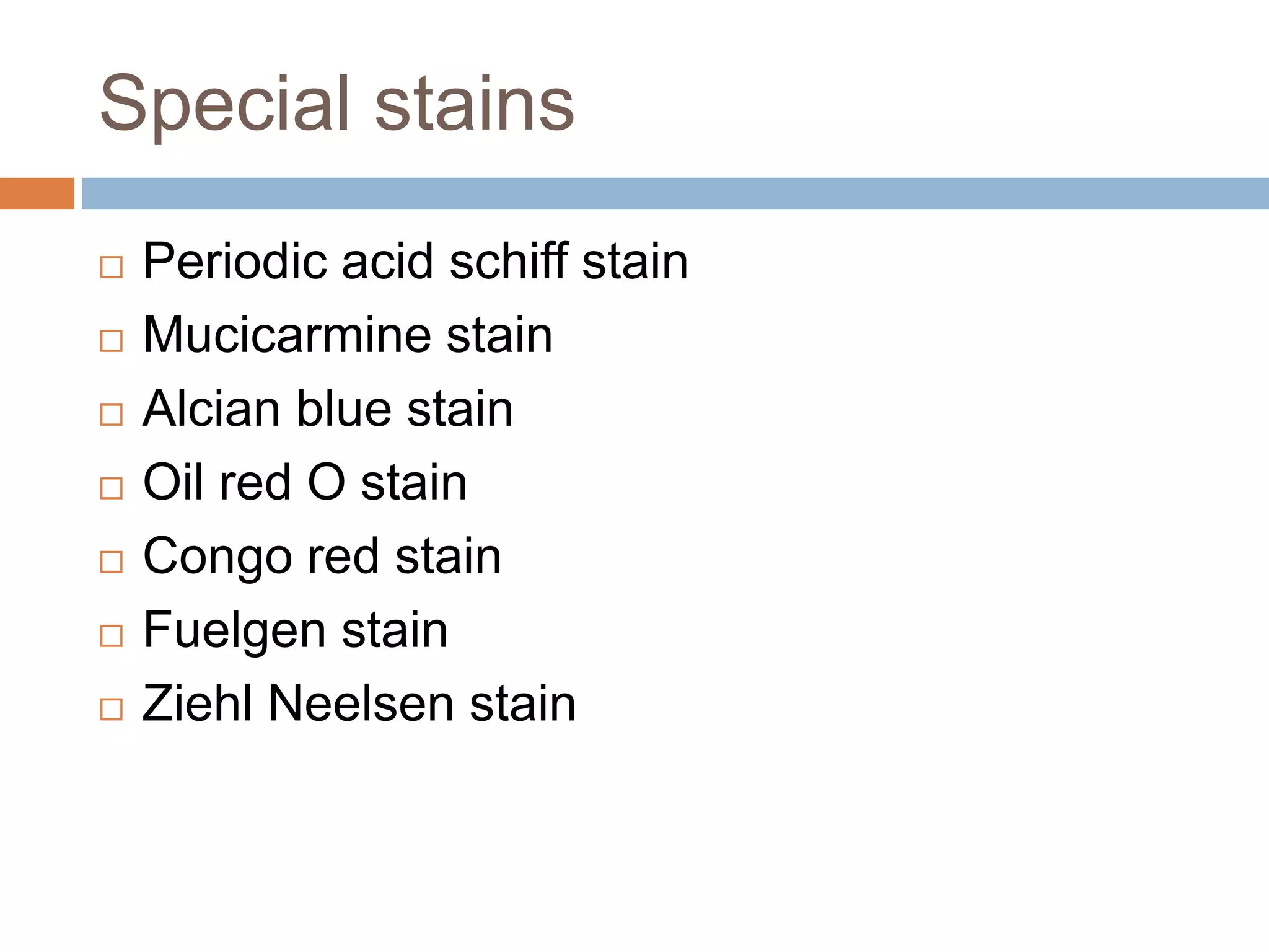
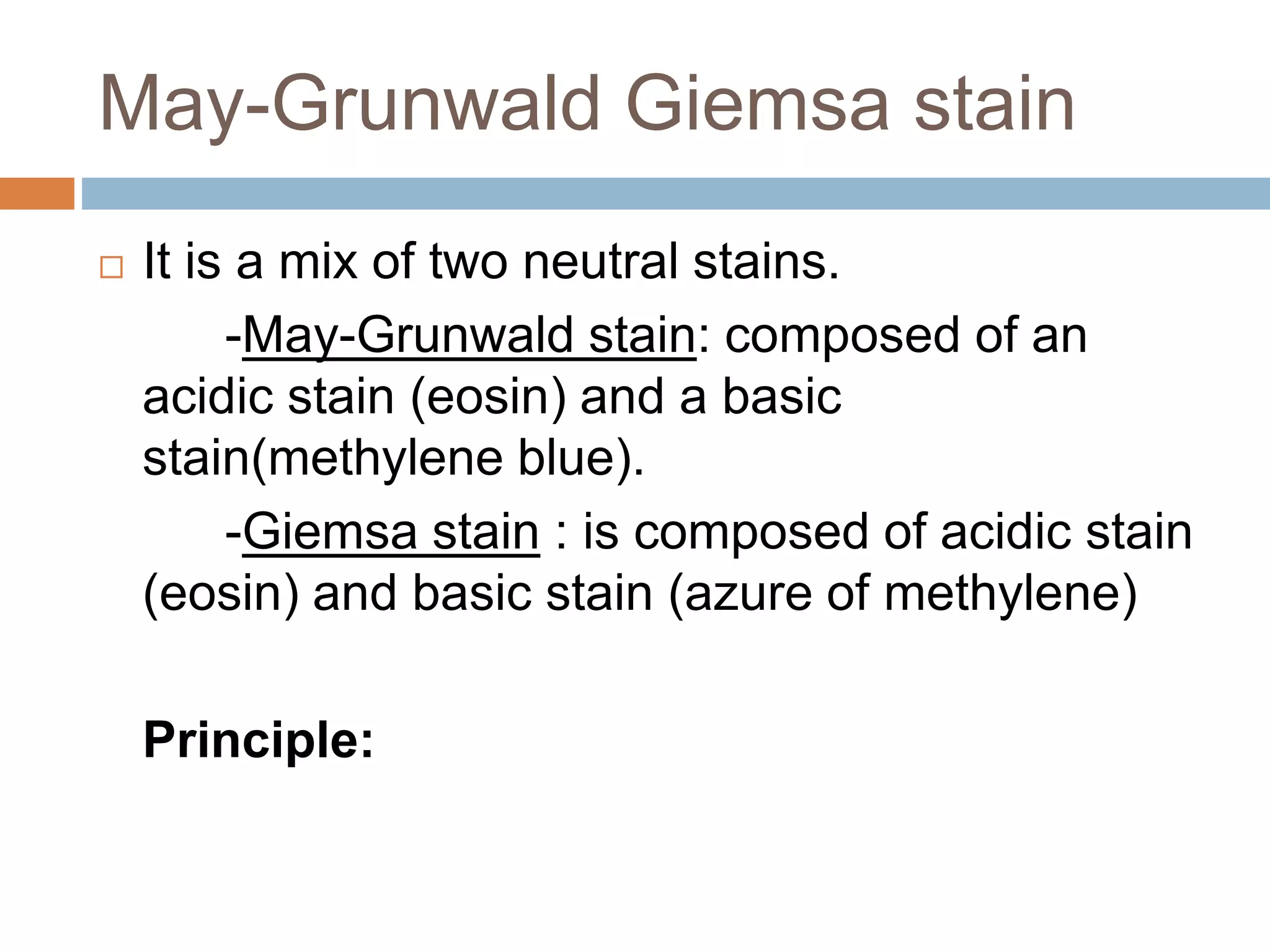
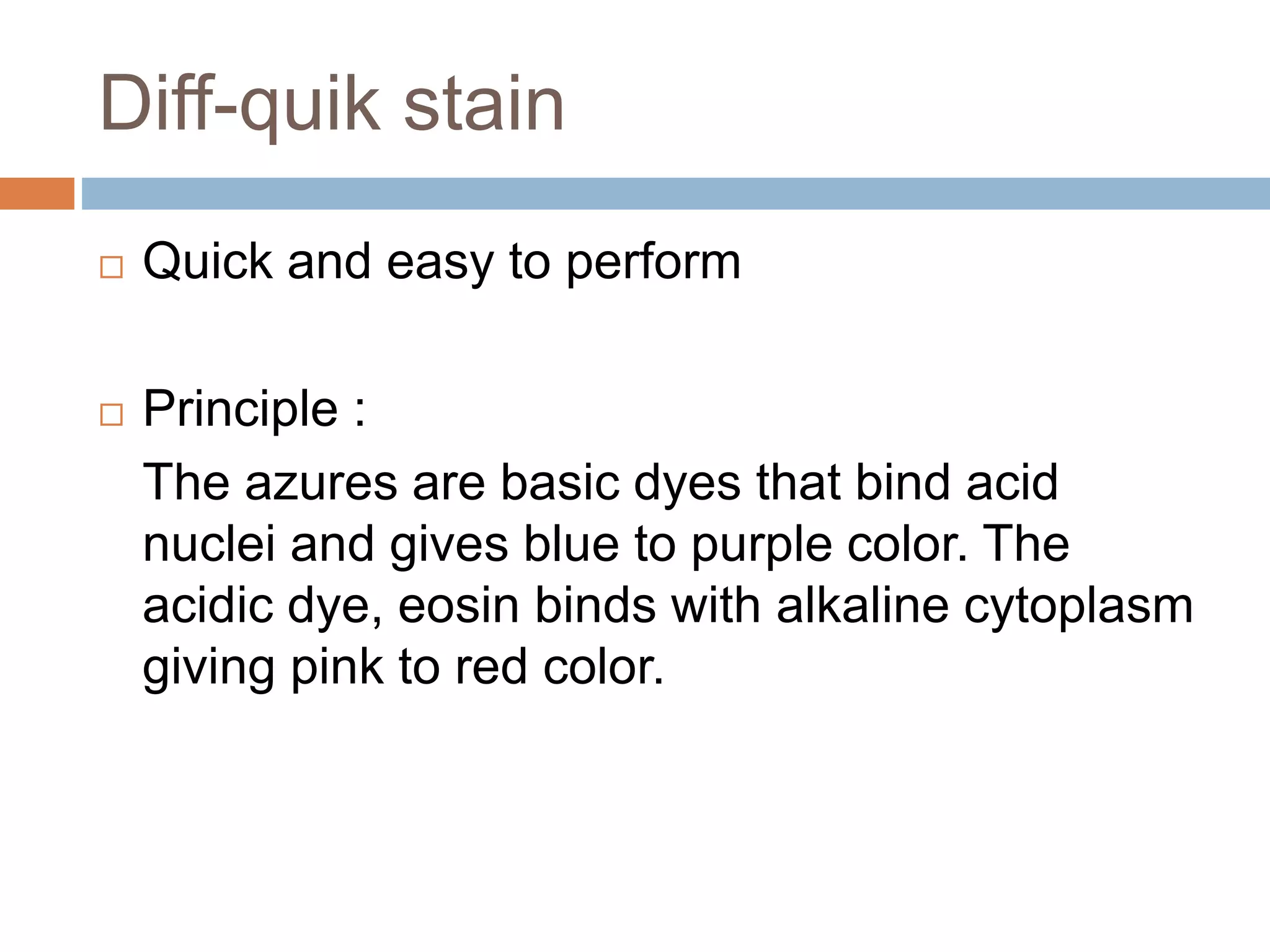
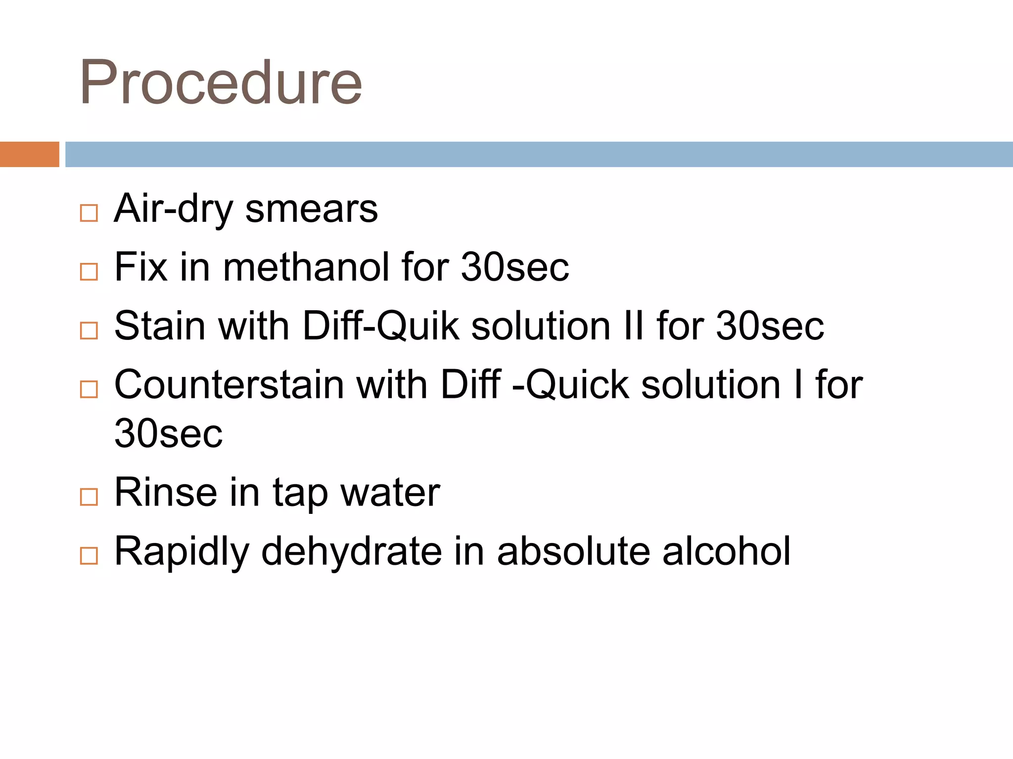
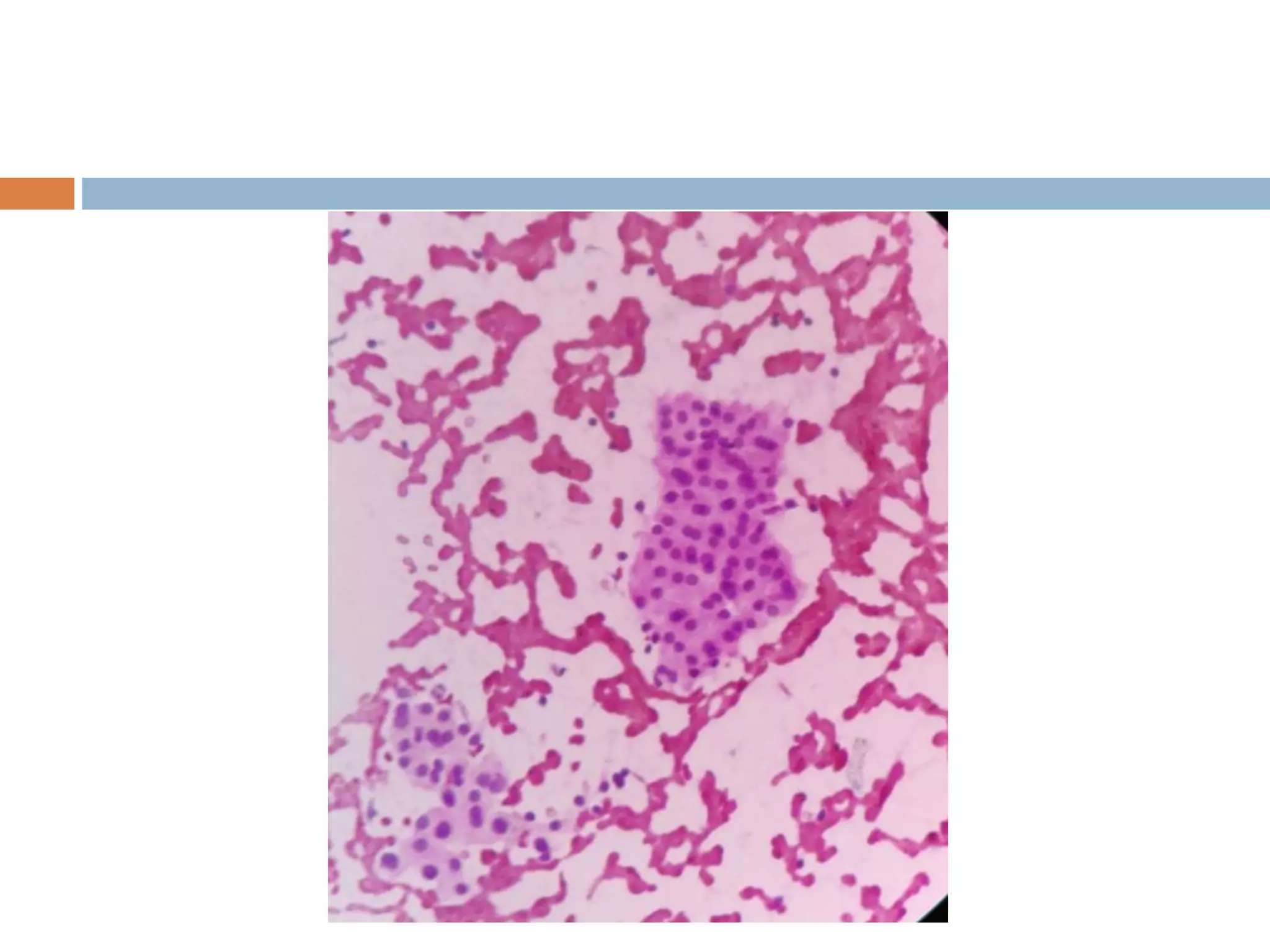
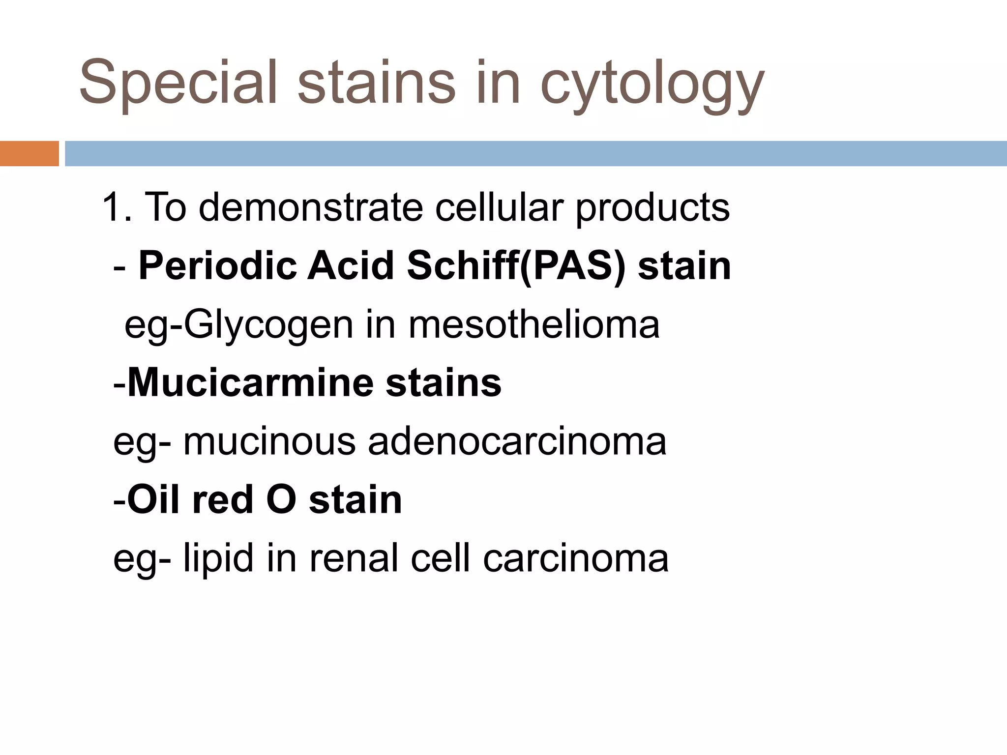
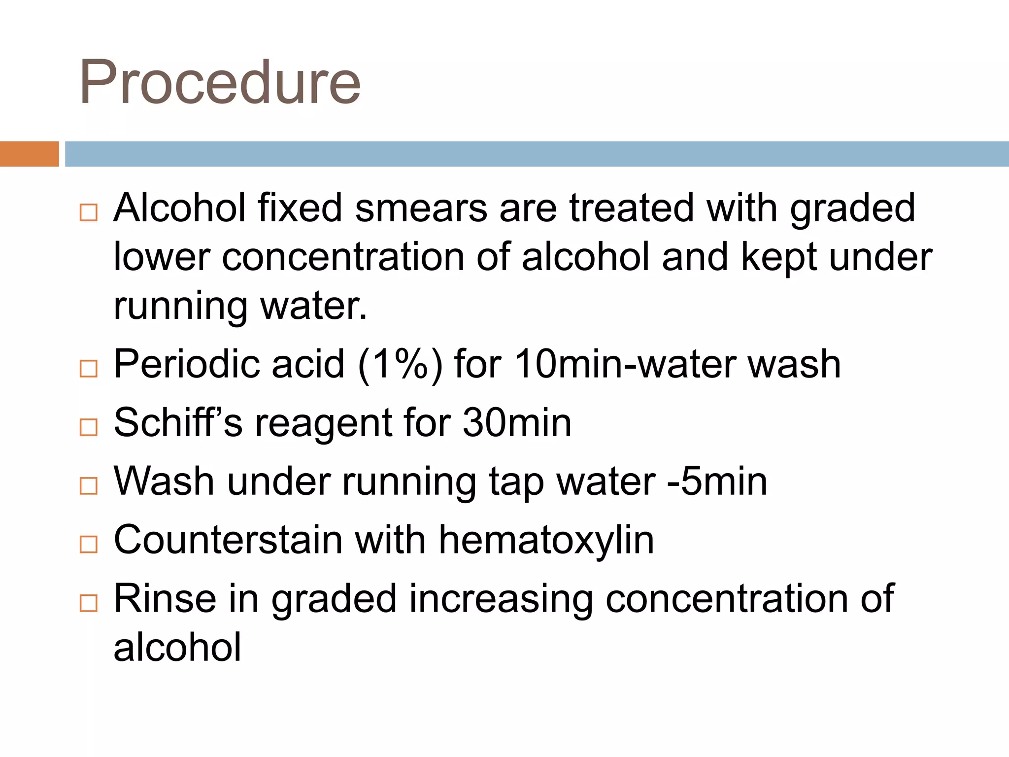
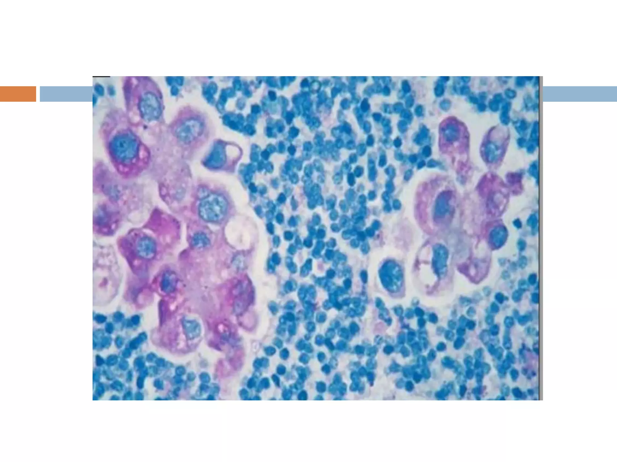
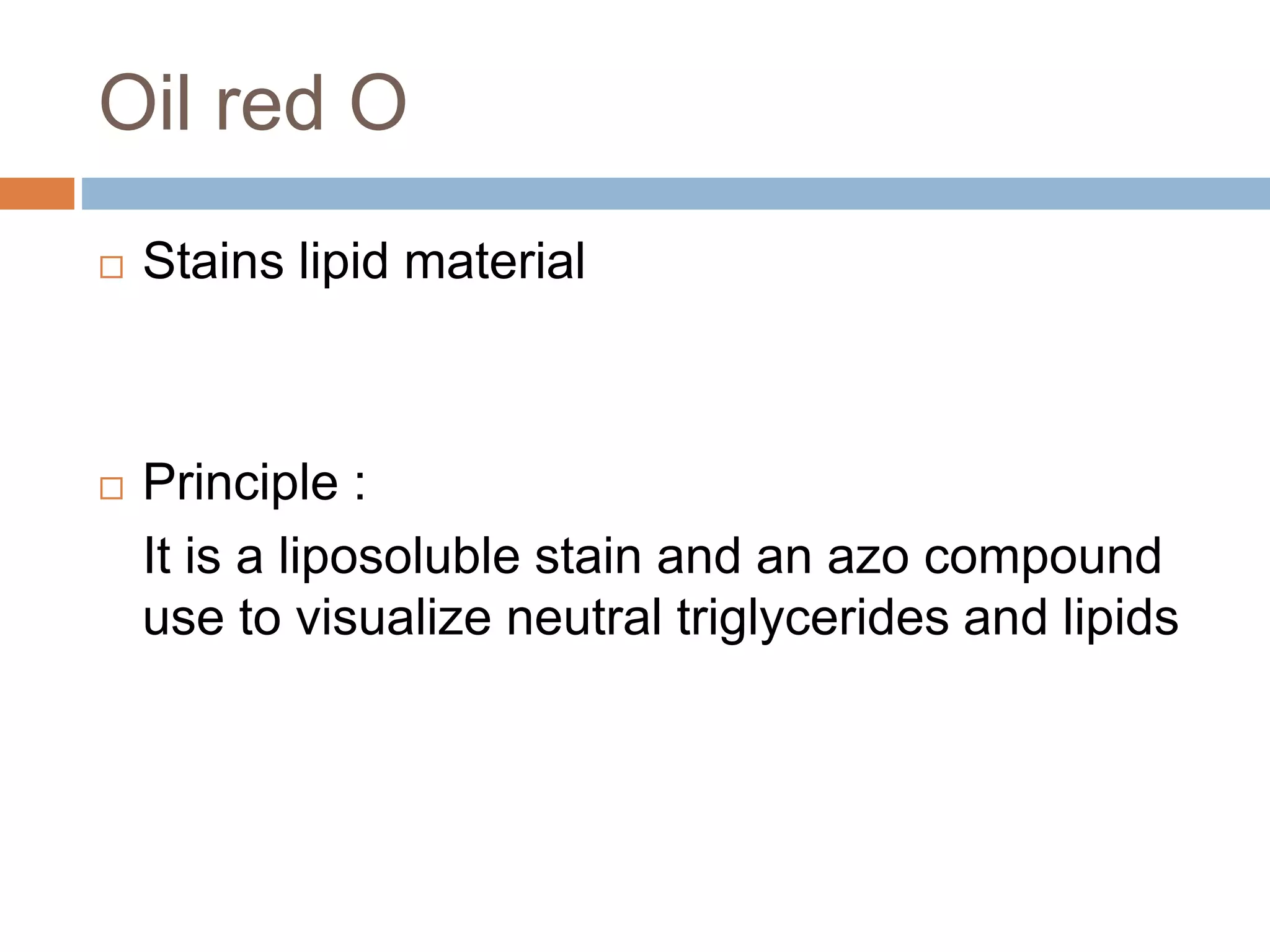
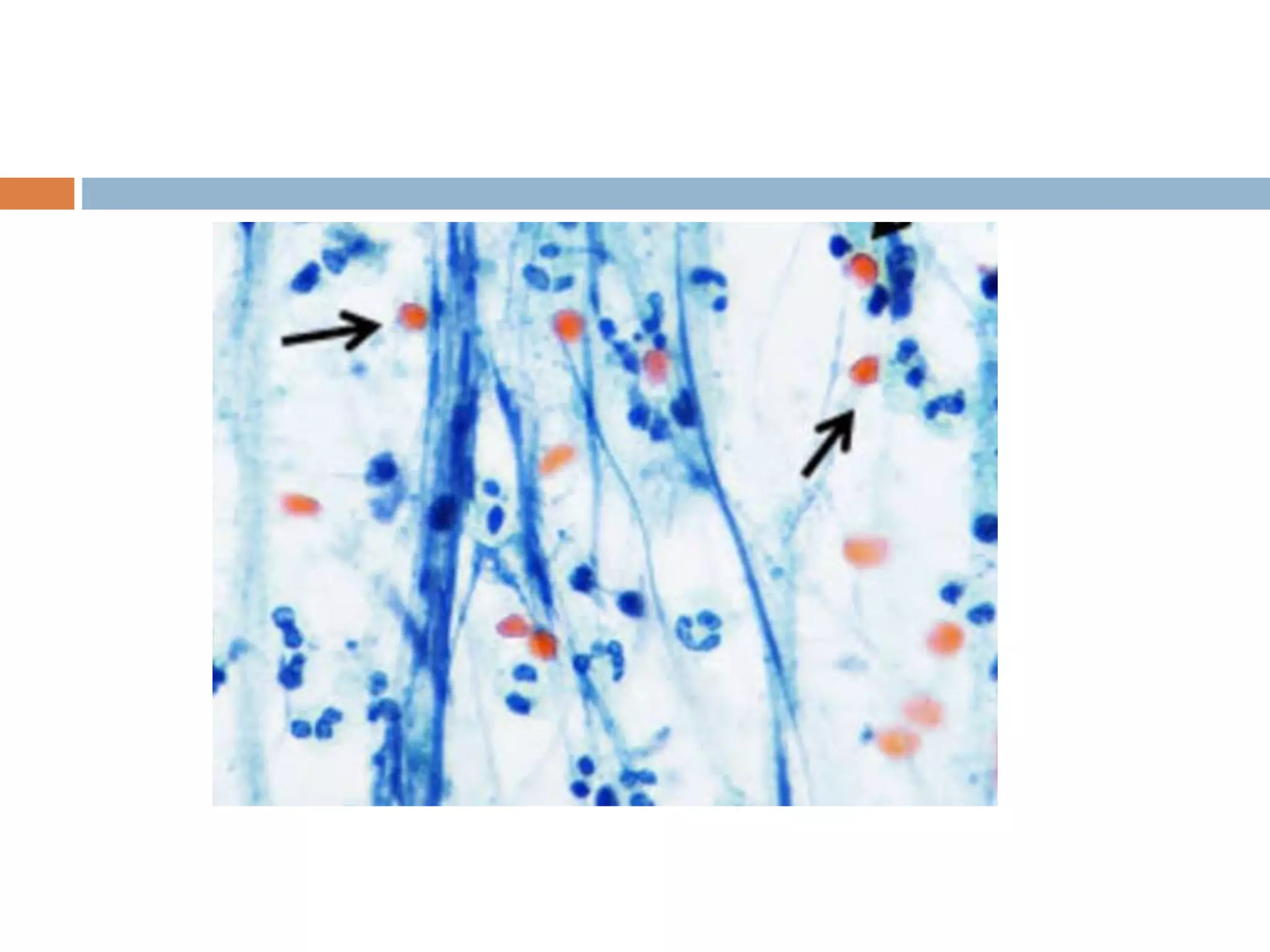
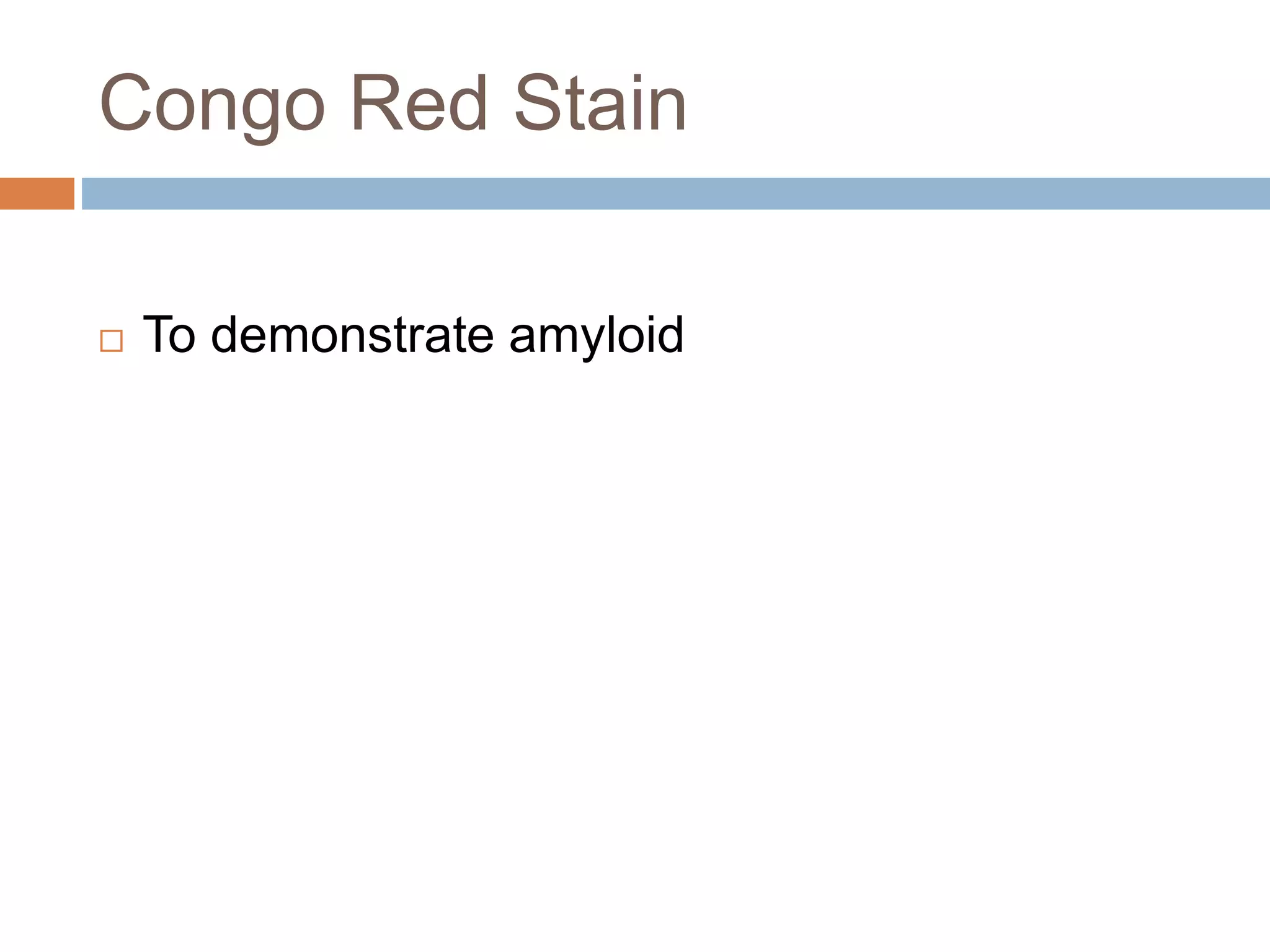
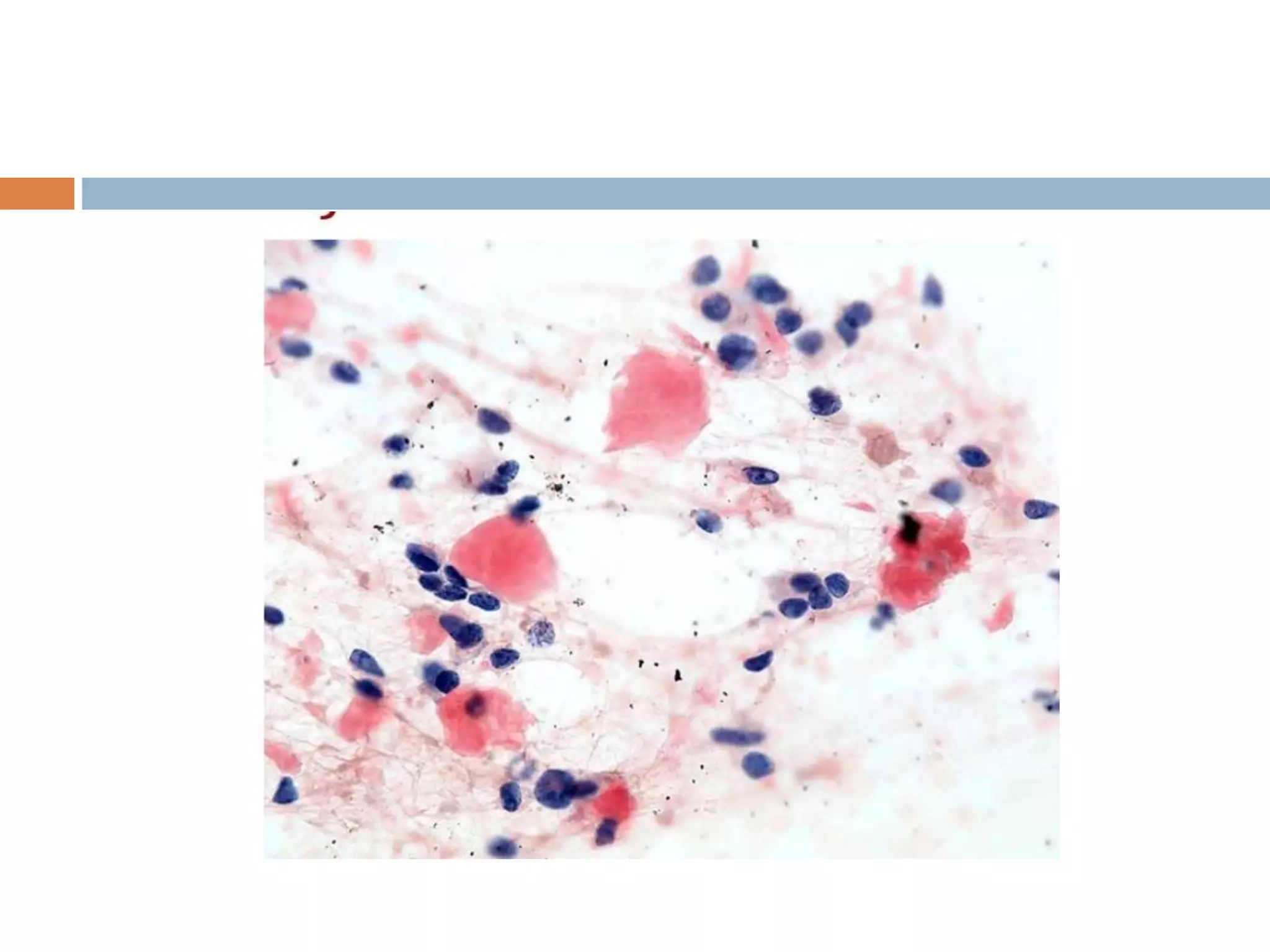
This document discusses various staining techniques used in cytology, including both routine and special stains. It provides details on the principles, procedures, and applications of stains such as May-Grunwald Giemsa, Diff-Quik, Papanicolaou, hematoxylin and eosin, periodic acid Schiff, mucicarmine, Alcian blue, Oil red O, Congo red, Feulgen, and Ziehl-Neelsen. The stains are used to demonstrate cellular and extracellular components, identify infectious organisms, and examine DNA content. Proper staining allows visualization of structures like glycogen, mucin, lipids, amyloid, and acid-fast bacteria under the microscope.