Histopathological staining techniques used in liver diseases
•Download as PPTX, PDF•
0 likes•23 views

1. Hematoxylin & Eosin staining 2. Masson's Trichrome staining 3. Sirius Red staining

Recommended
More Related Content
Similar to Histopathological staining techniques used in liver diseases
Similar to Histopathological staining techniques used in liver diseases (20)
More from PHARMA IQ EDUCATION
More from PHARMA IQ EDUCATION (20)
Recently uploaded
Recently uploaded (20)
Histopathological staining techniques used in liver diseases
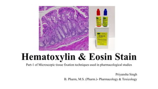
- 1. Hematoxylin & Eosin Stain Part-1 of Microscopic tissue fixation techniques used in pharmacological studies Priyansha Singh B. Pharm, M.S. (Pharm.)- Pharmacology & Toxicology
- 2. The sections of tissues as they are prepared are colourless & different components cannot be identified. Staining makes them differently coloured as various dyes have affinities towards specific components of tissues, making the identification & morphological studies possible. Hematoxylin & Eosin- most frequently used stain in histology
- 3. INTRODUCTION • Hematoxylin and eosin staining technique functions to recognize different types of tissues and their morphological changes, especially in cancer diagnosis. • It is mainly used as a gold standard stain in medical diagnosis • Hematoxylin has a deep blue-purple color and stains nucleic acids. • Eosin is pink and stains proteins non-specifically. • In a typical tissue, nuclei are stained blue, whereas the cytoplasm and extracellular matrix have varying degrees of pink staining. • Hematoxylin and eosin are both dyes, and naturally, dyes have a high affinity for tissues, depending on the Acidity and/or alkalinity of the dyes.
- 4. OBJECTIVE To demonstrate the morphologies of tissues and cells
- 5. PRINCIPLE • Haematoxylin is a dye which when combined with a mordant like potash alum starts to behave like a basic/ cationic dye. The staining of nucleus is blue due to binding of dye metal complex with the nuclear material. • Eosin dye is acidic dye hence it as a negative charge (eosinophilic). Therefore it stains the basic protein structures of a cell & extracellular matrix (acidophils or eosinophils), giving them a red or pink color, for example, the cytoplasm is positively charged, and therefore it will take up the eosin dye, and appear pink.
- 7. Main types of staining seen on H&E stain
- 9. PROCEDURE Materials Required • Xylene I & II- for deparaffinization & clearance • 70%, 80%, 100% alcohol- for rehydration & dehydration after counter staining • Haematoxylin- for nuclear staining • 1% acid alcohol- for differentiation • Li2CO3- for bluing • Eosin- counter staining • DPX- mounting
- 10. Procedure with principle behind it
- 11. • Deparaffinization- to remove wax embedded in the tissue. • Descending grades of alcohol- After thorough de-waxing, the slide is passed through several changes of alcohol to remove the xylene, then thoroughly rinsed in water. The section is now hydrated so that aqueous reagents will readily penetrate the cells and tissue elements. • Haematoxylin- for nuclear staining • Differentiation- to remove non-specific background staining and to improve contrast. • Bluing- changes the color of hematoxylin from red to blue by chemically altering the dye with a slightly basic solution. The bluing solution's alkaline pH stabilizes the stain and makes it insoluble in water and alcohol. The change in pH is what causes the color change. • Counterstaining- A counterstain is a stain with color contrasting to the principal stain, making the stained structure easily visible using a microscope. • Ascending grades of alcohol- for dehydration. To remove water from the tissue so that paraffin wax can completely penetrate it. Since wax is not water soluble, but is soluble in xylene, the water in the tissue must be replaced with xylene. • Clearance by Xylene I & II- to remove dehydrating agents and prepare tissues for impregnation with the embedding agent. Clearing makes the tissue transparent, or clear, making the slides easier to read. • DPX mounting- to increase the refractive index and improve image quality in microscope
- 12. RESULTS • Nuclei are stained blue, cytoplasm & ECM- varied degrees of pink stain • Well fixed cells/ tissues show considerable intracellular details • Nuclei stains with eosin • If polyribosomes are present then cytoplasm will have a distinct blue cast
- 13. Masson’s Trichrome Staining Part-2 of Microscopic tissue fixation techniques used in pharmacological studies Priyansha Singh B. Pharm, M.S. (Pharm.)- Pharmacology & Toxicology
- 14. Definition • Masson’s trichrome is a differential staining technique for selective staining of collagen fibres, fibrin, muscles & RBCs. • Trichrome stain- Wiegert's haematoxylin (nucleus) - Biebrich scarlet (muscle, cytoplasm & nucleus) - Aniline/ methylene blue/ light green (collagen)
- 15. Objectives of Masson’s Trichrome staining • To stain the collage fibers. • To stain collagen. • To stain keratin. • To stain fibrin. • To stain muscle fibers. • To differentiate between collagen & smooth muscle in tumors, increase in collagen in diseases like cirrhosis. Routine stain in kidney fibrosis & liver cirrhosis biopsies.
- 16. Principal behind the staining • As the name implies, 3 dyes are employed selectively staining muscle, collagen fibers, fibrin & erythrocytes. • The general rule in trichrome staining is that the less porous tissues are coloured by the smallest size dye molecule. • Wherever a dye of large molecular size is able to penetrate but the dye of smaller size passes off the larger pore size. • The tissue is first stained with the acid dye (Biebrich Scarlet red- Acid Fuchsin) binding with acidophilic tissue components. • Further treatment with the phospho acids, the less permeable components retain red stain and the red colour comes out of the collagen pores • Collagen gets bind with aniline blue.
- 18. Fix the sample using Bouin’s solution- mordant (microwave for 1 min) stand for 15 mins Add Wiegert's Haematoxylin to stain nucleus (10 mins) Add Biebrich scarlet red stain (5 mins) to stain cytoplasm, keratin, muscle & RBCs Add Phosphotungstic/ phosphomolybdic acid for decolorization (10 mins). Add aniline blue/ methylene blue/ light green + 1% acetic acid to stain collagen Wash (5 mins) Wash & rinse Rinse Procedure of staining
- 19. Results Nuclei black Cytoplasm, Muscle, Erythrocytes- red Collagen- blue ADVANTAGES 1. To measure & differentiate collagen deposition as compared to HE staining 2. Produces clear images of collagen compared to other stains 3. More accurate than other staining techniques DISADVANTAGES 1. Preparation of solutions to be used within 24 hrs 2. Corrosive stains
- 20. Sirius red staining Part-3 of Microscopic tissue fixation techniques used in pharmacological studies Priyansha Singh B. Pharm, M.S. (Pharm.)- Pharmacology & Toxicology