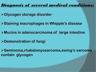
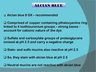
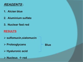
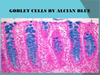
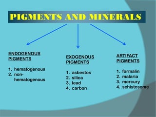
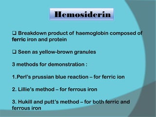
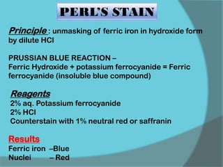
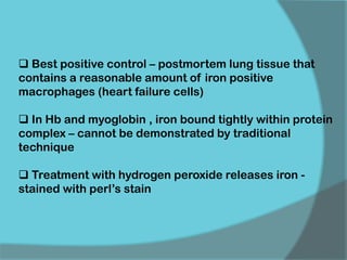
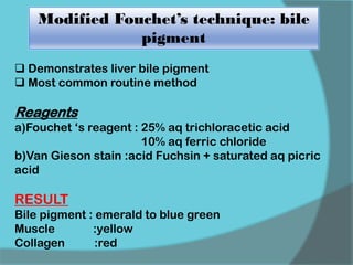
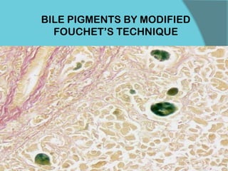
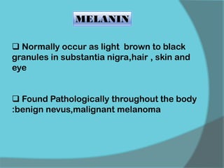
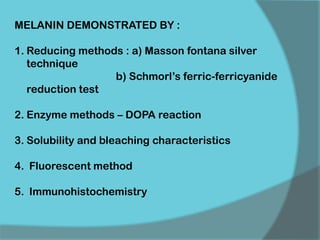
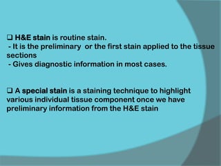
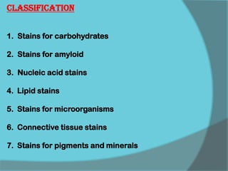
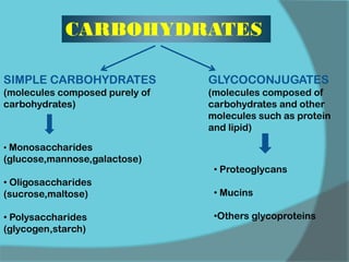
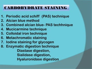
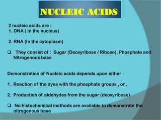
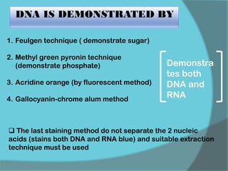
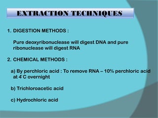
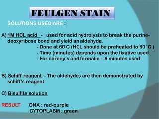
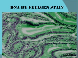
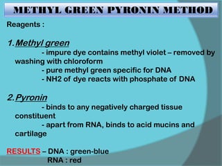
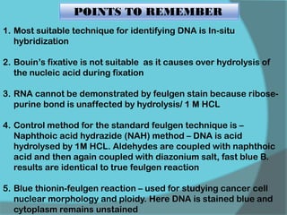
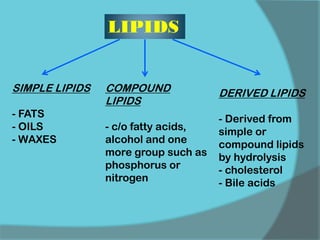
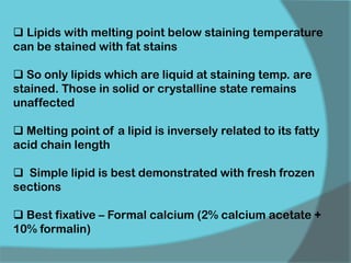
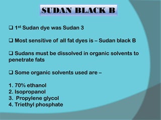
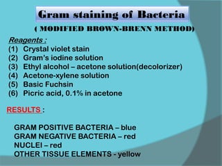
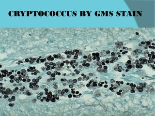
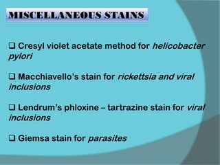
The document discusses various histological staining techniques. It begins by explaining hematoxylin and eosin staining, which provides basic diagnostic information. It then covers special stains that highlight specific tissue components, categorized by the structures they identify such as carbohydrates, amyloid, lipids, nucleic acids, and microorganisms. Carbohydrate stains discussed include periodic acid Schiff, alcian blue, mucicarmine, and others. Amyloid identification using Congo red and methyl violet is explained. Lipid stains using Sudan dyes are also summarized. The document provides details on techniques for staining nucleic acids and identifying bacteria by Gram staining.
This presentation deals tissue processing in histopathology, the detailed of presentation given blow:
Histology, study the organization of tissues at all levels, from the whole organ down to the molecular components of cells that are found in most multicellular plants and animals.
Animal tissues are classified as epithelium, with closely spaced cells and very little intercellular space; connective tissue, with large amounts of intercellular material; muscle, specialized for contraction; and nerve, specialized for conduction of electrical impulses. Blood is also sometimes considered a separate tissue type.
Plants are composed of relatively undifferentiated tissue known as meristematic tissue; storage tissue or parenchyma; vascular tissue; photosynthetic tissue or chlorenchyma and support tissue or sclerenchyma and collenchyma.
Hematoxylin and Eosin (H&E) staining is a routine staining technique that reveals exceptional detail of tissue structure and makeup of the cells. Stained cell structures (e.g. nucleus, cytoplasm, organelles, extra-cellular components) provide important information for tissue-based cancer diagnosis. Special stains refer to alternative staining techniques that are used when H&E stains do not provide all the cellular information required. These techniques use a variety of dyes and methods so that pathologists can visualize tissue morphology and detect the presence of particular cell types, structures or pathogens (e.g. bacteria). We have the broadest special stain† menu anywhere (over 30 special stains), including:
Grocott’s Methenamine Silver (GMS) Stain
Reticulin Stains
Trichrome Stains
Giemsa Stain
Periodic Acid-Schiff (PAS) Stains
For More information Contact Customer support at customer.service@biogenex.com or follow the link http://biogenex.com/us/applications/special-stains/special-stains-controls.html
Introduction, classification, isolation, purification, biological activity of alkaloids, general methods of structural determination of alkaloids, structural elucidation of Morphine, Reserpine and Emetine
ARTIFICIAL INTELLIGENCE IN HEALTHCARE.pdfAnujkumaranit
Artificial intelligence (AI) refers to the simulation of human intelligence processes by machines, especially computer systems. It encompasses tasks such as learning, reasoning, problem-solving, perception, and language understanding. AI technologies are revolutionizing various fields, from healthcare to finance, by enabling machines to perform tasks that typically require human intelligence.
Ethanol (CH3CH2OH), or beverage alcohol, is a two-carbon alcohol
that is rapidly distributed in the body and brain. Ethanol alters many
neurochemical systems and has rewarding and addictive properties. It
is the oldest recreational drug and likely contributes to more morbidity,
mortality, and public health costs than all illicit drugs combined. The
5th edition of the Diagnostic and Statistical Manual of Mental Disorders
(DSM-5) integrates alcohol abuse and alcohol dependence into a single
disorder called alcohol use disorder (AUD), with mild, moderate,
and severe subclassifications (American Psychiatric Association, 2013).
In the DSM-5, all types of substance abuse and dependence have been
combined into a single substance use disorder (SUD) on a continuum
from mild to severe. A diagnosis of AUD requires that at least two of
the 11 DSM-5 behaviors be present within a 12-month period (mild
AUD: 2–3 criteria; moderate AUD: 4–5 criteria; severe AUD: 6–11 criteria).
The four main behavioral effects of AUD are impaired control over
drinking, negative social consequences, risky use, and altered physiological
effects (tolerance, withdrawal). This chapter presents an overview
of the prevalence and harmful consequences of AUD in the U.S.,
the systemic nature of the disease, neurocircuitry and stages of AUD,
comorbidities, fetal alcohol spectrum disorders, genetic risk factors, and
pharmacotherapies for AUD.
HOT NEW PRODUCT! BIG SALES FAST SHIPPING NOW FROM CHINA!! EU KU DB BK substit...GL Anaacs
Contact us if you are interested:
Email / Skype : kefaya1771@gmail.com
Threema: PXHY5PDH
New BATCH Ku !!! MUCH IN DEMAND FAST SALE EVERY BATCH HAPPY GOOD EFFECT BIG BATCH !
Contact me on Threema or skype to start big business!!
Hot-sale products:
NEW HOT EUTYLONE WHITE CRYSTAL!!
5cl-adba precursor (semi finished )
5cl-adba raw materials
ADBB precursor (semi finished )
ADBB raw materials
APVP powder
5fadb/4f-adb
Jwh018 / Jwh210
Eutylone crystal
Protonitazene (hydrochloride) CAS: 119276-01-6
Flubrotizolam CAS: 57801-95-3
Metonitazene CAS: 14680-51-4
Payment terms: Western Union,MoneyGram,Bitcoin or USDT.
Deliver Time: Usually 7-15days
Shipping method: FedEx, TNT, DHL,UPS etc.Our deliveries are 100% safe, fast, reliable and discreet.
Samples will be sent for your evaluation!If you are interested in, please contact me, let's talk details.
We specializes in exporting high quality Research chemical, medical intermediate, Pharmaceutical chemicals and so on. Products are exported to USA, Canada, France, Korea, Japan,Russia, Southeast Asia and other countries.
Report Back from SGO 2024: What’s the Latest in Cervical Cancer?bkling
Are you curious about what’s new in cervical cancer research or unsure what the findings mean? Join Dr. Emily Ko, a gynecologic oncologist at Penn Medicine, to learn about the latest updates from the Society of Gynecologic Oncology (SGO) 2024 Annual Meeting on Women’s Cancer. Dr. Ko will discuss what the research presented at the conference means for you and answer your questions about the new developments.
Anti ulcer drugs and their Advance pharmacology ||
Anti-ulcer drugs are medications used to prevent and treat ulcers in the stomach and upper part of the small intestine (duodenal ulcers). These ulcers are often caused by an imbalance between stomach acid and the mucosal lining, which protects the stomach lining.
||Scope: Overview of various classes of anti-ulcer drugs, their mechanisms of action, indications, side effects, and clinical considerations.
TEST BANK for Operations Management, 14th Edition by William J. Stevenson, Ve...kevinkariuki227
TEST BANK for Operations Management, 14th Edition by William J. Stevenson, Verified Chapters 1 - 19, Complete Newest Version.pdf
Title: Sense of Taste
Presenter: Dr. Faiza, Assistant Professor of Physiology
Qualifications:
MBBS (Best Graduate, AIMC Lahore)
FCPS Physiology
ICMT, CHPE, DHPE (STMU)
MPH (GC University, Faisalabad)
MBA (Virtual University of Pakistan)
Learning Objectives:
Describe the structure and function of taste buds.
Describe the relationship between the taste threshold and taste index of common substances.
Explain the chemical basis and signal transduction of taste perception for each type of primary taste sensation.
Recognize different abnormalities of taste perception and their causes.
Key Topics:
Significance of Taste Sensation:
Differentiation between pleasant and harmful food
Influence on behavior
Selection of food based on metabolic needs
Receptors of Taste:
Taste buds on the tongue
Influence of sense of smell, texture of food, and pain stimulation (e.g., by pepper)
Primary and Secondary Taste Sensations:
Primary taste sensations: Sweet, Sour, Salty, Bitter, Umami
Chemical basis and signal transduction mechanisms for each taste
Taste Threshold and Index:
Taste threshold values for Sweet (sucrose), Salty (NaCl), Sour (HCl), and Bitter (Quinine)
Taste index relationship: Inversely proportional to taste threshold
Taste Blindness:
Inability to taste certain substances, particularly thiourea compounds
Example: Phenylthiocarbamide
Structure and Function of Taste Buds:
Composition: Epithelial cells, Sustentacular/Supporting cells, Taste cells, Basal cells
Features: Taste pores, Taste hairs/microvilli, and Taste nerve fibers
Location of Taste Buds:
Found in papillae of the tongue (Fungiform, Circumvallate, Foliate)
Also present on the palate, tonsillar pillars, epiglottis, and proximal esophagus
Mechanism of Taste Stimulation:
Interaction of taste substances with receptors on microvilli
Signal transduction pathways for Umami, Sweet, Bitter, Sour, and Salty tastes
Taste Sensitivity and Adaptation:
Decrease in sensitivity with age
Rapid adaptation of taste sensation
Role of Saliva in Taste:
Dissolution of tastants to reach receptors
Washing away the stimulus
Taste Preferences and Aversions:
Mechanisms behind taste preference and aversion
Influence of receptors and neural pathways
Impact of Sensory Nerve Damage:
Degeneration of taste buds if the sensory nerve fiber is cut
Abnormalities of Taste Detection:
Conditions: Ageusia, Hypogeusia, Dysgeusia (parageusia)
Causes: Nerve damage, neurological disorders, infections, poor oral hygiene, adverse drug effects, deficiencies, aging, tobacco use, altered neurotransmitter levels
Neurotransmitters and Taste Threshold:
Effects of serotonin (5-HT) and norepinephrine (NE) on taste sensitivity
Supertasters:
25% of the population with heightened sensitivity to taste, especially bitterness
Increased number of fungiform papillae
Pulmonary Thromboembolism - etilogy, types, medical- Surgical and nursing man...VarunMahajani
Disruption of blood supply to lung alveoli due to blockage of one or more pulmonary blood vessels is called as Pulmonary thromboembolism. In this presentation we will discuss its causes, types and its management in depth.
micro teaching on communication m.sc nursing.pdfAnurag Sharma
Microteaching is a unique model of practice teaching. It is a viable instrument for the. desired change in the teaching behavior or the behavior potential which, in specified types of real. classroom situations, tends to facilitate the achievement of specified types of objectives.
2. H&E stain is routine stain.
- It is the preliminary or the first stain applied to the tissue
sections
- Gives diagnostic information in most cases.
A special stain is a staining technique to highlight
various individual tissue component once we have
preliminary information from the H&E stain
3. Classification
1. Stains for carbohydrates
2. Stains for amyloid
3. Nucleic acid stains
4. Lipid stains
5. Stains for microorganisms
6. Connective tissue stains
7. Stains for pigments and minerals
4. CARBOHYDRATES
SIMPLE CARBOHYDRATES
(molecules composed purely of
carbohydrates)
• Monosaccharides
(glucose,mannose,galactose)
• Oligosaccharides
(sucrose,maltose)
• Polysaccharides
(glycogen,starch)
GLYCOCONJUGATES
(molecules composed of
carbohydrates and other
molecules such as protein
and lipid)
• Proteoglycans
• Mucins
•Others glycoproteins
5. 90-95% of their molecular weight is due to the carbohydrate
component
The carbohydrate component is known as glycosaminoglycans(GAG)
A GAG is composed of repeating disaccharide units , each made up
of 2 different monosaccharides
Each disaccharide is composed of a carboxylated uronic acid
(glucuronic or iduronic acid) and a hexosamine such as N-
acetylglucosamine or N-acetylgalactosamine
TYPES OF GYCOSAMINOGLYCANS
Chondroitin sulfate
Dermatan sulfate
Keratan sulfate
Heparin sulfate
Heparin
Hyaluronic acid
PROTEOGLYCANS
7. FIXATIVES
Tissue placed in fixative promptly after removal – capable
of autolytic reaction
If immediate fixation not possible, tissue refrigerated until
adequate fixation possible
Recommended fixatives : Rossman’s fluid
Alcoholic formalin with picric
acid
Glutaraldehyde fixatives avoidedfree aldehyde groups
are capable of undergoing schiff reaction – so increased
background staining
10. PERIODIC ACID SCHIFF METHOD
1st histochemical use was by McManus for
demonstration of mucin
Reagents – 1. periodic acid
2. schiff reagent
0.5-1% solution of periodic acid (oxidant) used for 5-10
minutes oxidation of hydroxyl group within the
formation of two free aldehyde groups
free aldehydes react with schiff reagent
carbohydrate
bright
red magenta end product
RESULT – glycogen and mucins : magenta
nuclei : blue
12. Other oxidants like potassium permanganate/ chromic
acid not used – further oxidise aldehyde groups to
carboxylic groups – not reactive to schiff reagent
MILD PAS TECHNIQUE –
0.01% periodic acid used for shorter period for N-acetyl
sialic acid containing mucins as the hydroxyl groups are
highly susceptible to periodic acid oxidation
13. PAS REACTIVE CELLS AND TISSUE
COMPONENTS
1.GLYCOGEN
2. STARCH
3. MUCIN
4. BASEMENT MEMBRANE
5. ALPHA ANTI TRYPSIN
6. RETICULIN
7. FUNGI(CAPSULES)
8. PANCREATIC ZYMOGEN GRANULES
9. THYROID COLLOID
10. CORPORA AMYLACEA
11. RUSSELL BODIES
14. Diagnosis of several medical conditions:
Glycogen storage disorder
Staining macrophages in Whipple's disease
Mucins in adenocarcinoma of large intestine
Demonstration of fungi
Seminoma,rhabdomyosarcoma,ewing’s sarcoma
contain glycogen
15. ALCIAN BLUE
Alcian blue 8 GX – recommended
Comprised of copper containing pthalocyanine ring
linked to 4 isothiouronium groups – strong bases -
account for cationic nature of the dye
Sulfate and carboxylate groups of proteoglycans
ionised at pH 2.5 and carry a negative charge
Sialo- and sulfo mucins also reactive at pH 2.5
So, they stain with alcian blue at pH 2.5
Neutral mucins are not reactive with alcian blue
16. REAGENTS :
1. Alcian blue
2. Aluminium sulfate
3. Nuclear fast red
RESULTS
sulfomucin,sialomucin
Proteoglycans
Hyaluronic acid
Nucleus red
Blue
18. COMBINED ALCIAN BLUE- PAS TECHNIQUE
PRINCIPLE
Demonstrate presence of mucin
Differentiate acid mucin from neutral mucin
1st stain all acid mucin with alcian blue
(blue)
Those acid mucin which are PAS +ve will not
be stained on PAS reaction only neutral
mucin will be stained(magenta)
19. ALCIAN BLUE WITH VARYING
ELECTROLYTE CONCENTRATIONS
This technique is based upon phenomenon known as
CRITICAL ELECTROLYTE CONCENTRATION (CEC)
CEC is defined as point at which amount of electrolyte
such as MgCl2 is sufficient to prevent staining from AB
Competition between cations of salt and dye occurs for
polyanionic sites within tissue
Different acidic carbohydrates have different CEC value
So can differentiate acidic mucins and proteoglycans
20. MUCICARMINE
Active dye molecule is aluminium – carminic acid
complex known as CARMINE
carminic acid produced from dried bodies of female
Coccus Cacti insects
Carmine complex has a positive charge and so attracts
polyanions such as sialomucins and sulfomucins
Useful for identification of adenocarcinoma ( especially
of GIT)
Capsule of fungus – cryptococcus neoformans is also
detected
21. REAGENTS :
1.Southgate’s mucicarmine solution
2.Alcoholic hematoxylin
3.Acidified ferric chloride solution
4.Weigert’s iron hematoxylin solution
5.Metanil yellow solution
RESULTS :
Acidic mucins – deep rose to red
Nuclei – black
Other tissue elements – light yellow
24. NUCLEIC ACIDS
2 nucleic acids are :
1. DNA ( In the nucleus)
2. RNA (In the cytoplasm)
They consist of : Sugar (Deoxyribose / Ribose), Phosphate and
Nitrogenous base
Demonstration of Nucleic acids depends upon either :
1. Reaction of the dyes with the phosphate groups , or ,
2. Production of aldehydes from the sugar (deoxyribose)
No histochemical methods are available to demonstrate the
nitrogenous base
25. 1. Feulgen technique ( demonstrate sugar)
2. Methyl green pyronin technique
(demonstrate phosphate)
3. Acridine orange (by fluorescent method)
4. Gallocyanin-chrome alum method
Demonstra
tes both
DNA and
RNA
The last staining method do not separate the 2 nucleic
acids (stains both DNA and RNA blue) and suitable extraction
technique must be used
DNA IS DEMONSTRATED BY
26. EXTRACTION TECHNIQUES
1. DIGESTION METHODS :
Pure deoxyribonuclease will digest DNA and pure
ribonuclease will digest RNA
2. CHEMICAL METHODS :
a) By perchloric acid : To remove RNA – 10% perchloric acid
at 4̊ C overnight
b) Trichloroacetic acid
c) Hydrochloric acid
27. FEULGEN STAIN
SOLUTIONS USED ARE :
A) 1M HCL acid - used for acid hydrolysis to break the purine-
deoxyribose bond and yield an aldehyde.
- Done at 60̊ C (HCL should be preheated to 60 ̊ C )
- Time (minutes) depends upon the fixative used
- For carnoy’s and formalin – 8 minutes used
B) Schiff reagent - The aldehydes are then demonstrated by
schiff’s reagent
C) Bisulfite solution
RESULT DNA : red-purple
CYTOPLASM : green
29. METHYL GREEN PYRONIN METHOD
Reagents :
1.Methyl green
- impure dye contains methyl violet – removed by
washing with chloroform
- pure methyl green specific for DNA
- NH2 of dye reacts with phosphate of DNA
2.Pyronin
- binds to any negatively charged tissue
constituent
- apart from RNA, binds to acid mucins and
cartilage
RESULTS – DNA : green-blue
RNA : red
31. 1. Most suitable technique for identifying DNA is In-situ
hybridization
2. Bouin’s fixative is not suitable as it causes over hydrolysis of
the nucleic acid during fixation
3. RNA cannot be demonstrated by feulgen stain because ribose-
purine bond is unaffected by hydrolysis/ 1 M HCL
4. Control method for the standard feulgen technique is –
Naphthoic acid hydrazide (NAH) method – DNA is acid
hydrolysed by 1M HCL. Aldehydes are coupled with naphthoic
acid and then again coupled with diazonium salt, fast blue B.
results are identical to true feulgen reaction
5. Blue thionin-feulgen reaction – used for studying cancer cell
nuclear morphology and ploidy. Here DNA is stained blue and
cytoplasm remains unstained
POINTS TO REMEMBER
32. LIPIDS
SIMPLE LIPIDS
- FATS
- OILS
- WAXES
COMPOUND
LIPIDS
- c/o fatty acids,
alcohol and one
more group such as
phosphorus or
nitrogen
DERIVED LIPIDS
- Derived from
simple or
compound lipids
by hydrolysis
- cholesterol
- Bile acids
33. Lipids with melting point below staining temperature
can be stained with fat stains
So only lipids which are liquid at staining temp. are
stained. Those in solid or crystalline state remains
unaffected
Melting point of a lipid is inversely related to its fatty
acid chain length
Simple lipid is best demonstrated with fresh frozen
sections
Best fixative – Formal calcium (2% calcium acetate +
10% formalin)
34. 1st Sudan dye was Sudan 3
Most sensitive of all fat dyes is – Sudan black B
Sudans must be dissolved in organic solvents to
penetrate fats
Some organic solvents used are –
1. 70% ethanol
2. Isopropanol
3. Propylene glycol
4. Triethyl phosphate
SUDAN BLACK B
35. Sudan black b has 2 fractions –
1st stains neutral fats blue-black
2nd stains phospholipids gray
This gray reaction can be enhanced as a bronze
dichroism if section is viewed in polarised light
It fails to stain crystalline cholesterol, lecithin and free
fatty acids
Bromine pre treatment converts crystalline
cholesterol to oily derivatives and hence permeable to
Sudan dye
36. AMYLOID
Extracellular , amorphous , eosinophilic
material
Composed of protein in an antiparallel -
pleated sheet configuration
In H&E stain , can be confused with hyaline
and fibrinoid substances
Earliest special stain used for amyloid was
Iodine by Virchow
37. CONGO RED STAIN
Acidic dye and composed of 2 identical halves
Each half has a phenyl ring bound to a naphthalene
moiety by a diazo group
2 phenyl groups bound by a diphenyl bond - gives a
linear dye molecule
It stains amyloid by hydrogen bonding and other tissue
components by electrochemical bonds
Electrochemical staining of other tissues can be
suppressed by –
using alkaline-alcoholic solvents
using competitive inhibition by salt solution
38. 2 factors are important to the congo red-
amyloid reaction
1.Linearity of the dye molecule
2.-pleated sheet configuration of the amyloid
If the spatial configuration of either is altered,
the reaction fails
39. Fixation – Not critical
Solution- 0.5 % Congo red in 50% alcohol
0.2% Potassium Hydroxide in 80% alcohol
Results- Amyloid - red
Nuclei - Blue
TECHNIQUE
41. ALKALINE CONGO-RED TECHNIQUE
High concentration of NaCl is used
Background electrochemical staining is
reduced
hydrogen bonding of congo-red to amyloid is
enhanced
42. POLARIZING MICROSCOPY AND
CONGO-RED
Under polarized light, congo red stained
amyloid exhibits apple-green birefringence
Most reliable diagnostic test for amyloid
currently
43. POINTS TO REMEMBER
Thickness of section is critical – 8-10 micro meter is
ideal
Too thin section – show faint red color
Too thick section – show yellow birefringent
Other structures giving apple-green birefringence :
1. neurofibrillary tangles of alzheimer’s
2. intracellular inclusions seen in adrenal cortical cells
3. cellulose and chitin
4. dense collagen
Thioflavin T – flourescent method
44. To differentiate AA and AL amyloid :
Section pretreatment with trypsin or
potassium permanganate done
AA amyloid lose their affinity for congo-red
but AL amyloid is resistant
45. METHYL /CRYSTAL VIOLET METHOD
Methyl violet contains a mixture of tetra- , penta- , and
hexa- methyl pararosaniline
Amyloid stained due to selective affinity for one of the
colored fractions
Hence, polychromasia seen
Ammonium oxalate accentuates polychromatic effect
RESULT :
AMYLOID, MUCIN , HYALINE – red-purple
BACKGROUND - blue
47. Gram staining of Bacteria
( MODIFIED BROWN-BRENN METHOD)
Reagents :
(1) Crystal violet stain
(2) Gram’s iodine solution
(3) Ethyl alcohol – acetone solution(decolorizer)
(4) Acetone-xylene solution
(5) Basic Fuchsin
(6) Picric acid, 0.1% in acetone
RESULTS :
GRAM POSITIVE BACTERIA – blue
GRAM NEGATIVE BACTERIA – red
NUCLEI – red
OTHER TISSUE ELEMENTS - yellow
48. Dry picric acid is explosive – recommended to
purchase picric acid – acetone solution pre made
Do not allow sections to dry at any point in the
staining process – decolorization will be difficult
and uneven
49. Acid Fast Staining for Bacteria
Mycobacteria cannot be demonstrated by gram’s stain
– possess a capsule containing long chain fatty acid
(mycolic acid) that makes them hydrophobic
Can be stained by a strong stain like carbol fuchsin
Fatty capsule resist the removal of stain by acid-
alcohol solution (acid and alcohol fastness)
Mycobacteria are PAS positive due to carbohydrate
content of their cell wall
50. Ziehl Neelson (ZN) stain
Reagents
(1) Carbol fuchsin solution
(2) 1% acid alcohol
(3) 0.1%Methylene blue solution
RESULTS
Acid fast bacilli bright red
Other tissue Pale blue
Caseous material very pale grayish blue
51. Blue counterstain may be patchy if extensive
caseation is present
Avoid over counterstaining – scant organism
can easily be obscured
Decalcification using strong acids may
destroy acid-fastness - so formic acid
recommended
52. MODIFIED FITE TECHNIQUE
REAGENTS :
1. Carbol fuchsin solution
2. 5% sulphuric acid in 25% alcohol
3. Methylene blue solution
RESULTS:
M.leprae – bright red
nuclei and other tissue elements – pale blue
53. Fite stain Modified ZN
Uses mixture of xylol & liquid
paraffin prior to stain
Does not use
Incubation in ZN carbolfuchsin at
37ºc for 45 min
Incubation In preheated ZN carbol
fuchsin at 56ºc for 60min
Decolorize with 5% H2SO4 1% acid alcohol. Acid :20%
H2SO4
Demonstrates M leprae M tuberculosis
AFB : Fite vs modified ZN
54. Warthin Starry Method for Spirochetes
REAGENTS :
1. Acetate buffer pH-3.6
2. 1% silver nitrate
RESULTS :
SPIROCHETES – black
BACKGROUND – golden -yellow
56. FUNGAL STAINS
Fungal cell walls are rich in polysaccharides
which can be converted by oxidation to
dialdehydes
Dialdehydes are then detected by silver
solution
58. Results
Fungi , Pneumocystis, melanin - Black
Mucin & Glycogen - dark grey
Background - Pale green
Hyphae & yeast form - sharply delineated in black
against green background
60. MISCELLANEOUS STAINS
Cresyl violet acetate method for helicobacter
pylori
Macchiavello’s stain for rickettsia and viral
inclusions
Lendrum’s phloxine – tartrazine stain for viral
inclusions
Giemsa stain for parasites
61. CONNECTIVE TISSUES
Provide a matrix that connects and binds the
cells and organs and ultimately gives support
to the body.
Parent cell is embryonic mesenchyme
62.
63. COLLAGEN FIBRES
1. Masson ‘s trichrome technique
2. Van Gieson’s stain
3. Mallory’s Phosphotungstic Acid
Hematoxylin
4. MSB Technique
5. PAS
6. Heidenhain’s Azan stain
7. lillie’s allochrome method
8. Luxol fast blue G
64. FACTORS AFFECTING TRICHROME
STAINING
1. TISSUE PERMEABILITY AND DYE
MOLECULAR SIZE
When protein component of a tissue exposed to a fixative –
insoluble protein network formed
Structure of the protein network directly related to the
staining reactions
Erythrocyte protein – dense network with small pores
Muscle cells – larger pores
Collagen – least dense network and quite porous
65. 2. Heat :
Increases rate of staining and penetration
3. pH :
Low pH ( 1.5 – 3)
4. Nuclear stain for trichrome
Iron hematoxylin preferred
More resistant to acidity of dye solutions
Alum hematoxylins are decolorized
Can use Celestin blue- alum hematoxylin sequence
66. EFFECT OF FIXATION
10% NBF will not yield optimal results
Treatment of formaldehyde fixed tissue with picric
acid /mercuric chloride solution enhances intensity
and radiance of trichrome
Recommended fixatives are: Bouin’s
Zenker’s,
Formal-mercury
Zinc formalin
67. Demonstrate collagen and muscle in normal tissue
Differentiate collagen and Muscle in tumors
Identify an increase in collagenous tissue
Indicate fibrotic change in cirrhosis of liver
Indicate fibrotic change in pyelonephritis
Distinguish tumors that have arisen from muscle cells
and fibroblasts
Masson ‘s trichrome technique
68. REAGENTS
1. Weigert’s iron hematoxylin
2. Acid fuchsin
3. Glacial acetic acid
4. Phosphomolybdic acid
5. Methyl blue
RESULT
Nuclei – Blue/ Black
Cytoplasm, muscle , RBC → Red
Collagen → Blue
72. Hemosiderin
Breakdown product of haemoglobin composed of
ferric iron and protein
Seen as yellow-brown granules
3 methods for demonstration :
1.Perl’s prussian blue reaction – for ferric ion
2. Lillie’s method – for ferrous iron
3. Hukill and putt’s method – for both ferric and
ferrous iron
73. PERL’S STAIN
Principle : unmasking of ferric iron in hydroxide form
by dilute HCl
PRUSSIAN BLUE REACTION –
Ferric Hydroxide + potassium ferrocyanide = Ferric
ferrocyanide (insoluble blue compound)
Results
Ferric iron –Blue
Nuclei – Red
Reagents
2% aq. Potassium ferrocyanide
2% HCl
Counterstain with 1% neutral red or saffranin
74. Best positive control – postmortem lung tissue that
contains a reasonable amount of iron positive
macrophages (heart failure cells)
In Hb and myoglobin , iron bound tightly within protein
complex – cannot be demonstrated by traditional
technique
Treatment with hydrogen peroxide releases iron -
stained with perl’s stain
75. Modified Fouchet’s technique: bile
pigment
Demonstrates liver bile pigment
Most common routine method
Reagents
a)Fouchet ‘s reagent : 25% aq trichloracetic acid
10% aq ferric chloride
b)Van Gieson stain :acid Fuchsin + saturated aq picric
acid
RESULT
Bile pigment : emerald to blue green
Muscle :yellow
Collagen :red
77. MELANIN
Normally occur as light brown to black
granules in substantia nigra,hair , skin and
eye
Found Pathologically throughout the body
:benign nevus,malignant melanoma
78. MELANIN DEMONSTRATED BY :
1. Reducing methods : a) Masson fontana silver
technique
b) Schmorl’s ferric-ferricyanide
reduction test
2. Enzyme methods – DOPA reaction
3. Solubility and bleaching characteristics
4. Fluorescent method
5. Immunohistochemistry
79. MASSON FONTANA STAIN
ARGENTAFFIN REACTION – reduction of ammoniacal
silver solution to form metallic silver without the use of
extraneous reducer.
Masson’s method( using fontana’s silver solution) rely
on melanin’s argentaffin property
Melanins are blackened by acid silver nitrate solution
RESULT :
Melanin – black
Nuclei - red
80. Schmorl’s ferric-ferricyanide
reduction test
Schmorl reaction – Melanin reduce ferricyanide to
ferrocyanide with production of prussian blue in the
presence of ferric salts
RESULT :
Melanin – dark blue
Nuclei - red
81. Special stains enhance detection &
localization of individual tissue component
But should not be substituted for routine H&E
CONCLUSION