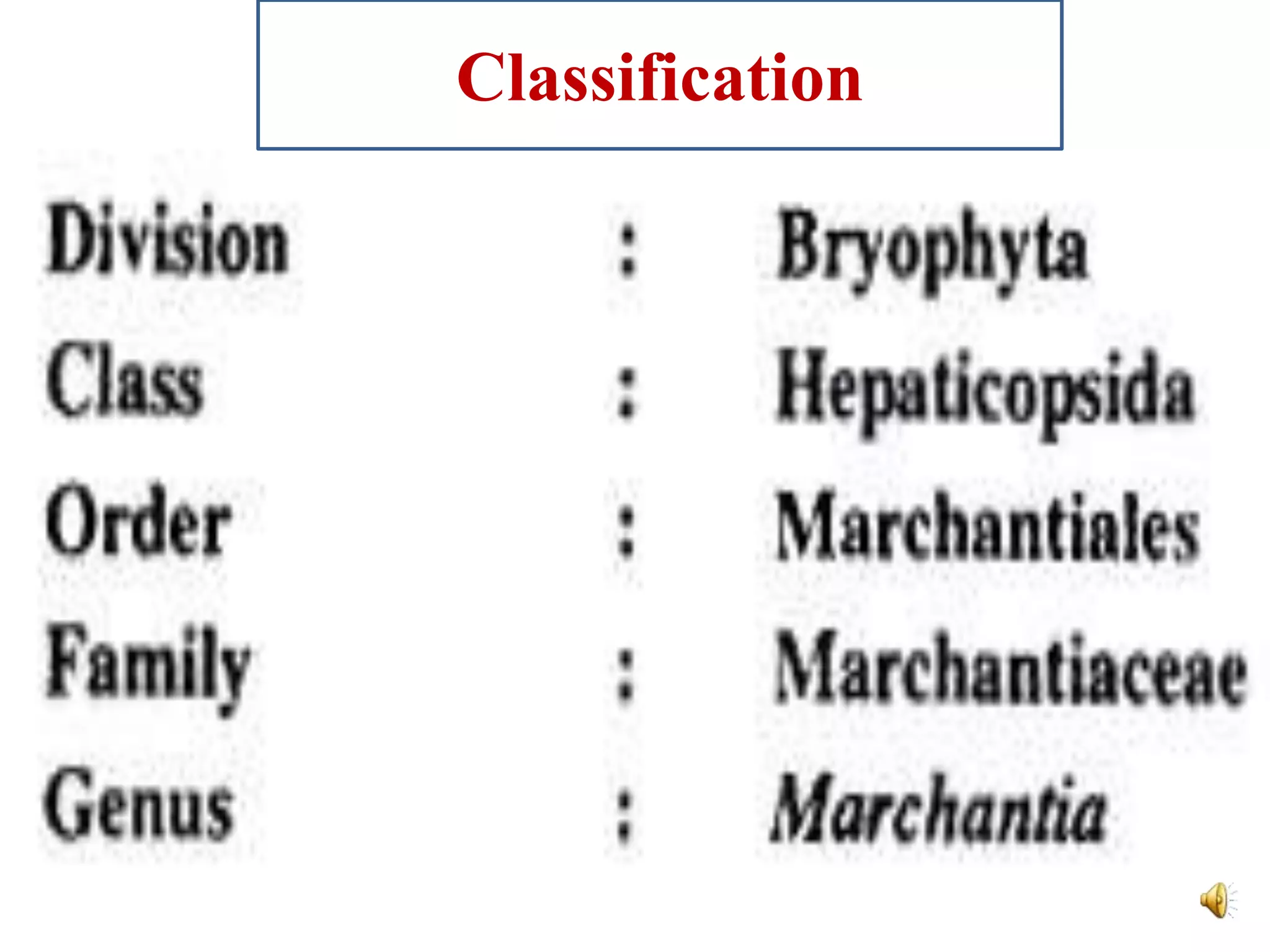
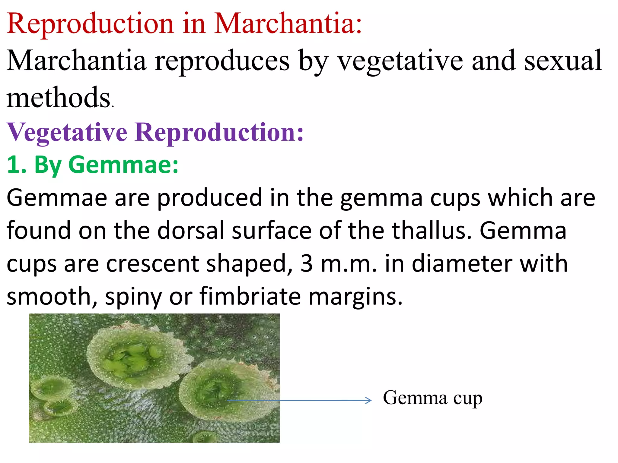
1. Marchantia is a genus of liverworts that reproduces both sexually and asexually. It has a flat, thalloid gametophyte body that is dichotomously branched.
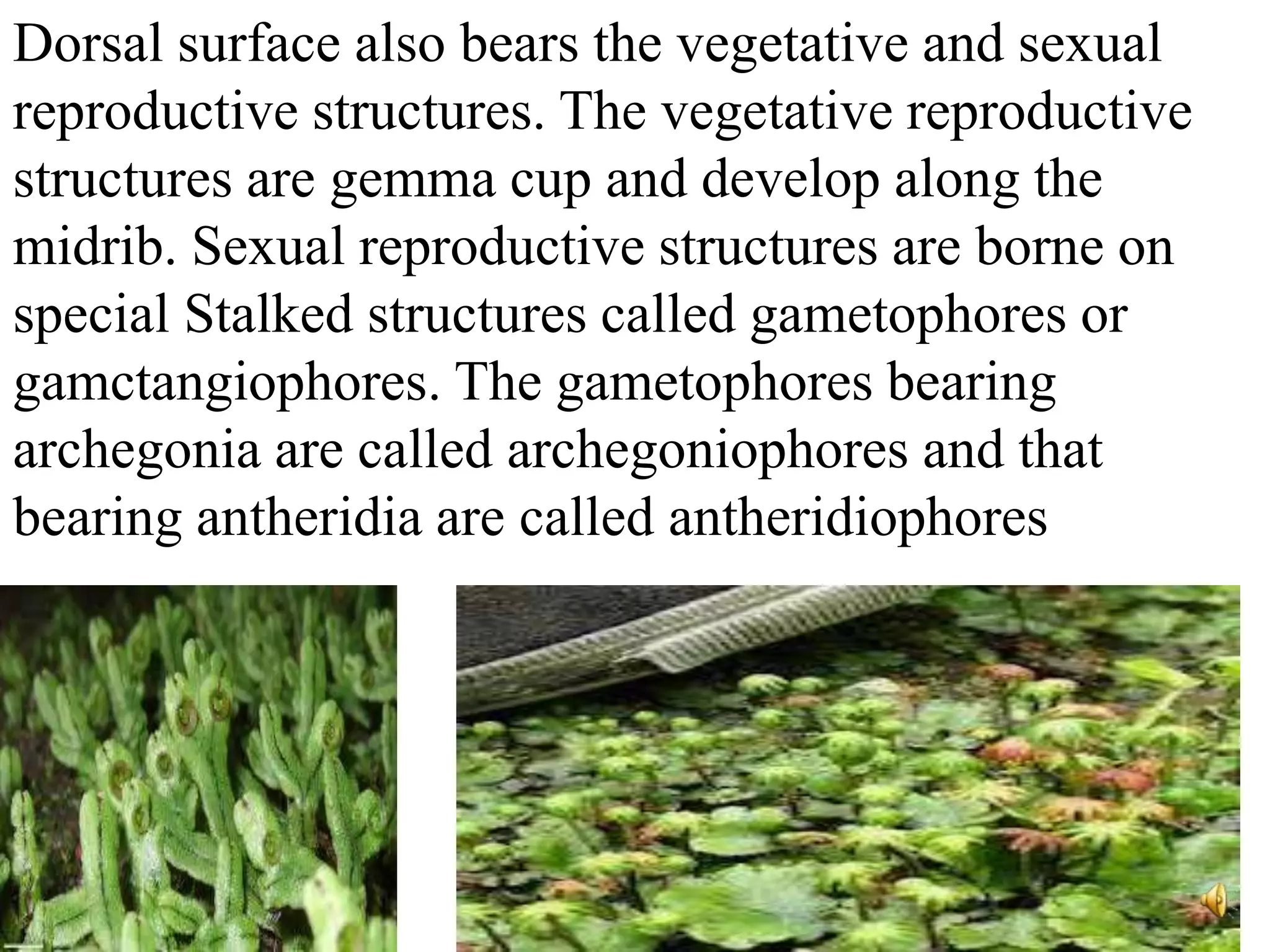
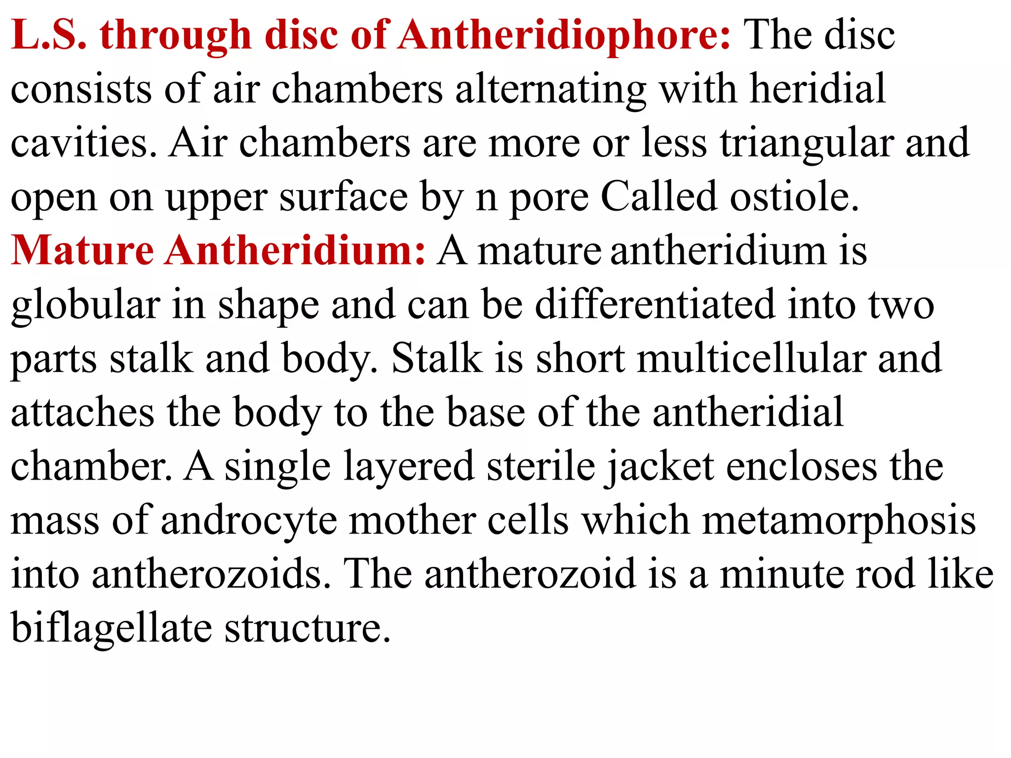
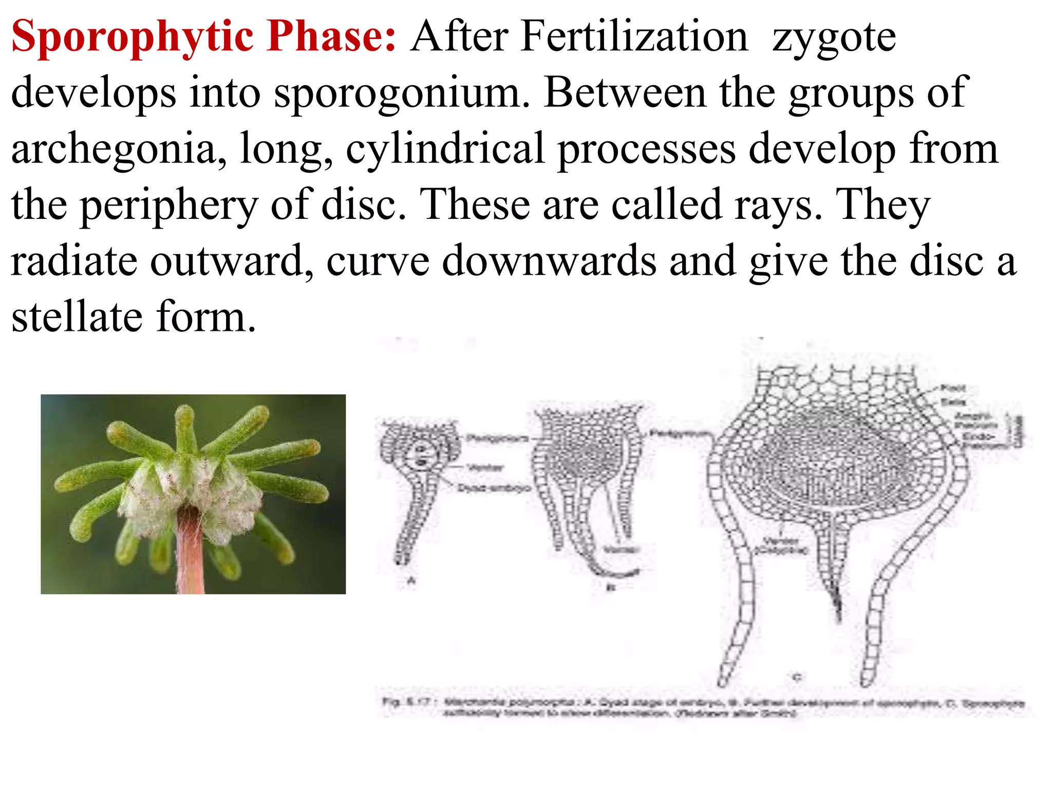
2. The gametophyte produces male and female sex organs called antheridia and archegonia on specialized stalks. Fertilization results in a diploid zygote that develops into a sporophyte.
3. The sporophyte produces spores through meiosis in capsules. The spores germinate to form new gametophytes, completing the life cycle with alternation between haploid and diploid generations.


![bryophytes.pptxforbotany [Autosaved].pptx](https://cdn.slidesharecdn.com/ss_thumbnails/bryophytes-241024055212-3ccb7683-thumbnail.jpg?width=640&height=640&fit=bounds)


