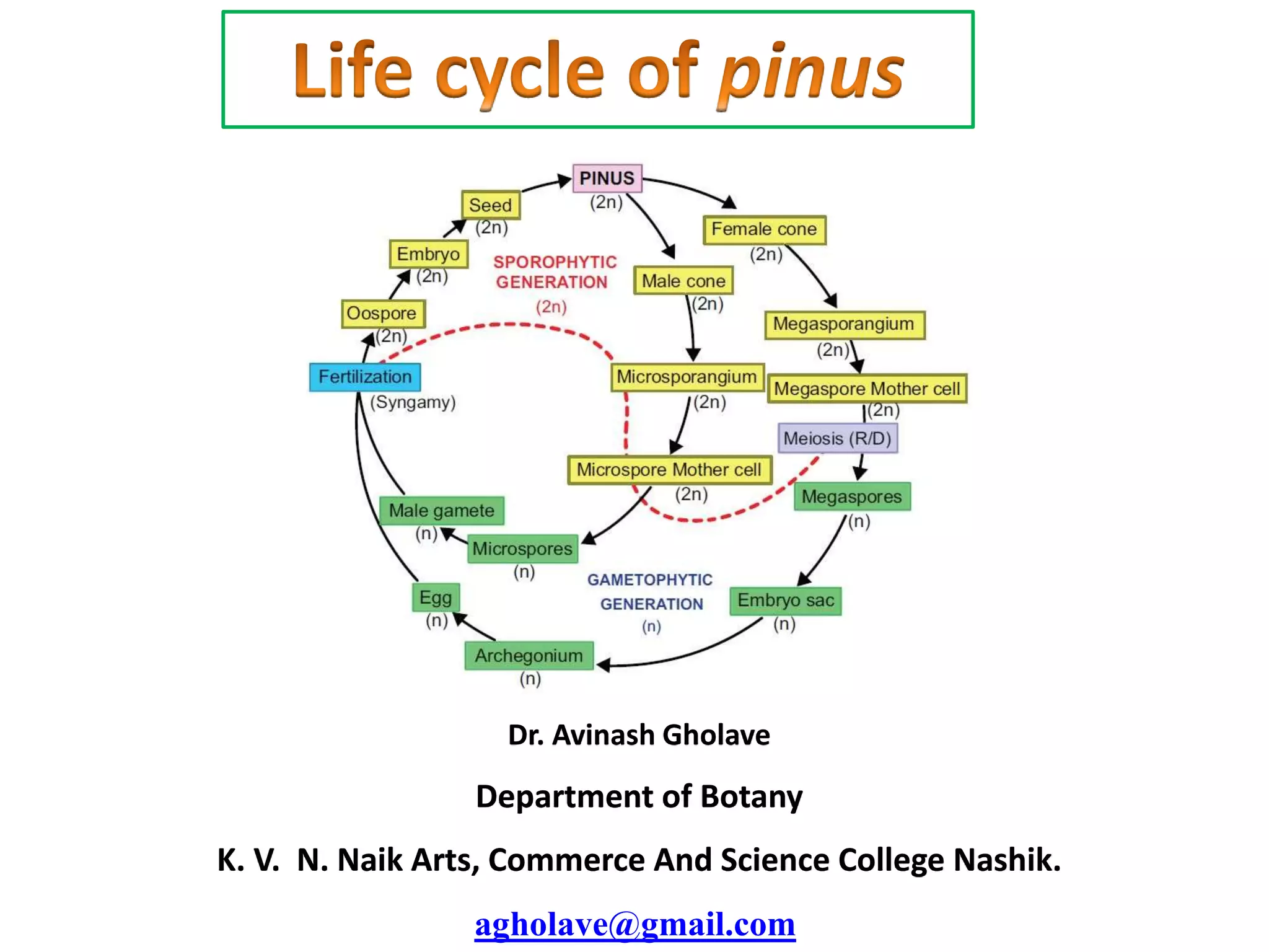
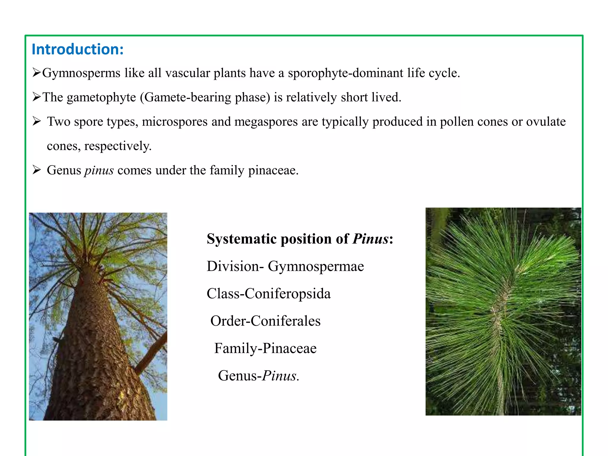
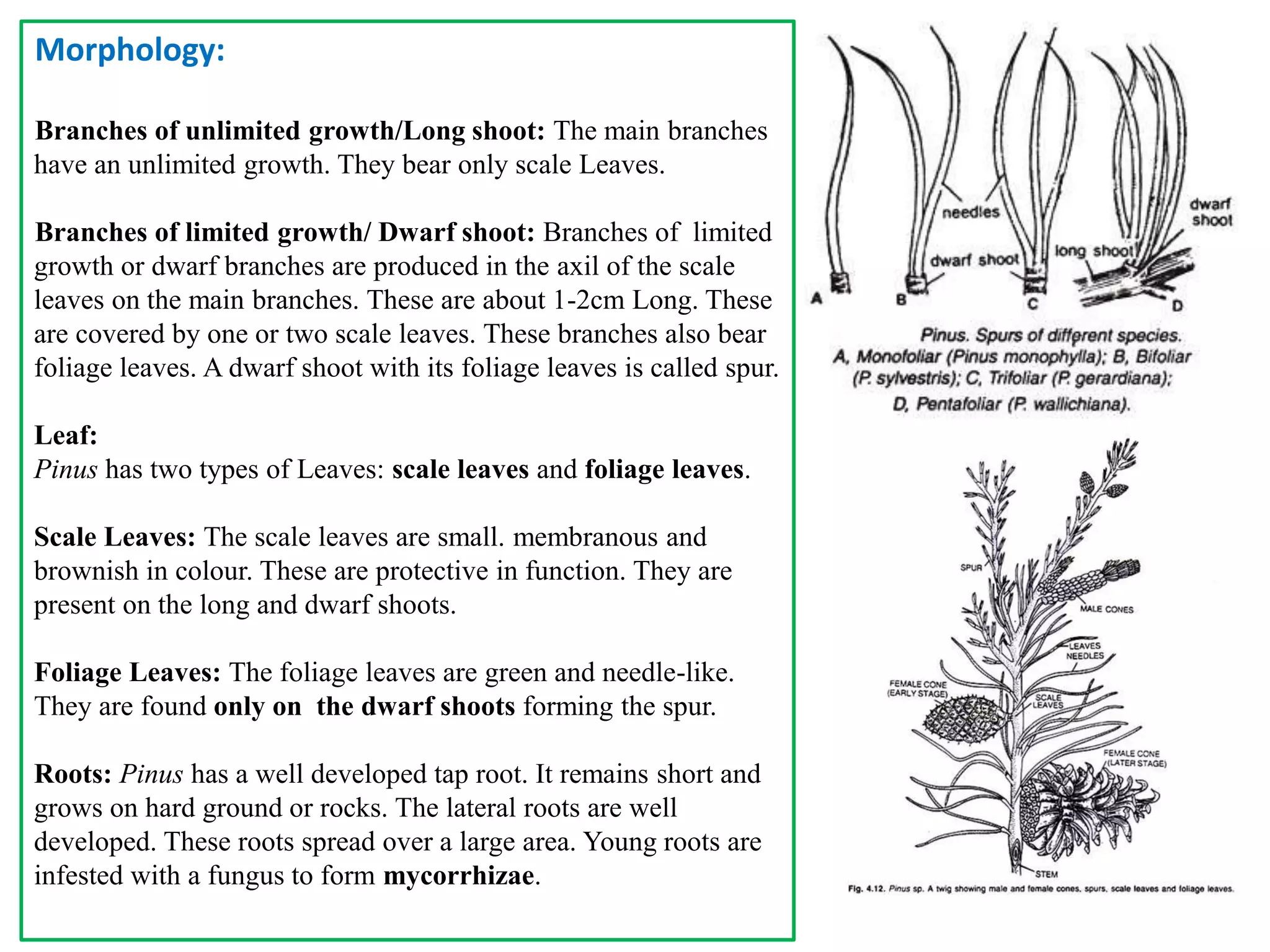
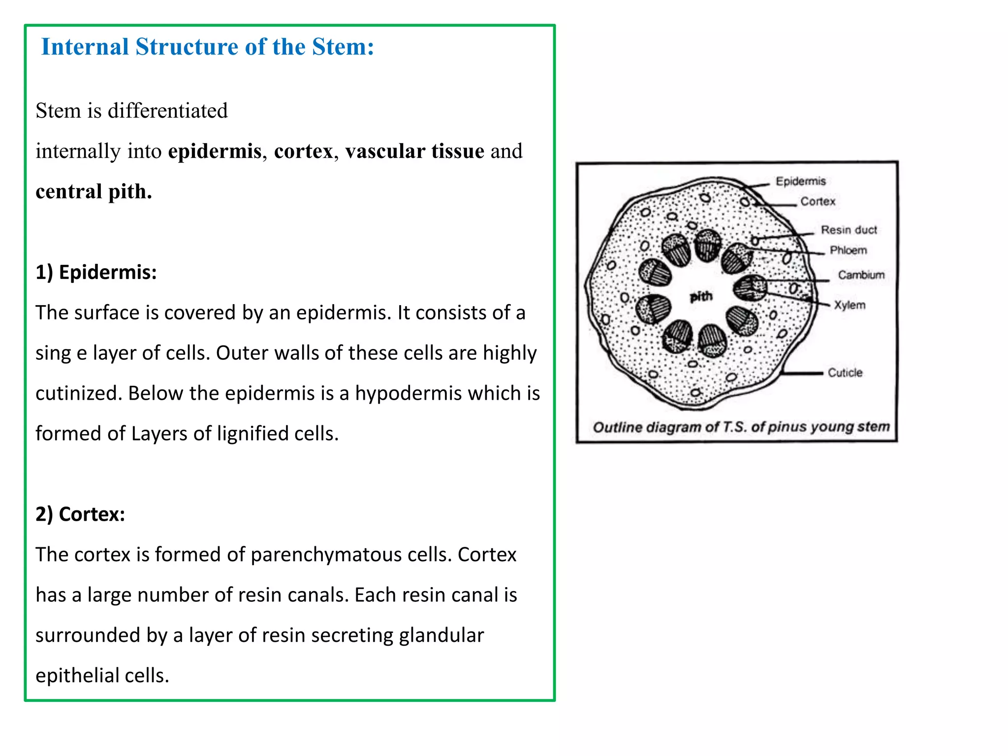
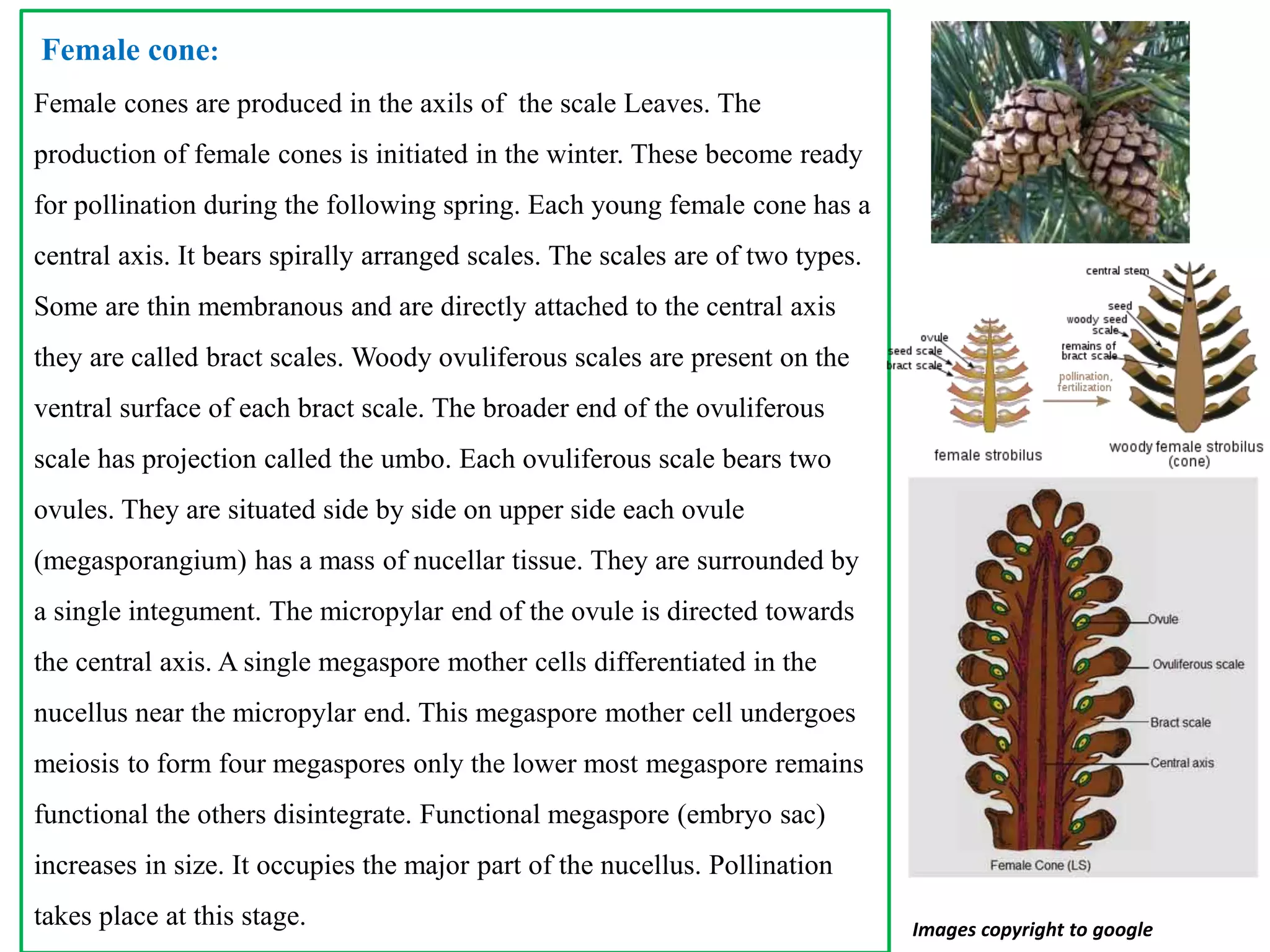
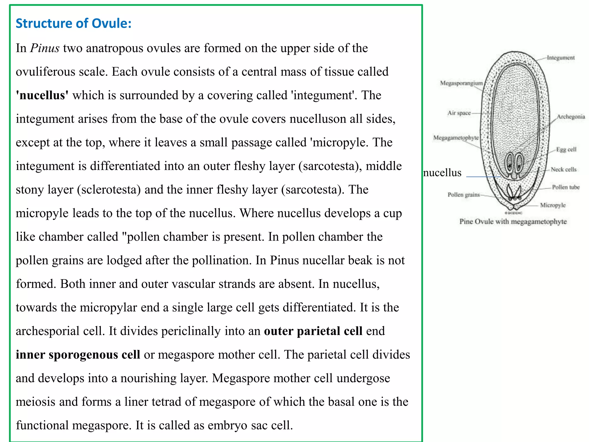
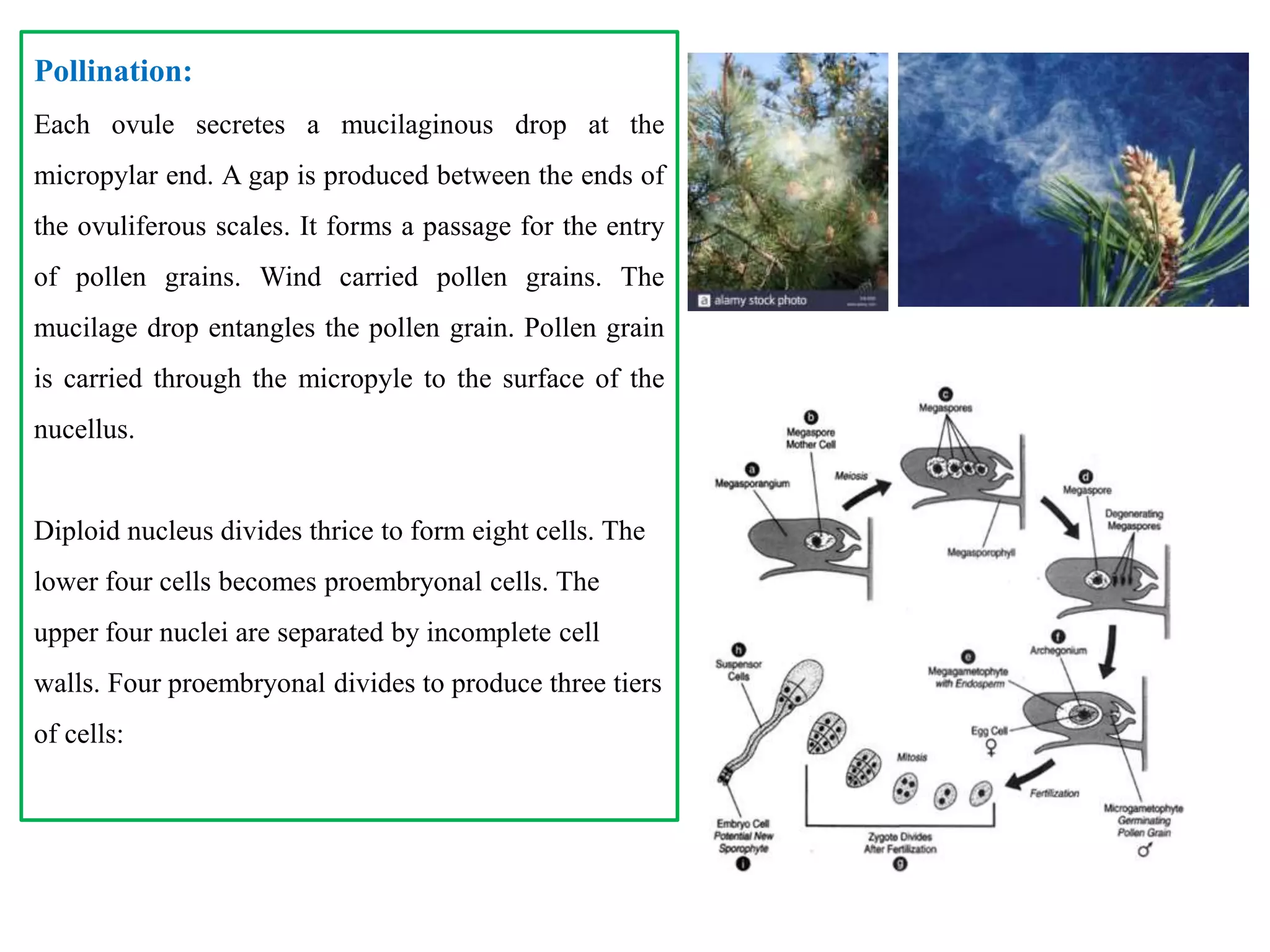
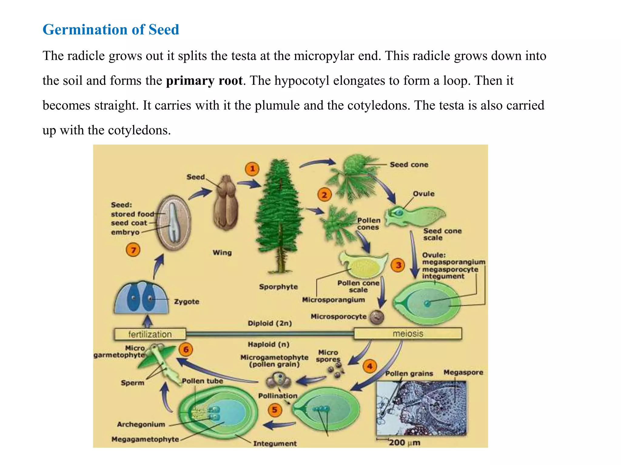
This document provides information about the genus Pinus. It discusses the systematic position of Pinus, describing that it is in the division Gymnospermae, class Coniferopsida, order Coniferales, and family Pinaceae. It then discusses the geographical distribution, morphology, internal stem and leaf structure, reproduction, development of microsporangium and female cone, structure of ovule, pollination, embryology, and germination of Pinus. Key details include that Pinus is widely distributed in the Northern hemisphere, has scale and needle-like foliage leaves, and reproduces through monoecious cones that produce microspores and megaspores through meiosis.
Vascular Cambium & Seasonal activity & its Role in Stem & RootFatima Ramay
Vascular Cambium & Seasonal activity & its Role in Stem & Root:
The vascular cambium (pl. cambia or cambiums) is a lateral meristem in the vascular tissue of plants.
The vascular cambium is a cylindrical layer of cambium that runs through the stem of a plant that undergoes secondary growth.
In Dicots:
The vascular cambium is in dicot stems and roots, located between the xylem and the phloem in the stem and root of a vascular plant, and is the source of both the secondary xylem growth (inwards, towards the pith) and the secondary phloem growth (outwards).
In Monocots:
Monocot stems, such as corn, palms and bamboos, do not have a vascular cambium and do not exhibit secondary growth by the production of concentric annual rings. They cannot increase in girth by adding lateral layers of cells as in conifers and woody dicots.
Cambium of some plants remains active for the entire period of their life, i.e., cambial cells divide and resulting cells mature to form xylem and phloem elements.
This type of seasonal activity usually found in the plants present in the tropical regions, and not all plants show cambial activity.
Percentage of ringless trees in the rain forests of;India : 75%Amazon : 43%Malaysia : 15%
In regions with definite seasonal climate; seasonal activity of cambium ceased with onset of unfavorable conditions; In Autumn, it enters the dormant state and lasts for the end of summer; In Spring, cambium again becomes active.
Duration of cambial activity is also affected by day-length, e.g., In Robinia pseudoacacia, cambium is dormant under short-day condition.
The cambium cells formed in circular in cross section from the beginning onwards.
The cambial ring is partially primary (fascicular cambium) and partially secondary (interfascicular cambium).
Periderm originates from the cortical cells (extra stelar in origin).
In Dicot stem, for mechanical support xylem is with comparatively smaller vessels, greater fibers and less parenchyma.
More amount of cork is produces for protection.
Lenticels on periderm are very prominent.
The cambial ring formed is wavy in the beginning and later becomes circular.
The cambium ring is completely secondary in origin.
Periderm originates from the pericycle (intra stelar in origin).
In Dicot root, xylem is with big thin walled vessels with few fibers and more parenchyma.
Less amount of cork is produced as root is underground.
Lenticels on periderm are not very prominent.
This is a detailed presentation on Morphology, anatomy and reproduction of Marchantia spp. with high quality pics and eye capturing transitions and animations
• Gymnosperms (Gymnos = naked, Sperma = seed) include the small group of plants with naked seeds.
• The Gymnosperms originated in the Devonian period of the Paleozoic Era and formed the supreme vegetation in the Mesozoic Era.
economic importance of gymnosperms.Gymnosperms are simple and primitive seed-bearing plants without flowers.
The plant body is sporophytic and is differentiated into root,stem and leaves.
All gymnosperms are usually wind-pollinated.
Leaves have thick cuticle and sunken stomata.
Gymnosperms are heterosporous.magasporangia and microsporangia occur on mega and microsporophylls respectively.
The "Telome theory" of Walter Zimmermann (1930, 1952) is the most accepted theory that is based on fossil record and synthesizes the major steps in the evolution of vascular plants.
It describes how the primitive type of vascular plants developed from Rhynia like plants.
Ginkgo is known as a Living Fossil.Anatomy of Ginkgo clearly shows primary and secondary structures. sex in Ginkgo is determined by sex chromosomes (XY in male and XX in female). Reproductive bodies of Ginkgo are most primitive among living seed plants except some Cycadales. Ginkgos are dioecious, with separate sexes, some trees being female and others being male. Male plants produce small pollen cones with sporophylls, each bearing two microsporangia spirally arranged around a central axis. Female plants do not produce cones. Two ovules are formed at the end of a stalk, and after pollination, one or both develop into seeds. The fertilization of ginkgo seeds occurs via motile sperm, as in cycads, ferns, mosses and algae.
Class 12||Chapter 2|| Sexual Reproduction in flowering plantsPrathamBiology
This chapter includes flowers, their detailed structure and developmental processess which took place durin sexual reproduction. Helpful for Board and NEET students.
Fell free for any query or suggestion
Mail us on: biologypratham@gmail.com
Website : www.prathambiology.in
Gymnosperms definition
Morphological characters of Gymnosperms
Anatomy of Gymnosperms
Cycas
General character of cycas
Sexual reproduction in cycas
Asexual reproduction in cycas
Economical importance of cycas
Pinus
Characters of pinus
Sexual reproduction in pinus
Asexual reproduction in pinus
Importance of pinus
Roots anatomy
Stem anatomy
Xylem
Phloem
the top three theories of root apical meristem in plants. The theories are: 1. Apical Cell Theory 2. Histogen Theory 3. Korper-Kappe Theory.The root apical meristem, or root apex, is a small region at the tip of a root in which all cells are capable of repeated division and from which all primary root tissues are derived. The root apical meristem is protected as it passes through the soil by an outer region of living parenchyma cells called the root cap.
Operation “Blue Star” is the only event in the history of Independent India where the state went into war with its own people. Even after about 40 years it is not clear if it was culmination of states anger over people of the region, a political game of power or start of dictatorial chapter in the democratic setup.
The people of Punjab felt alienated from main stream due to denial of their just demands during a long democratic struggle since independence. As it happen all over the word, it led to militant struggle with great loss of lives of military, police and civilian personnel. Killing of Indira Gandhi and massacre of innocent Sikhs in Delhi and other India cities was also associated with this movement.
Francesca Gottschalk - How can education support child empowerment.pptxEduSkills OECD
Francesca Gottschalk from the OECD’s Centre for Educational Research and Innovation presents at the Ask an Expert Webinar: How can education support child empowerment?
Synthetic Fiber Construction in lab .pptxPavel ( NSTU)
Synthetic fiber production is a fascinating and complex field that blends chemistry, engineering, and environmental science. By understanding these aspects, students can gain a comprehensive view of synthetic fiber production, its impact on society and the environment, and the potential for future innovations. Synthetic fibers play a crucial role in modern society, impacting various aspects of daily life, industry, and the environment. ynthetic fibers are integral to modern life, offering a range of benefits from cost-effectiveness and versatility to innovative applications and performance characteristics. While they pose environmental challenges, ongoing research and development aim to create more sustainable and eco-friendly alternatives. Understanding the importance of synthetic fibers helps in appreciating their role in the economy, industry, and daily life, while also emphasizing the need for sustainable practices and innovation.
Introduction to AI for Nonprofits with Tapp NetworkTechSoup
Dive into the world of AI! Experts Jon Hill and Tareq Monaur will guide you through AI's role in enhancing nonprofit websites and basic marketing strategies, making it easy to understand and apply.
How to Make a Field invisible in Odoo 17Celine George
It is possible to hide or invisible some fields in odoo. Commonly using “invisible” attribute in the field definition to invisible the fields. This slide will show how to make a field invisible in odoo 17.
June 3, 2024 Anti-Semitism Letter Sent to MIT President Kornbluth and MIT Cor...Levi Shapiro
Letter from the Congress of the United States regarding Anti-Semitism sent June 3rd to MIT President Sally Kornbluth, MIT Corp Chair, Mark Gorenberg
Dear Dr. Kornbluth and Mr. Gorenberg,
The US House of Representatives is deeply concerned by ongoing and pervasive acts of antisemitic
harassment and intimidation at the Massachusetts Institute of Technology (MIT). Failing to act decisively to ensure a safe learning environment for all students would be a grave dereliction of your responsibilities as President of MIT and Chair of the MIT Corporation.
This Congress will not stand idly by and allow an environment hostile to Jewish students to persist. The House believes that your institution is in violation of Title VI of the Civil Rights Act, and the inability or
unwillingness to rectify this violation through action requires accountability.
Postsecondary education is a unique opportunity for students to learn and have their ideas and beliefs challenged. However, universities receiving hundreds of millions of federal funds annually have denied
students that opportunity and have been hijacked to become venues for the promotion of terrorism, antisemitic harassment and intimidation, unlawful encampments, and in some cases, assaults and riots.
The House of Representatives will not countenance the use of federal funds to indoctrinate students into hateful, antisemitic, anti-American supporters of terrorism. Investigations into campus antisemitism by the Committee on Education and the Workforce and the Committee on Ways and Means have been expanded into a Congress-wide probe across all relevant jurisdictions to address this national crisis. The undersigned Committees will conduct oversight into the use of federal funds at MIT and its learning environment under authorities granted to each Committee.
• The Committee on Education and the Workforce has been investigating your institution since December 7, 2023. The Committee has broad jurisdiction over postsecondary education, including its compliance with Title VI of the Civil Rights Act, campus safety concerns over disruptions to the learning environment, and the awarding of federal student aid under the Higher Education Act.
• The Committee on Oversight and Accountability is investigating the sources of funding and other support flowing to groups espousing pro-Hamas propaganda and engaged in antisemitic harassment and intimidation of students. The Committee on Oversight and Accountability is the principal oversight committee of the US House of Representatives and has broad authority to investigate “any matter” at “any time” under House Rule X.
• The Committee on Ways and Means has been investigating several universities since November 15, 2023, when the Committee held a hearing entitled From Ivory Towers to Dark Corners: Investigating the Nexus Between Antisemitism, Tax-Exempt Universities, and Terror Financing. The Committee followed the hearing with letters to those institutions on January 10, 202
Read| The latest issue of The Challenger is here! We are thrilled to announce that our school paper has qualified for the NATIONAL SCHOOLS PRESS CONFERENCE (NSPC) 2024. Thank you for your unwavering support and trust. Dive into the stories that made us stand out!
Biological screening of herbal drugs: Introduction and Need for
Phyto-Pharmacological Screening, New Strategies for evaluating
Natural Products, In vitro evaluation techniques for Antioxidants, Antimicrobial and Anticancer drugs. In vivo evaluation techniques
for Anti-inflammatory, Antiulcer, Anticancer, Wound healing, Antidiabetic, Hepatoprotective, Cardio protective, Diuretics and
Antifertility, Toxicity studies as per OECD guidelines
2024.06.01 Introducing a competency framework for languag learning materials ...Sandy Millin
http://sandymillin.wordpress.com/iateflwebinar2024
Published classroom materials form the basis of syllabuses, drive teacher professional development, and have a potentially huge influence on learners, teachers and education systems. All teachers also create their own materials, whether a few sentences on a blackboard, a highly-structured fully-realised online course, or anything in between. Despite this, the knowledge and skills needed to create effective language learning materials are rarely part of teacher training, and are mostly learnt by trial and error.
Knowledge and skills frameworks, generally called competency frameworks, for ELT teachers, trainers and managers have existed for a few years now. However, until I created one for my MA dissertation, there wasn’t one drawing together what we need to know and do to be able to effectively produce language learning materials.
This webinar will introduce you to my framework, highlighting the key competencies I identified from my research. It will also show how anybody involved in language teaching (any language, not just English!), teacher training, managing schools or developing language learning materials can benefit from using the framework.
2. Introduction:
Gymnosperms like all vascular plants have a sporophyte-dominant life cycle.
The gametophyte (Gamete-bearing phase) is relatively short lived.
Two spore types, microspores and megaspores are typically produced in pollen cones or ovulate
cones, respectively.
Genus pinus comes under the family pinaceae.
Systematic position of Pinus:
Division- Gymnospermae
Class-Coniferopsida
Order-Coniferales
Family-Pinaceae
Genus-Pinus.
3. Geographical distribution of Pinus:
The genus Pinus is widely distributed in the Northern hemisphere. There are about 75 species of
this genus. About six species have been recorded from different parts of our country. The blue
pine, Pinus wallichiana (Syn. P. excelsa) is largely found in North-West Himalayan region at
1,800 metres to 7,000 metres elevation.
In India the Pinus is represented by the following species.
1) Pinus gerardiana: Found in Kashmir and Himachal.
2) P. roxburghii: Found in Himalayas, kashmir, Himachal, Punjab and Uttaranchal.
3) P. wallichiana: Found in outer Himalayas, Kashmir, Punjab and Uttaranchal.
4) P. insularis: Found in Khasi hills, Nagaland and Burma.
5) P. merkusi: Found in east India and Burma.
6) P. armandi: Found in Arunachal Pradesh.
4. Morphology:
Branches of unlimited growth/Long shoot: The main branches
have an unlimited growth. They bear only scale Leaves.
Branches of limited growth/ Dwarf shoot: Branches of limited
growth or dwarf branches are produced in the axil of the scale
leaves on the main branches. These are about 1-2cm Long. These
are covered by one or two scale leaves. These branches also bear
foliage leaves. A dwarf shoot with its foliage leaves is called spur.
Leaf:
Pinus has two types of Leaves: scale leaves and foliage leaves.
Scale Leaves: The scale leaves are small. membranous and
brownish in colour. These are protective in function. They are
present on the long and dwarf shoots.
Foliage Leaves: The foliage leaves are green and needle-like.
They are found only on the dwarf shoots forming the spur.
Roots: Pinus has a well developed tap root. It remains short and
grows on hard ground or rocks. The lateral roots are well
developed. These roots spread over a large area. Young roots are
infested with a fungus to form mycorrhizae.
5. Internal Structure of the Stem:
Stem is differentiated
internally into epidermis, cortex, vascular tissue and
central pith.
1) Epidermis:
The surface is covered by an epidermis. It consists of a
sing e layer of cells. Outer walls of these cells are highly
cutinized. Below the epidermis is a hypodermis which is
formed of Layers of lignified cells.
2) Cortex:
The cortex is formed of parenchymatous cells. Cortex
has a large number of resin canals. Each resin canal is
surrounded by a layer of resin secreting glandular
epithelial cells.
6. 3) Vascular bundle.
The vascular bundles are conjoint collateral and open. They
form a ring around the pith. In each vascular bundle the
xylem is towards the inner side and phloem towards the
outerside.
A narrow strip of cambium is present between xylem and
phloem. Pericycle is present outer to the ring of vascular
bundles. A single layered thin walled endodermis is present
outside the pericycle. Secondary growth takes place by the
activity of cambium ring. There are distinct annual growth
rings in the wood.
4) Bark
Phellogen originates in the deeper layers of the cortex. It is
present in the form of strips. It produces characteristic scaly
bark.
Fig. T. S. of pinus stem
7. Internal Structure of Leaf
A transverse section shows following internal parts of the
leaf:
1) Epidermis:
Leaf s covered by thick walled epidermis. Epidermis s covered by a
thick layer of cuticle. Sunken stomata are present below the general
surface. Two or three Layered hypodermis is present underneath the
epidermis. Hypodermis is composed of sclerenchymatous tissues.
This hypodermis is the main strengthening tissue of the leaf.
2) Mesophyll:
The mesophyll of the leaf is parenchymatous. It is not
differentiated in to palisade and spongy parenchyma. Resin canals
are present below the hypodermis. Each resin canal is lined by a
layer of small epithelial cells. Each leaf is supplied by two
unbranched veins.
3) Endodermis and vascular tissues:
Endodermis is present outside the pericycle. Pericycle surrounds the
xylem Pericycle is formed of parenchymatous cells. Its cell adjacent
to the phloem are called aluminous cells. The cells adjacent to the
xylem are called tracheidal cells. These specialized cells form the
transfusion tissue. They help in the lateral flow of nutrients.
8. Internal Structure of Root:
The internal structure of root resembles to that of a
dicotyledonous root. In transverse section the root shows
a piliferous layer bearing unicellular root hairs. The root
hairs are found only in the young roots and root tips. In
young roots there is fungal growth of ectophytic
mycorrhiza. With the appearance of this fungus the root
hairs of the root disappear.
Just beneath the piliferous layer there lies a broad
cortex which consists of 4 to 5 layers of thin-walled
parenchymatous cells. The inner-most layer of the cortex
is single- layered endodermis consisting of brown
suberized cells containing tannin in them. Just below the
endodermis there is multi-layered pericycle containing
tannin and starch grains.
Lateral roots are developed from the second layer of the
pericycle. The outermost layer of the pericycle helps in
the formation of the digestive sac which enables the
lateral roots to penetrate through the cortex to the
outside.
In the center of the stele there are two to six Y-shaped
xylem bundles alternating with them. The xylem has no
true vessels and consists of tracheids. The phloem
consists of sieve tubes and phloem parenchyma.
9. Reproduction:
Pinus is monoecious. Plant develops both male and female strobili
on the same plant. The strobili are monosporous. There is no
vegetative reproduction in Pinus.
Male Cone:
The male cones are much smaller. They are produced in clusters
near the tip of the long shoots. The male cones are produced in the
spring. Each male cone has a central axis. It bears a number of
spirally arranged microsporophyll's . Each microsporophyll has
saclike microsporangia on the ventral side. Each Microsporangium
produces a large number of microspores (pollen grains).The wall of
each microspore (pollen grain) consists of inner intine and an outer
exine. It has balloon like wings. The wings help in the dispersal of
spores by wind.
10. Development of Microsporangium (Stamen)
1) A number of hypodermal cells act sporangial initials. The sporangial initials divides to form outer
wall initials and the inner archesporial initial.
2) The wall initials divide to form a many layered wall of the sporangium. The archesporial initials
also increase in number by the repeated divisions. The peripheral cells of the archesporium form the
tapetum.
3.Some of the archesporial cells are transformed into microspore mother cells. The remaining
archesporial cells and the tapetal layer provide nourishment to the developing microspore
mother cells.
4. The microspore mother cell divides by meiosis to form four microspores or pollen grains. The
exine of spore forms wings. The pollen grain divides in to smaller and larger cells.
The smaller cell again divides to form two small prothalial cells.
The larger cell becomes antheridial cell the sporangium splits and microspores are released from the
microsporangia at this stage.
11.
12. Female cone:
Female cones are produced in the axils of the scale Leaves. The
production of female cones is initiated in the winter. These become ready
for pollination during the following spring. Each young female cone has a
central axis. It bears spirally arranged scales. The scales are of two types.
Some are thin membranous and are directly attached to the central axis
they are called bract scales. Woody ovuliferous scales are present on the
ventral surface of each bract scale. The broader end of the ovuliferous
scale has projection called the umbo. Each ovuliferous scale bears two
ovules. They are situated side by side on upper side each ovule
(megasporangium) has a mass of nucellar tissue. They are surrounded by
a single integument. The micropylar end of the ovule is directed towards
the central axis. A single megaspore mother cells differentiated in the
nucellus near the micropylar end. This megaspore mother cell undergoes
meiosis to form four megaspores only the lower most megaspore remains
functional the others disintegrate. Functional megaspore (embryo sac)
increases in size. It occupies the major part of the nucellus. Pollination
takes place at this stage. Images copyright to google
13. Structure of Ovule:
In Pinus two anatropous ovules are formed on the upper side of the
ovuliferous scale. Each ovule consists of a central mass of tissue called
'nucellus' which is surrounded by a covering called 'integument'. The
integument arises from the base of the ovule covers nucelluson all sides,
except at the top, where it leaves a small passage called 'micropyle. The
integument is differentiated into an outer fleshy layer (sarcotesta), middle
stony layer (sclerotesta) and the inner fleshy layer (sarcotesta). The
micropyle leads to the top of the nucellus. Where nucellus develops a cup
like chamber called "pollen chamber is present. In pollen chamber the
pollen grains are lodged after the pollination. In Pinus nucellar beak is not
formed. Both inner and outer vascular strands are absent. In nucellus,
towards the micropylar end a single large cell gets differentiated. It is the
archesporial cell. It divides periclinally into an outer parietal cell end
inner sporogenous cell or megaspore mother cell. The parietal cell divides
and develops into a nourishing layer. Megaspore mother cell undergose
meiosis and forms a liner tetrad of megaspore of which the basal one is the
functional megaspore. It is called as embryo sac cell.
nucellus
14. Pollination:
Each ovule secretes a mucilaginous drop at the
micropylar end. A gap is produced between the ends of
the ovuliferous scales. It forms a passage for the entry
of pollen grains. Wind carried pollen grains. The
mucilage drop entangles the pollen grain. Pollen grain
is carried through the micropyle to the surface of the
nucellus.
Diploid nucleus divides thrice to form eight cells. The
lower four cells becomes proembryonal cells. The
upper four nuclei are separated by incomplete cell
walls. Four proembryonal divides to produce three tiers
of cells:
15. Embryonal cell; The cells of the lower tier become embryonal cells. The four
embryonal cells separate from each other. Each develops into a separate embryo
independently. Each embryonal cell forms secondary suspensor cells. The formation of
more than one embryo from a single fertilized oosphere is called polyembryony. Only
one embryo reaches maturity. The rest are aborted.
Suspensor cells: The cells of the middle tier become suspensor cells. suspensor cells
elongate very much. It pushes the developing embryos into the prothalial tissue for
nutrition.
Rosette cells: The cells of the upper most tiers are called the rosette cells. These cells
do not take part in the development of the embryo.
A fully developed embryo is in the form a short straight axis. Its radicle is present
towards the micropylar end. Plumule is present towards the inner side. Plumule is
surrounded by the cotyledons.The unutilized prothalial tissue forms the endosperm. The
persistant nucellus tissues near the micropylar end form the perisperm. The integument
becomes hard testa. Some part of the ovuliferous scale fuses with the developing seed.
It makes a large wing for dispersal of seed. The axis of the female cone rapidly
increases. It produces gaps in ovuliferous scales. The cone becomes woody for the
dispersal of seeds
16. Germination of Seed
The radicle grows out it splits the testa at the micropylar end. This radicle grows down into
the soil and forms the primary root. The hypocotyl elongates to form a loop. Then it
becomes straight. It carries with it the plumule and the cotyledons. The testa is also carried
up with the cotyledons.