Download as PDF, PPTX
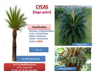


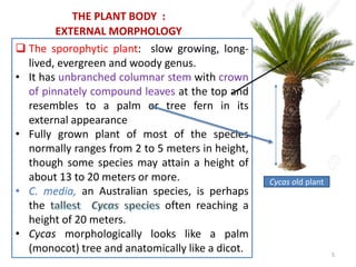


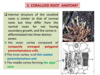


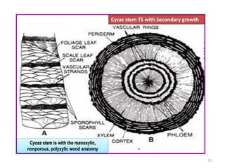


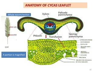


Cycas is a genus of cycads distributed throughout tropical and subtropical regions. It has unbranched woody stems topped with a crown of large pinnately compound leaves. Species such as C. revoluta are commonly cultivated as ornamental plants. The document describes the anatomy of Cycas roots, stems, and leaves. Roots have a taproot system and some develop coralloid roots through symbiosis with algae and fungi. The stem has a eustele vascular cylinder and undergoes both primary and secondary growth. Leaves are pinnately compound.