- Mastigomycotina is a former taxonomic grouping of fungi that included classes like Chytridiomycetes.
- Chytridiomycetes, commonly called chytrids, are mostly aquatic fungi found in soils and aquatic habitats. They can be unicellular or filamentous.
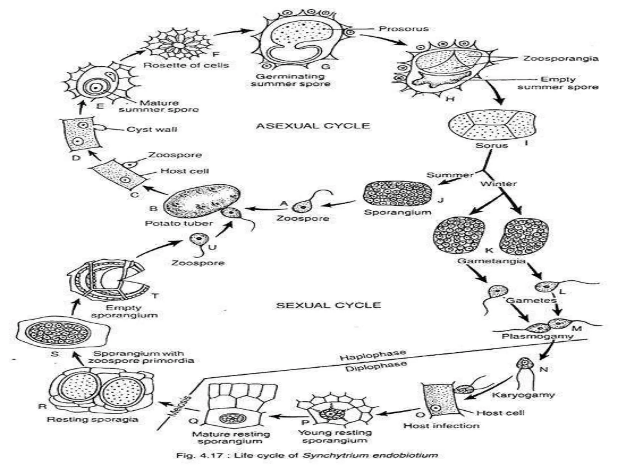
- Synchytrium endobioticum is a chytrid fungus that causes the black wart disease of potato. It has both asexual and sexual life cycles involving zoospores, gametes and resting spores that allow it to infect and overwinter on potato plants.