FUNARIA
SYSTEMATICPOSITION..
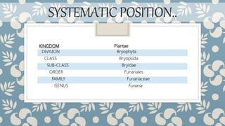
KINGDOM Plantae
DIVISION Bryophyta
CLASS Bryopsida
SUB-CLASS Bryidae
ORDER Funariales
FAMILY Funariaceae
GENUS Funaria
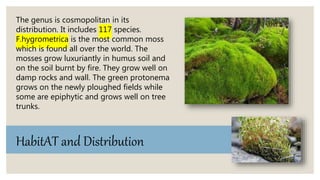
The genus is cosmopolitan in its
distribution. It includes 117 species.
F.hygrometrica is the most common moss
which is found all over the world. The
mosses grow luxuriantly in humus soil and
on the soil burnt by fire. They grow well on
damp rocks and wall. The green protonema
grows on the newly ploughed fields while
some are epiphytic and grows well on tree
trunks.
HabitAT and Distribution
The plant body is gametophytic and consists of two
different stages :
External structure of the gametophyte
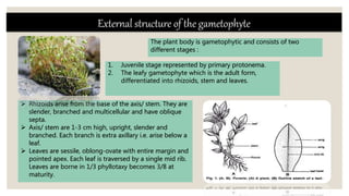
 Rhizoids arise from the base of the axis/ stem. They are
slender, branched and multicellular and have oblique
septa.
 Axis/ stem are 1-3 cm high, upright, slender and
branched. Each branch is extra axillary i.e. arise below a
leaf.
 Leaves are sessile, oblong-ovate with entire margin and
pointed apex. Each leaf is traversed by a single mid rib.
Leaves are borne in 1/3 phyllotaxy becomes 3/8 at
maturity.
1. Juvenile stage represented by primary protonema.
2. The leafy gametophyte which is the adult form,
differentiated into rhizoids, stem and leaves.
Internal Morphology
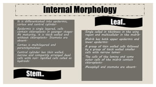
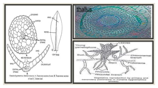
o It is differentiated into epidermis,
cortex and central cylinder.
o Epidermis is single layered, cells
contain chloroplasts in younger stages.
At maturity, it is thick walled and
without chloroplasts. Stomata are
absent.
o Cortex is multilayered and
parenchymatous.
o Central cylinder has thin walled,
narrow and compactly arranged dead
cells with non- lignified cells called as
hydroids.
◦ Single celled in thickness in the wing
region and multicellular in the midrib.
◦ Midrib has both upper epidermis and
lower epidermis.
◦ A group of thin walled cells followed
by a group of thick walled smaller
cells with narrow lumen.
◦ The cells of the lamina and some
outer cells of the midrib contain
chloroplasts.
◦ Mesophyll and stomata are absent.
Stem..
Leaf..
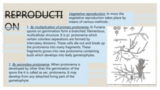
thallus
REPRODUCTI
ON
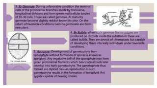
2. By secondary protonema- When protonema is
developed by other than the germination of the
spore the it is called as sec. protonema. It may
develop from any detached living part of the
gametophyte.
Vegetative reproduction- In moss the
vegetative reproduction takes place by
means of various methods :
1. By multiplication of primary protonema- In Funaria
spores on germination form a branched, filamentous,
multicellular structure. It is pr. protonema which
certain colorless separations are formed by
intercalary divisions. These cells die out and break up
the protonema into many fragments. These
fragments grows into new protonema containing
buds which develops into leafy gametophytes.
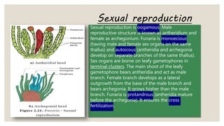
3. By Gemmae- During unfavorable condition the terminal
cells of the protonemal branches divide by transverse,
longitudinal divisions and form green multicellular bodies
of 10-30 cells. These are called gemmae. At maturity
gemmae become slightly reddish brown in color. On the
return of favorable conditions Gemma germinate and form
new plant.
5. Apospory- Development of gametophyte from
sporophyte without formation of spores is known as
apospory. Any vegetative cell of the sporophyte may form
green protonemal filaments which bears lateral buds later
develop into leafy gametophyte. The gametophyte thus
formed are diploid. Sexual reproduction in such
gametophyte results in the formation of tetraploid (4n)
zygote capable of bearing spores.
4. By Bulbils- When such gemmae like structures are
produced on rhizoids inside the substratum these are
called bulbils. They are devoid of chloroplasts but capable
of developing them into leafy individuals under favorable
conditions.
Sexual reproduction is oogamous. Male
reproductive structure is known as antheridium and
female as archegonium. Funaria is monoecious
(having male and female sex organs on the same
thallus) and autoicous (antheridia and archegonia
develop on separate branches of the same thallus).
Sex organs are borne on leafy gametophores in
terminal clusters. The main shoot of the leafy
gametophore bears antheridia and act as male
branch. Female branch develops as a lateral
outgrowth from the base of the male branch and
bears archegonia. It grows higher than the male
branch. Funaria is protandrous (antheridia mature
before the archegonia). It ensures the cross
fertilization.
Sexual reproduction
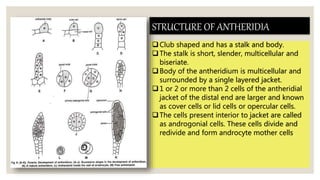
STRUCTURE OF ANTHERIDIA
Club shaped and has a stalk and body.
The stalk is short, slender, multicellular and
biseriate.
Body of the antheridium is multicellular and
surrounded by a single layered jacket.
1 or 2 or more than 2 cells of the antheridial
jacket of the distal end are larger and known
as cover cells or lid cells or opercular cells.
The cells present interior to jacket are called
as androgonial cells. These cells divide and
redivide and form androcyte mother cells.
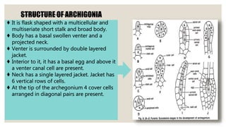
STRUCTURE OF ARCHIGONIA
♦ It is flask shaped with a multicellular and
multiseriate short stalk and broad body.
♦ Body has a basal swollen venter and a
projected neck.
♦ Venter is surrounded by double layered
jacket.
♦ Interior to it, it has a basal egg and above it
a venter canal cell are present.
♦ Neck has a single layered jacket. Jacket has
6 vertical rows of cells.
♦ At the tip of the archegonium 4 cover cells
arranged in diagonal pairs are present.
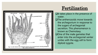
It takes place in the presence of
water.
The antherozoids move towards
the archegonium in response to
the sugars of archegonial
secretion. This phenomenon is
known as Chemotaxy.
One of the male gametes that
enter into the archegonial venter
unites with the egg cell to form
diploid zygote.
Fertilization
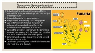
Sporophyte (Sporogonium) (2n)
♠ It divides in the archegonial venter and forms
multicellular embryo. It develops into sporophyte
or sporogonium.
♠ It is partial parasite on gametophore.
♠ It reproduces asexually by producing spores.
♠ Due to fertilization stimulus, the jacket of
archegonial venter divides and forms the
protective structure known as Calyptra.
♠ During the growth of sporophyte, the calyptra is
ruptured transversely and the upper part remains
as a hood like structure over the capsule.
♠ Calyptra functions as a transpiring shield around
the immature capsule.
♠ Developed sporophyte of Funaria is differentiated
into Foot, Seta and Capsule.
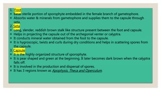
1. Foot
¤ Basal sterile portion of sporophyte embedded in the female branch of gametophore.
¤ Absorbs water & minerals from gametophore and supplies them to the capsule through
seta.
2. Seta
¤ Long, slender, reddish brown stalk like structure present between the foot and capsule.
¤ Helps in projecting the capsule out of the archegonial venter or calyptra.
¤ It conducts mineral water obtained from the foot to the capsule.
¤ It is hygroscopic, twists and curls during dry conditions and helps in scattering spores from
the capsule.
3. Capsule
¤ It is the highly organized structure of sporophyte.
¤ It is pear shaped and green at the beginning. It later becomes dark brown when the calyptra
falls off.
¤ It is involved in the production and dispersal of spores.
¤ It has 3 regions known as Apophysis, Theca and Operculum.
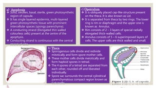
a) Apophysis
 Solid, swollen, basal, sterile, green photosynthetic
region of capsule.
 It has single layered epidermis, multi-layered
green photosynthetic tissue with prominent
intercellular spaces (spongy parenchyma).
 A conducting strand (Elongated thin walled
colourless cells) present at the centre of the
apophysis.
 Conducting strand is continuous with the central
strand of seta.
b) Theca
 Sporogenous cells divide and redivide
mitotically and form spore mother cells.
 These mother cells divide meiotically and
form haploid spores in tetrad.
 The 4 spores of a tetrad are separated from
each other, rounded off and liberated
individually.
 Spore sac surrounds the central cylindrical
parenchymatous compact region known as
Columella.
c) Operculum
 It is obliquely placed cap-like structure present
on the theca. It is also known as Lid.
 It is separated from theca by two rings. The lower
ring is rim or diaphragm and the upper one is
known as Annulus.
 Rim consists of 2 – 3 layers of special radially
elongated thick-walled cells.
 Annulus consists of 5 – 6 superimposed layers of
cells. The upper cells are thick walled and small.
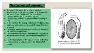
a. At maturity the lower tier of cells of annulus
degenerate and the operculum proper is removed in
the form of cap. This exposes the peristome.
b. The thin walled cells of columella dry off.
c. The capsule is inverted due to downward twisting of
seta.
d. In dry weather, the outer peristomal teeth loose water,
curl, bend backwards and promote the easy liberation
of spores.
e. The jerking movements of peristomal teeth also aid in
the removal of operculum.
f. The inner peristomal teeth do not exhibit hygroscopic
movements but act as a further check over the spore
sac.
g. The mechanical jerk of the capsule caused by twisting
and untwisting of seta also aid in the dispersal of
spores.
Dehiscence of capsule..
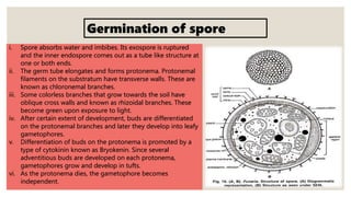
i. Spore absorbs water and imbibes. Its exospore is ruptured
and the inner endospore comes out as a tube like structure at
one or both ends.
ii. The germ tube elongates and forms protonema. Protonemal
filaments on the substratum have transverse walls. These are
known as chloronemal branches.
iii. Some colorless branches that grow towards the soil have
oblique cross walls and known as rhizoidal branches. These
become green upon exposure to light.
iv. After certain extent of development, buds are differentiated
on the protonemal branches and later they develop into leafy
gametophores.
v. Differentiation of buds on the protonema is promoted by a
type of cytokinin known as Bryokenin. Since several
adventitious buds are developed on each protonema,
gametophores grow and develop in tufts.
vi. As the protonema dies, the gametophore becomes
independent.
Germination of spore
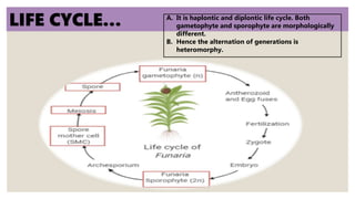
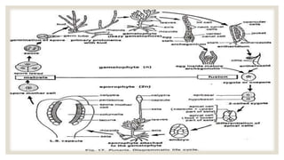
LIFE CYCLE… A. It is haplontic and diplontic life cycle. Both
gametophyte and sporophyte are morphologically
different.
B. Hence the alternation of generations is
heteromorphy.
THANK YOU!!!
MANSI YADAV
SECTION-C
ROLL NO.-204