Downloaded 300 times
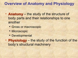

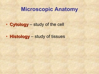


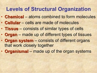
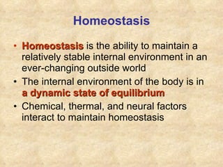
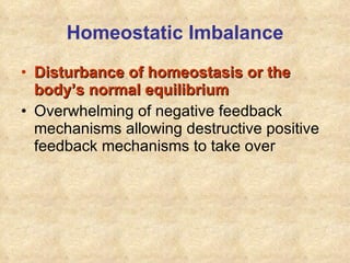
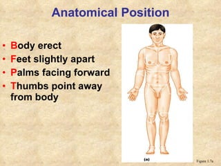


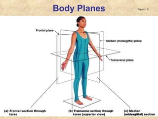
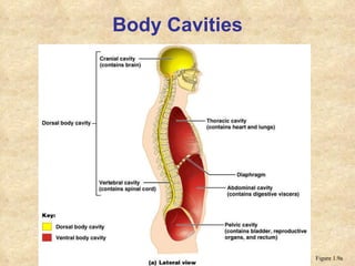


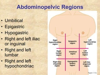


The document provides an overview of anatomy and physiology, describing their relationship and key areas of focus. It discusses the different levels of structural organization in the body from atoms to organ systems. Homeostasis and factors that maintain the stable internal environment are also summarized. The main body cavities and regions are defined, including the dorsal, ventral, thoracic and abdominopelvic cavities. Direction terms and anatomical planes are also introduced.


![04 [chapter 4 the tissue level of organization][11e]](https://cdn.slidesharecdn.com/ss_thumbnails/04chapter4thetissueleveloforganization11e-170828035609-thumbnail.jpg?width=640&height=640&fit=bounds)


![13 [chapter 13 the spinal cord and spinal nerves]](https://cdn.slidesharecdn.com/ss_thumbnails/13chapter13thespinalcordandspinalnerves-170828040950-thumbnail.jpg?width=640&height=640&fit=bounds)