Downloaded 400 times

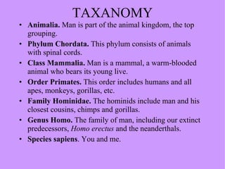
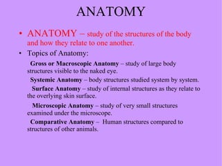
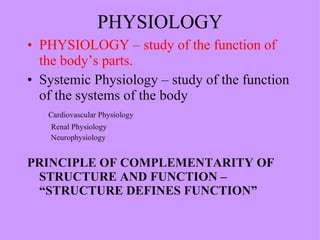
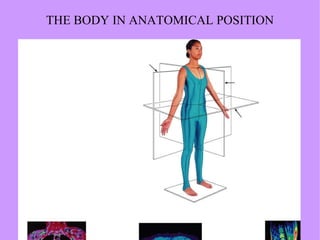

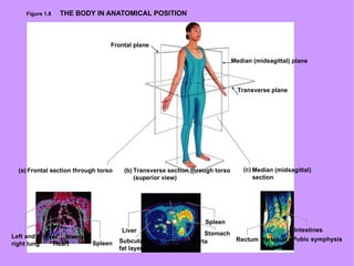
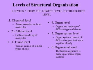
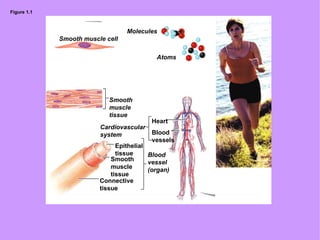
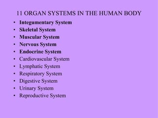
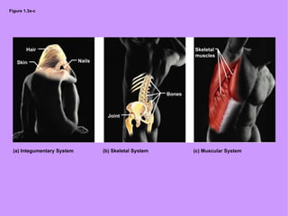
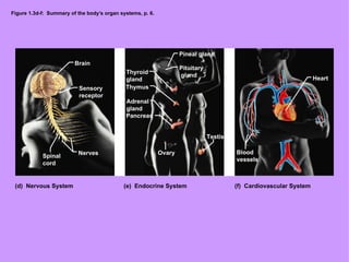
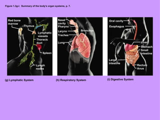
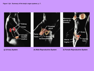

The document provides an overview of human anatomy and physiology. It discusses the taxonomy of humans and defines key terms like anatomy, gross anatomy, microscopic anatomy, and comparative anatomy. It also outlines the six levels of structural organization in the human body from chemical to organismal. Finally, it summarizes the 11 organ systems that make up the human body and introduces key concepts like homeostasis and anatomical positioning.


![Anatomy introduction[1]](https://cdn.slidesharecdn.com/ss_thumbnails/9rhvq4jzrwacyv5bjs6b-signature-460517c25b85fc4e63c8080c3e27df73c8dfae9e0c6544cc7ea6d9e8b5e79cc7-poli-180213064029-thumbnail.jpg?width=640&height=640&fit=bounds)


