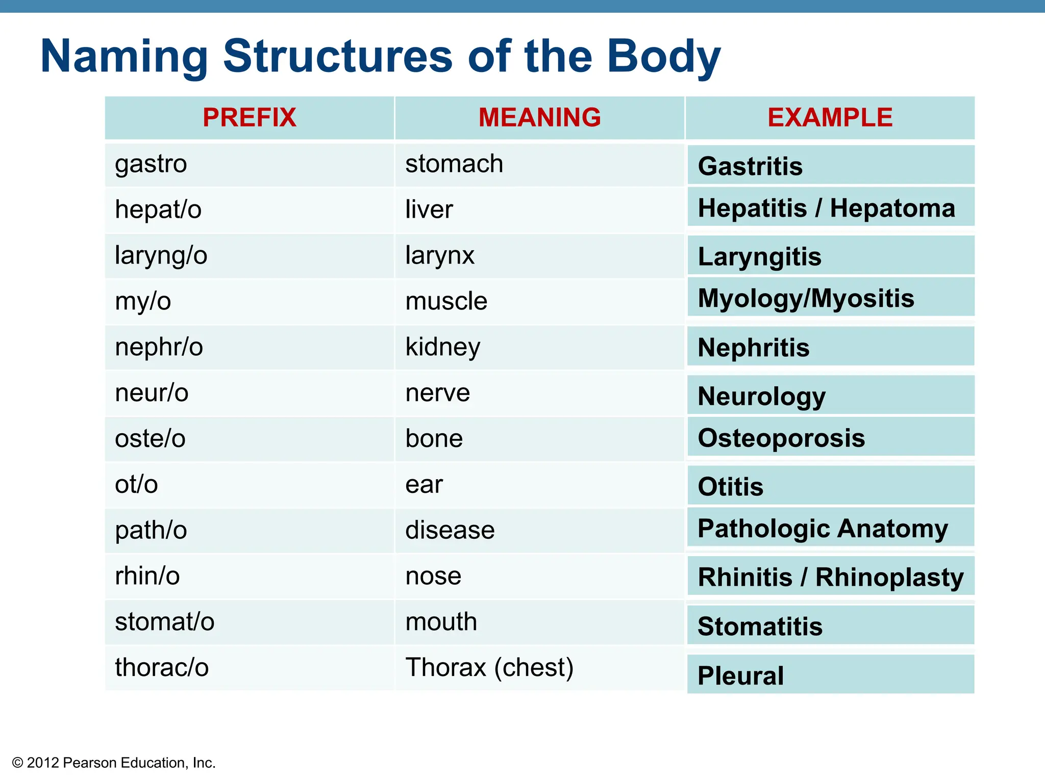
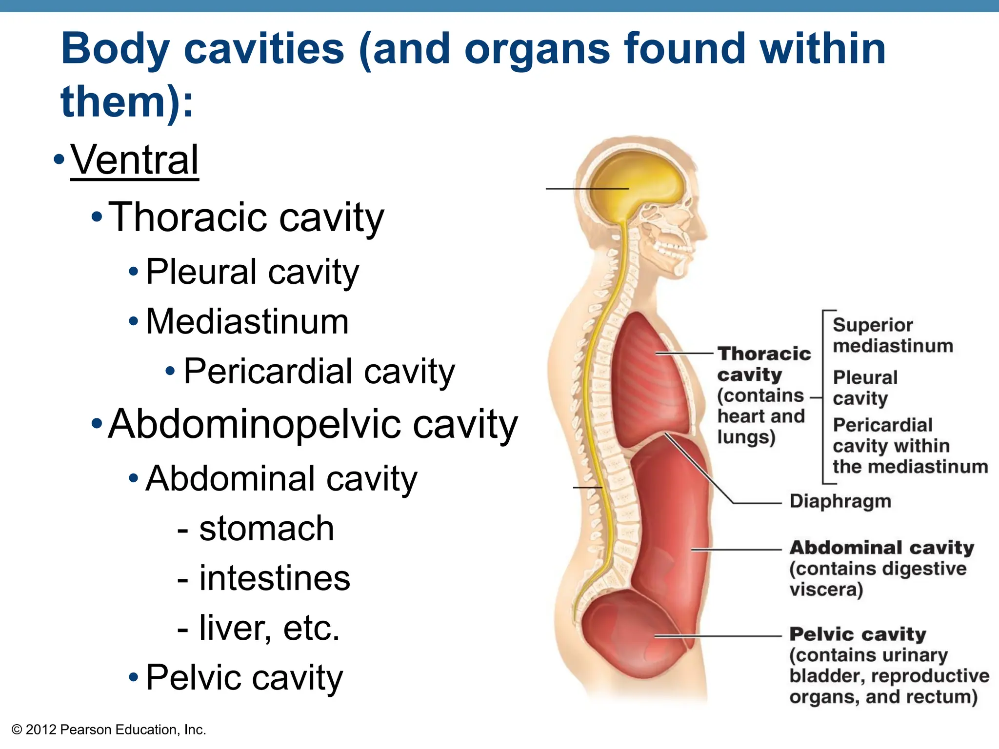
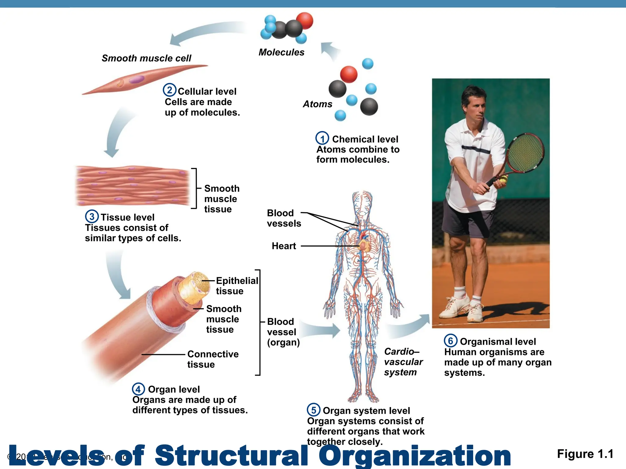
© 2012 Pearson Education, Inc.
Human Anatomy
and
Physiology
Chapter 1
The Human Body:
An Orientation
Lecture Presentation by
Patty Bostwick-Taylor
Florence-Darlington Technical
College
© 2018 Pearson Education, Inc.
© 2012 Pearson Education, Inc.
The Human Body—An Orientation
Anatomy
•Studies the structure of body parts and their
relationships.
Physiology
•Study of how the body and its parts work or
function
•Study of functional changes associated with
disease and aging.
Pathophysiology
© 2012 Pearson Education, Inc.
Gross or Macroscopic anatomy
•Larger structures
•Can be seen with the naked eye
•Approached of study: Regional and
Systemic Anatomy
Anatomy—Levels of Study
© 2012 Pearson Education, Inc.
Anatomy—Levels of Study
Surface anatomy
•Study of internal structures as they relate to
the overlying skin surface.
© 2012 Pearson Education, Inc.
Anatomy—Levels of Study
Microscopic anatomy
•Structures cannot be seen with the
naked eye
•Structures can only be viewed with a
microscope
•Subdivisions include cytology and
histology
© 2012 Pearson Education, Inc.
Anatomy—Levels of Study
Developmental anatomy
•Traces structural changes that occur in
the body throughout the life span.
•Embryology – concerns developmental
changes that occur before birth.
© 2012 Pearson Education, Inc.
The Language of Anatomy
•Special terminology is used to prevent
misunderstanding
•Exact terms are used for
•Position
•Direction
•Regions
•Structures
© 2012 Pearson Education, Inc.
Anatomical Position
© 2012 Pearson Education, Inc.
• prone position - anatomical position is
placed face-down
© 2012 Pearson Education, Inc.
• supine position- anatomical position is
placed face-up
© 2012 Pearson Education, Inc.
• posterosuperior view combines the posterior
and superior
© 2012 Pearson Education, Inc.
• B) anterosuperior
© 2012 Pearson Education, Inc.
Cervical
Umbilical
Pubic (genital)
Pelvic
Inguinal
(groin)
(a) Anterior/Ventral
KEY:
Deltoid
Digital
Lower limb
Coxal (hip)
Patellar
Fibular
Pedal (foot)
Digital
Upper limb
Acromial
Brachial (arm)
Antecubital
Antebrachial
Carpal (wrist)
Manus (hand)
Crural (leg)
Tarsal (ankle)
Femoral (thigh)
Cephalic
Frontal
Orbital
Nasal
Buccal
Oral
Mental
Thorax
Abdomen
Back (Dorsum)
Thoracic
Sternal
Axillary
Abdominal
(forearm)
Figure 1.5a
Regional Terms
•Anterior body
landmarks
© 2012 Pearson Education, Inc.
(b) Posterior/Dorsal
Gluteal
Sacral
Lumbar
Vertebral
Back (dorsal)
Scapular
Cervical
Cephalic
Occipital (back
of head)
Acromial
Brachial (arm)
Olecranal
Antebrachial
Manus (hand)
Digital
Femoral (thigh)
Popliteal
Sural (calf)
Fibular
Pedal (foot)
Calcaneal
Plantar
Upper limb
KEY:
Thorax
Abdomen
Back (Dorsum)
(forearm)
Lower limb
Figure 1.5b
Regional Terms
•Posterior body
landmarks
© 2012 Pearson Education, Inc.
Directional Terms
•Superior (cranial or cephalad)
toward the head end or upper part of a
structure or the body; above
•Inferior (caudal)
away from the head end or toward the
lower part of a structure
the body; below
© 2012 Pearson Education, Inc.
Directional Terms
•Ventral (anterior)
toward or at the front of the body; in front of
•Dorsal (posterior)
toward or at the backside of the
body; behind
© 2012 Pearson Education, Inc.
Directional Terms
•Medial:
toward or at the midline of the
body; on the inner side of
•Lateral:
away from the midline of the
body; on the outer side of the
body
•Intermediate:
between a more medial and a
more lateral structure
© 2012 Pearson Education, Inc.
Directional Terms
•Proximal:
close to the origin of the body
part or point of attachment to a
limb to the body trunk
•Distal:
farther from the origin of a body
part or the point of attachment of
a limb to the body trunk
© 2012 Pearson Education, Inc.
Directional Terms
•Superficial:
toward or at the body surface
•Deep:
away from the body surface;
more internal
•Ipsilateral: on the same side
•Contralateral: opposite side
© 2012 Pearson Education, Inc.
(a) Median (midsagittal)
Vertebral
column
(b) Frontal (coronal) plane
Right
lung Heart
Left
lung
(c) Transverse plane
Liver Aorta Spleen
Spinal
cord
Rectum Intestines Liver Stomach Spleen Subcutaneous
fat layer
Stomach
Figure 1.6
© 2012 Pearson Education, Inc.
PREFIX MEANING EXAMPLE
abdomin/o abdomen
acr/o extremity
aden/o gland
angi/o vessel
arthr/o joint
cardi/o heart
chondr/o cartilage
cyst/o bladder
cyt/o cell
den/o tooth
dermat/o skin
derm/o skin
duoden/o duodenum
Naming Structures of the Body
Peritoneal
Acromegaly
Adenoma
Angiogram
Arthritis
Cystitis
cytology
© 2012 Pearson Education, Inc.
PREFIX MEANING EXAMPLE
gastro stomach
hepat/o liver
laryng/o larynx
my/o muscle
nephr/o kidney
neur/o nerve
oste/o bone
ot/o ear
path/o disease
rhin/o nose
stomat/o mouth
thorac/o Thorax (chest)
Naming Structures of the Body
Gastritis
Hepatitis / Hepatoma
Laryngitis
Myology/Myositis
Nephritis
Otitis
Pathologic Anatomy
Neurology
Osteoporosis
Rhinitis / Rhinoplasty
Stomatitis
Pleural
© 2012 Pearson Education, Inc.
Body Cavities
•Dorsal body cavity
•Cranial cavity houses the brain
•Spinal cavity houses the spinal cord
•Ventral body cavity
•Thoracic cavity houses heart, lungs, and
others
•Abdominopelvic cavity houses digestive
system and most urinary system organs
© 2012 Pearson Education, Inc.
Body cavities (and organs found within
them):
•Dorsal
Cranial cavity
- within the rigid
skull, contains the
brain.
Spinal cavity
- which runs within
the bony vertebral
column, protects the
spinal cord.
© 2012 Pearson Education, Inc.
Body cavities (and organs found within
them):
•Ventral
•Thoracic cavity
• Pleural cavity
• Mediastinum
• Pericardial cavity
•Abdominopelvic cavity
• Abdominal cavity
- stomach
- intestines
- liver, etc.
• Pelvic cavity
© 2012 Pearson Education, Inc.
Ventral Body Cavity Membranes
•Called serous membranes or serosa
•Parietal serosa lines internal body walls
•Visceral serosa covers the internal organs
•Serous fluid separates the serosae
© 2012 Pearson Education, Inc.
Heart Serosae
Figure 1.10b
© 2012 Pearson Education, Inc.
Serous Membrane Relationship
Figure 1.10a
© 2012 Pearson Education, Inc.
Abdominopelvic quadrants (and organs found in each
region):
•Right upper
•Right lower
•Left upper
•Left lower
© 2012 Pearson Education, Inc.
Nine abdominopelvic regions (and organs in
each regions):
•Right hypochondriac
•Epigastric
•Left hypochondriac
•Right lumbar
•Umbilical
•Left lumbar
•Right iliac (inguinal)
•Hypogastric
•Left iliac (inguinal)
© 2012 Pearson Education, Inc.
Other Body Cavities
•Oral and digestive cavities
•Nasal Cavity
•Orbital cavities
•Middle ear cavities
•Synovial cavities
© 2012 Pearson Education, Inc. Figure 1.1
Organismal level
Human organisms are
made up of many organ
systems.
Organ system level
Organ systems consist of
different organs that work
together closely.
Organ level
Organs are made up of
different types of tissues.
Chemical level
Atoms combine to
form molecules.
Smooth muscle cell
Cellular level
Cells are made
up of molecules.
Molecules
2
Atoms
1
Tissue level
Tissues consist of
similar types of cells.
3
Smooth
muscle
tissue
Epithelial
tissue
Smooth
muscle
tissue
Connective
tissue
Blood
vessel
(organ)
4
5
Cardio–
vascular
system
6
Blood
vessels
Heart
Levels of Structural Organization
© 2012 Pearson Education, Inc. Figure 1.1, step 2
© 2012 Pearson Education, Inc. Figure 1.1, step 3
© 2012 Pearson Education, Inc. Figure 1.1, step 4
© 2012 Pearson Education, Inc. Figure 1.1, step 5
© 2012 Pearson Education, Inc. Figure 1.1, step 6
© 2012 Pearson Education, Inc.
Organ System Overview
•Integumentary
•Protects deeper organs from
injury due to bumps,
chemicals, bacteria, and
dehydration.
•Excretes salts and urea
•Helps regulate body
temperature
•Produces vitamin D
• Components
• Skin
• Nails
• Hair
• Cutaneous sense
organs and glands
© 2012 Pearson Education, Inc.
•Skeletal
•Protects and supports internal
organs
•Provides levers for muscular
action
•Cavities provide a site of
blood cell formation
•Stores minerals
Organ System Overview
• Components
• Bones
• Cartilage
• Tendons
• Ligaments
© 2012 Pearson Education, Inc.
•Muscular
•Produces movement
•Maintains posture
•Produces heat
Organ System Overview
© 2012 Pearson Education, Inc.
Organ System Overview
•Nervous
•Fast-acting control system
•Responds to internal and
external change
•Activates muscles and
glands
•Helps maintain short-term
homeostasis of the body
via rapid transmission of
electrical signals
© 2012 Pearson Education, Inc.
Organ System Overview
•Endocrine system
•Secretes chemical
molecules, called hormones,
into the blood
•Body functions controlled by
hormones include:
•Growth
•Reproduction
•Use of nutrients
•Plays a role in regulating
long-term homeostasis
© 2012 Pearson Education, Inc.
Organ System Overview
•Cardiovascular
•Transports materials in
body via blood pumped
by heart
•Oxygen
•Carbon dioxide
•Nutrients
•Wastes
© 2012 Pearson Education, Inc.
Organ System Overview
•Lymphatic
•Returns fluids to blood
vessels
•Cleanses the blood
•Involved in immunity
© 2012 Pearson Education, Inc.
Organ System Overview
•Respiratory
•Keeps blood supplied
with oxygen
•Removes carbon dioxide
•Contributes to the acid-
base balance of the
blood
© 2012 Pearson Education, Inc.
Organ System Overview
•Digestive
•Breaks down food
•Allows for nutrient
absorption into blood
•Eliminates indigestible
material as feces
© 2012 Pearson Education, Inc.
Organ System Overview
•Urinary
•Eliminates nitrogenous
wastes
•Maintains acid-base
balance
•Regulates water and
electrolytes
© 2012 Pearson Education, Inc.
•Reproductive
•Produces offspring
•Testes produce
sperm and male
hormone
•Ovaries produce
eggs and female
hormones
Organ System Overview
© 2012 Pearson Education, Inc.
Necessary Life Functions
•Maintain boundaries
•Internal environment remains distinct from
the external environment
•Movement
•Locomotion
•Movement of substances
•Responsiveness or excitability
•Ability to sense changes and react
•Digestion
•Breakdown and absorption of nutrients
© 2012 Pearson Education, Inc.
Necessary Life Functions
•Metabolism—chemical reactions within the
body
•Break down complex molecules into smaller
ones
•Build larger molecules from smaller ones
•Produces energy
•Regulated by hormones
•Excretion
•Eliminates waste from metabolic reactions
•Wastes may be removed in urine or feces
© 2012 Pearson Education, Inc.
Necessary Life Functions
•Reproduction
•Occurs on cellular level or organismal level
•Produces future generation
•Growth
•Increases cell size and number of cells
© 2012 Pearson Education, Inc.
Survival Needs
•Nutrients
•Chemicals for energy and cell building
•Includes carbohydrates, proteins, lipids,
vitamins, and minerals
•Oxygen
•Required for chemical reactions
© 2012 Pearson Education, Inc.
Survival Needs
•Water
•60 to 80 percent of body weight
•Most abundant chemical in the human body
•Provides for metabolic reaction
•Stable body temperature
•37°C (98°F)
•Atmospheric pressure
•Must be appropriate for gas exchange
© 2012 Pearson Education, Inc.
Maintaining Homeostasis
•The body communicates through neural and
hormonal control systems
© 2012 Pearson Education, Inc.
Homeostasis
•Homeostasis—maintenance of relatively
stable internal conditions
•A dynamic state of equilibrium, or balance
•Necessary for normal body functioning and
to sustain life
•Homeostatic imbalance
•A disturbance in homeostasis results in
disease
© 2012 Pearson Education, Inc. Figure 1.4
Input: Information
sent along afferent
pathway to control
center.
Receptor
Control
Center
Effector
Receptor
detects change.
Afferent
pathway
Efferent
pathway
VARIABLE (in homeostasis)
Stimulus
produces
change in
variable.
Output: Information
sent along efferent
pathway to effector.
Response
of effector feeds
back to reduce
the effect of
stimulus and
returns variable
to homeostatic
level.
1
2
3 4
5
© 2012 Pearson Education, Inc. Figure 1.4, step 1
VARIABLE (in homeostasis)
Stimulus
produces
change in
variable.
1
© 2012 Pearson Education, Inc. Figure 1.4, step 2
Receptor
Receptor
detects change.
Stimulus
produces
change in
variable.
1
2
VARIABLE (in homeostasis)
•Receptor
•Responds to changes in the
environment (stimuli)
•Sends information to control
center
© 2012 Pearson Education, Inc. Figure 1.4, step 3
Input: Information
sent along afferent
pathway to control
center.
Receptor
Control
Center
Receptor
detects change.
Afferent
pathway
VARIABLE (in homeostasis)
Stimulus
produces
change in
variable.
1
3
2
•Control center
•Determines set
point
•Analyzes
information
•Determines
appropriate
response
© 2012 Pearson Education, Inc. Figure 1.4, step 4
Input: Information
sent along afferent
pathway to control
center.
Receptor
Control
Center
Effector
Receptor
detects change.
Afferent
pathway
Efferent
pathway
VARIABLE (in homeostasis)
Stimulus
produces
change in
variable.
Output: Information
sent along efferent
pathway to effector.
1
3 4
2
•Effector
•Provides a
means for
response to the
stimulus
© 2012 Pearson Education, Inc. Figure 1.4, step 5
Input: Information
sent along afferent
pathway to control
center.
Receptor
Control
Center
Effector
Receptor
detects change.
Afferent
pathway
Efferent
pathway
VARIABLE (in homeostasis)
Stimulus
produces
change in
variable.
Output: Information
sent along efferent
pathway to effector.
Response
of effector feeds
back to reduce
the effect of
stimulus and
returns variable
to homeostatic
level.
1
3 4
5
2
© 2012 Pearson Education, Inc.
Feedback Mechanisms
•Negative feedback
•Includes most homeostatic control
mechanisms
•Shuts off the original stimulus, or reduces its
intensity
•Works like a household thermostat
© 2012 Pearson Education, Inc.
Feedback Mechanisms
•Positive feedback
•Increases the original stimulus to push the
variable farther
•In the body this only occurs in blood clotting
and during the birth of a baby
© 2012 Pearson Education, Inc.
Homeostatic imbalance +
•SEROSITIS – inflammation of the serous
membrane
3 TYPES:
•Pericarditis – inflammation of pericardium
(heart)
•Pleuritis – also pleurisy
- inflammation of pleura (lungs)
•Peritonitis – peritoneum

CHAPTER-1-The-Human-Body-An-Orientation.pdf

  • 1.
    © 2012 PearsonEducation, Inc. Human Anatomy and Physiology
  • 2.
    Chapter 1 The HumanBody: An Orientation Lecture Presentation by Patty Bostwick-Taylor Florence-Darlington Technical College © 2018 Pearson Education, Inc.
  • 3.
    © 2012 PearsonEducation, Inc. The Human Body—An Orientation Anatomy •Studies the structure of body parts and their relationships. Physiology •Study of how the body and its parts work or function •Study of functional changes associated with disease and aging. Pathophysiology
  • 4.
    © 2012 PearsonEducation, Inc. Gross or Macroscopic anatomy •Larger structures •Can be seen with the naked eye •Approached of study: Regional and Systemic Anatomy Anatomy—Levels of Study
  • 5.
    © 2012 PearsonEducation, Inc. Anatomy—Levels of Study Surface anatomy •Study of internal structures as they relate to the overlying skin surface.
  • 6.
    © 2012 PearsonEducation, Inc. Anatomy—Levels of Study Microscopic anatomy •Structures cannot be seen with the naked eye •Structures can only be viewed with a microscope •Subdivisions include cytology and histology
  • 7.
    © 2012 PearsonEducation, Inc. Anatomy—Levels of Study Developmental anatomy •Traces structural changes that occur in the body throughout the life span. •Embryology – concerns developmental changes that occur before birth.
  • 8.
    © 2012 PearsonEducation, Inc. The Language of Anatomy •Special terminology is used to prevent misunderstanding •Exact terms are used for •Position •Direction •Regions •Structures
  • 9.
    © 2012 PearsonEducation, Inc. Anatomical Position
  • 10.
    © 2012 PearsonEducation, Inc. • prone position - anatomical position is placed face-down
  • 11.
    © 2012 PearsonEducation, Inc. • supine position- anatomical position is placed face-up
  • 12.
    © 2012 PearsonEducation, Inc. • posterosuperior view combines the posterior and superior
  • 13.
    © 2012 PearsonEducation, Inc. • B) anterosuperior
  • 14.
    © 2012 PearsonEducation, Inc. Cervical Umbilical Pubic (genital) Pelvic Inguinal (groin) (a) Anterior/Ventral KEY: Deltoid Digital Lower limb Coxal (hip) Patellar Fibular Pedal (foot) Digital Upper limb Acromial Brachial (arm) Antecubital Antebrachial Carpal (wrist) Manus (hand) Crural (leg) Tarsal (ankle) Femoral (thigh) Cephalic Frontal Orbital Nasal Buccal Oral Mental Thorax Abdomen Back (Dorsum) Thoracic Sternal Axillary Abdominal (forearm) Figure 1.5a Regional Terms •Anterior body landmarks
  • 15.
    © 2012 PearsonEducation, Inc. (b) Posterior/Dorsal Gluteal Sacral Lumbar Vertebral Back (dorsal) Scapular Cervical Cephalic Occipital (back of head) Acromial Brachial (arm) Olecranal Antebrachial Manus (hand) Digital Femoral (thigh) Popliteal Sural (calf) Fibular Pedal (foot) Calcaneal Plantar Upper limb KEY: Thorax Abdomen Back (Dorsum) (forearm) Lower limb Figure 1.5b Regional Terms •Posterior body landmarks
  • 16.
    © 2012 PearsonEducation, Inc. Directional Terms •Superior (cranial or cephalad) toward the head end or upper part of a structure or the body; above •Inferior (caudal) away from the head end or toward the lower part of a structure the body; below
  • 17.
    © 2012 PearsonEducation, Inc. Directional Terms •Ventral (anterior) toward or at the front of the body; in front of •Dorsal (posterior) toward or at the backside of the body; behind
  • 18.
    © 2012 PearsonEducation, Inc. Directional Terms •Medial: toward or at the midline of the body; on the inner side of •Lateral: away from the midline of the body; on the outer side of the body •Intermediate: between a more medial and a more lateral structure
  • 19.
    © 2012 PearsonEducation, Inc. Directional Terms •Proximal: close to the origin of the body part or point of attachment to a limb to the body trunk •Distal: farther from the origin of a body part or the point of attachment of a limb to the body trunk
  • 20.
    © 2012 PearsonEducation, Inc. Directional Terms •Superficial: toward or at the body surface •Deep: away from the body surface; more internal •Ipsilateral: on the same side •Contralateral: opposite side
  • 21.
    © 2012 PearsonEducation, Inc.
  • 22.
    © 2012 PearsonEducation, Inc.
  • 23.
    © 2012 PearsonEducation, Inc.
  • 24.
    © 2012 PearsonEducation, Inc.
  • 25.
    © 2012 PearsonEducation, Inc.
  • 26.
    © 2012 PearsonEducation, Inc.
  • 27.
    © 2012 PearsonEducation, Inc.
  • 28.
    © 2012 PearsonEducation, Inc. (a) Median (midsagittal) Vertebral column (b) Frontal (coronal) plane Right lung Heart Left lung (c) Transverse plane Liver Aorta Spleen Spinal cord Rectum Intestines Liver Stomach Spleen Subcutaneous fat layer Stomach Figure 1.6
  • 29.
    © 2012 PearsonEducation, Inc. PREFIX MEANING EXAMPLE abdomin/o abdomen acr/o extremity aden/o gland angi/o vessel arthr/o joint cardi/o heart chondr/o cartilage cyst/o bladder cyt/o cell den/o tooth dermat/o skin derm/o skin duoden/o duodenum Naming Structures of the Body Peritoneal Acromegaly Adenoma Angiogram Arthritis Cystitis cytology
  • 30.
    © 2012 PearsonEducation, Inc. PREFIX MEANING EXAMPLE gastro stomach hepat/o liver laryng/o larynx my/o muscle nephr/o kidney neur/o nerve oste/o bone ot/o ear path/o disease rhin/o nose stomat/o mouth thorac/o Thorax (chest) Naming Structures of the Body Gastritis Hepatitis / Hepatoma Laryngitis Myology/Myositis Nephritis Otitis Pathologic Anatomy Neurology Osteoporosis Rhinitis / Rhinoplasty Stomatitis Pleural
  • 31.
    © 2012 PearsonEducation, Inc. Body Cavities •Dorsal body cavity •Cranial cavity houses the brain •Spinal cavity houses the spinal cord •Ventral body cavity •Thoracic cavity houses heart, lungs, and others •Abdominopelvic cavity houses digestive system and most urinary system organs
  • 32.
    © 2012 PearsonEducation, Inc. Body cavities (and organs found within them): •Dorsal Cranial cavity - within the rigid skull, contains the brain. Spinal cavity - which runs within the bony vertebral column, protects the spinal cord.
  • 33.
    © 2012 PearsonEducation, Inc.
  • 34.
    © 2012 PearsonEducation, Inc. Body cavities (and organs found within them): •Ventral •Thoracic cavity • Pleural cavity • Mediastinum • Pericardial cavity •Abdominopelvic cavity • Abdominal cavity - stomach - intestines - liver, etc. • Pelvic cavity
  • 35.
    © 2012 PearsonEducation, Inc.
  • 36.
    © 2012 PearsonEducation, Inc.
  • 37.
    © 2012 PearsonEducation, Inc. Ventral Body Cavity Membranes •Called serous membranes or serosa •Parietal serosa lines internal body walls •Visceral serosa covers the internal organs •Serous fluid separates the serosae
  • 38.
    © 2012 PearsonEducation, Inc. Heart Serosae Figure 1.10b
  • 39.
    © 2012 PearsonEducation, Inc. Serous Membrane Relationship Figure 1.10a
  • 40.
    © 2012 PearsonEducation, Inc. Abdominopelvic quadrants (and organs found in each region): •Right upper •Right lower •Left upper •Left lower
  • 41.
    © 2012 PearsonEducation, Inc. Nine abdominopelvic regions (and organs in each regions): •Right hypochondriac •Epigastric •Left hypochondriac •Right lumbar •Umbilical •Left lumbar •Right iliac (inguinal) •Hypogastric •Left iliac (inguinal)
  • 42.
    © 2012 PearsonEducation, Inc.
  • 43.
    © 2012 PearsonEducation, Inc.
  • 44.
    © 2012 PearsonEducation, Inc. Other Body Cavities •Oral and digestive cavities •Nasal Cavity •Orbital cavities •Middle ear cavities •Synovial cavities
  • 45.
    © 2012 PearsonEducation, Inc. Figure 1.1 Organismal level Human organisms are made up of many organ systems. Organ system level Organ systems consist of different organs that work together closely. Organ level Organs are made up of different types of tissues. Chemical level Atoms combine to form molecules. Smooth muscle cell Cellular level Cells are made up of molecules. Molecules 2 Atoms 1 Tissue level Tissues consist of similar types of cells. 3 Smooth muscle tissue Epithelial tissue Smooth muscle tissue Connective tissue Blood vessel (organ) 4 5 Cardio– vascular system 6 Blood vessels Heart Levels of Structural Organization
  • 46.
    © 2012 PearsonEducation, Inc. Figure 1.1, step 2
  • 47.
    © 2012 PearsonEducation, Inc. Figure 1.1, step 3
  • 48.
    © 2012 PearsonEducation, Inc. Figure 1.1, step 4
  • 49.
    © 2012 PearsonEducation, Inc. Figure 1.1, step 5
  • 50.
    © 2012 PearsonEducation, Inc. Figure 1.1, step 6
  • 51.
    © 2012 PearsonEducation, Inc. Organ System Overview •Integumentary •Protects deeper organs from injury due to bumps, chemicals, bacteria, and dehydration. •Excretes salts and urea •Helps regulate body temperature •Produces vitamin D • Components • Skin • Nails • Hair • Cutaneous sense organs and glands
  • 52.
    © 2012 PearsonEducation, Inc. •Skeletal •Protects and supports internal organs •Provides levers for muscular action •Cavities provide a site of blood cell formation •Stores minerals Organ System Overview • Components • Bones • Cartilage • Tendons • Ligaments
  • 53.
    © 2012 PearsonEducation, Inc. •Muscular •Produces movement •Maintains posture •Produces heat Organ System Overview
  • 54.
    © 2012 PearsonEducation, Inc. Organ System Overview •Nervous •Fast-acting control system •Responds to internal and external change •Activates muscles and glands •Helps maintain short-term homeostasis of the body via rapid transmission of electrical signals
  • 55.
    © 2012 PearsonEducation, Inc. Organ System Overview •Endocrine system •Secretes chemical molecules, called hormones, into the blood •Body functions controlled by hormones include: •Growth •Reproduction •Use of nutrients •Plays a role in regulating long-term homeostasis
  • 56.
    © 2012 PearsonEducation, Inc. Organ System Overview •Cardiovascular •Transports materials in body via blood pumped by heart •Oxygen •Carbon dioxide •Nutrients •Wastes
  • 57.
    © 2012 PearsonEducation, Inc. Organ System Overview •Lymphatic •Returns fluids to blood vessels •Cleanses the blood •Involved in immunity
  • 58.
    © 2012 PearsonEducation, Inc. Organ System Overview •Respiratory •Keeps blood supplied with oxygen •Removes carbon dioxide •Contributes to the acid- base balance of the blood
  • 59.
    © 2012 PearsonEducation, Inc. Organ System Overview •Digestive •Breaks down food •Allows for nutrient absorption into blood •Eliminates indigestible material as feces
  • 60.
    © 2012 PearsonEducation, Inc. Organ System Overview •Urinary •Eliminates nitrogenous wastes •Maintains acid-base balance •Regulates water and electrolytes
  • 61.
    © 2012 PearsonEducation, Inc. •Reproductive •Produces offspring •Testes produce sperm and male hormone •Ovaries produce eggs and female hormones Organ System Overview
  • 62.
    © 2012 PearsonEducation, Inc.
  • 63.
    © 2012 PearsonEducation, Inc. Necessary Life Functions •Maintain boundaries •Internal environment remains distinct from the external environment •Movement •Locomotion •Movement of substances •Responsiveness or excitability •Ability to sense changes and react •Digestion •Breakdown and absorption of nutrients
  • 64.
    © 2012 PearsonEducation, Inc. Necessary Life Functions •Metabolism—chemical reactions within the body •Break down complex molecules into smaller ones •Build larger molecules from smaller ones •Produces energy •Regulated by hormones •Excretion •Eliminates waste from metabolic reactions •Wastes may be removed in urine or feces
  • 65.
    © 2012 PearsonEducation, Inc. Necessary Life Functions •Reproduction •Occurs on cellular level or organismal level •Produces future generation •Growth •Increases cell size and number of cells
  • 66.
    © 2012 PearsonEducation, Inc. Survival Needs •Nutrients •Chemicals for energy and cell building •Includes carbohydrates, proteins, lipids, vitamins, and minerals •Oxygen •Required for chemical reactions
  • 67.
    © 2012 PearsonEducation, Inc. Survival Needs •Water •60 to 80 percent of body weight •Most abundant chemical in the human body •Provides for metabolic reaction •Stable body temperature •37°C (98°F) •Atmospheric pressure •Must be appropriate for gas exchange
  • 68.
    © 2012 PearsonEducation, Inc. Maintaining Homeostasis •The body communicates through neural and hormonal control systems
  • 69.
    © 2012 PearsonEducation, Inc. Homeostasis •Homeostasis—maintenance of relatively stable internal conditions •A dynamic state of equilibrium, or balance •Necessary for normal body functioning and to sustain life •Homeostatic imbalance •A disturbance in homeostasis results in disease
  • 70.
    © 2012 PearsonEducation, Inc. Figure 1.4 Input: Information sent along afferent pathway to control center. Receptor Control Center Effector Receptor detects change. Afferent pathway Efferent pathway VARIABLE (in homeostasis) Stimulus produces change in variable. Output: Information sent along efferent pathway to effector. Response of effector feeds back to reduce the effect of stimulus and returns variable to homeostatic level. 1 2 3 4 5
  • 71.
    © 2012 PearsonEducation, Inc. Figure 1.4, step 1 VARIABLE (in homeostasis) Stimulus produces change in variable. 1
  • 72.
    © 2012 PearsonEducation, Inc. Figure 1.4, step 2 Receptor Receptor detects change. Stimulus produces change in variable. 1 2 VARIABLE (in homeostasis) •Receptor •Responds to changes in the environment (stimuli) •Sends information to control center
  • 73.
    © 2012 PearsonEducation, Inc. Figure 1.4, step 3 Input: Information sent along afferent pathway to control center. Receptor Control Center Receptor detects change. Afferent pathway VARIABLE (in homeostasis) Stimulus produces change in variable. 1 3 2 •Control center •Determines set point •Analyzes information •Determines appropriate response
  • 74.
    © 2012 PearsonEducation, Inc. Figure 1.4, step 4 Input: Information sent along afferent pathway to control center. Receptor Control Center Effector Receptor detects change. Afferent pathway Efferent pathway VARIABLE (in homeostasis) Stimulus produces change in variable. Output: Information sent along efferent pathway to effector. 1 3 4 2 •Effector •Provides a means for response to the stimulus
  • 75.
    © 2012 PearsonEducation, Inc. Figure 1.4, step 5 Input: Information sent along afferent pathway to control center. Receptor Control Center Effector Receptor detects change. Afferent pathway Efferent pathway VARIABLE (in homeostasis) Stimulus produces change in variable. Output: Information sent along efferent pathway to effector. Response of effector feeds back to reduce the effect of stimulus and returns variable to homeostatic level. 1 3 4 5 2
  • 76.
    © 2012 PearsonEducation, Inc. Feedback Mechanisms •Negative feedback •Includes most homeostatic control mechanisms •Shuts off the original stimulus, or reduces its intensity •Works like a household thermostat
  • 77.
    © 2012 PearsonEducation, Inc. Feedback Mechanisms •Positive feedback •Increases the original stimulus to push the variable farther •In the body this only occurs in blood clotting and during the birth of a baby
  • 78.
    © 2012 PearsonEducation, Inc. Homeostatic imbalance + •SEROSITIS – inflammation of the serous membrane 3 TYPES: •Pericarditis – inflammation of pericardium (heart) •Pleuritis – also pleurisy - inflammation of pleura (lungs) •Peritonitis – peritoneum