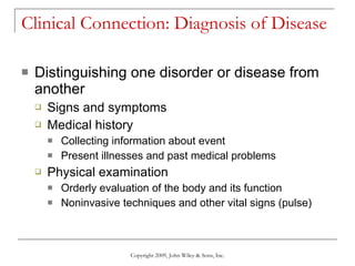
This document provides an overview of anatomy and physiology, including:
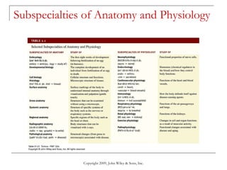
- It defines anatomy and physiology as the study of body structures and functions.
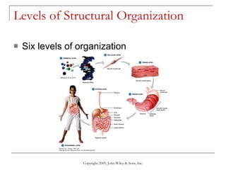
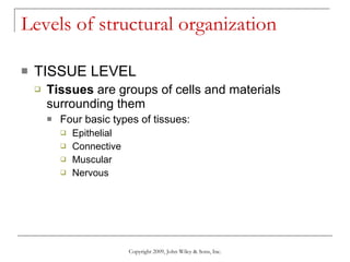
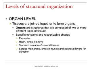
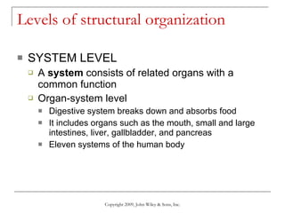
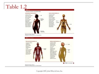
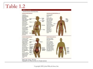
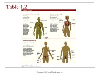
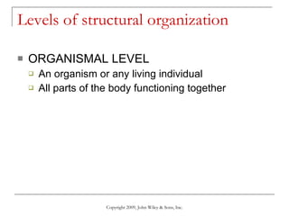
- It describes the six levels of structural organization from chemical to organism.
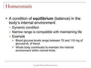
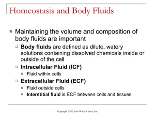
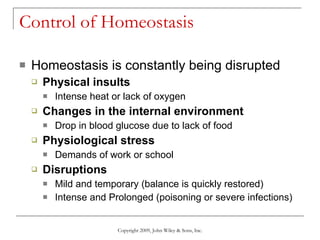
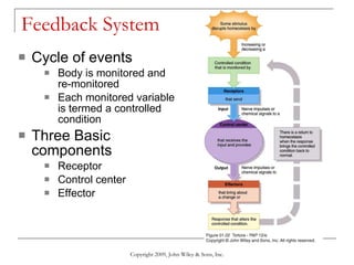
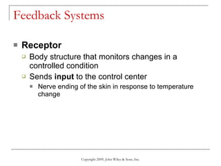
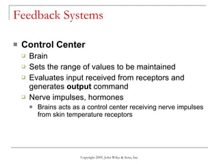
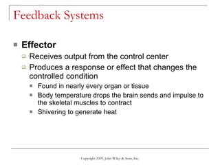
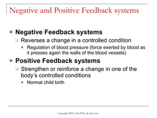
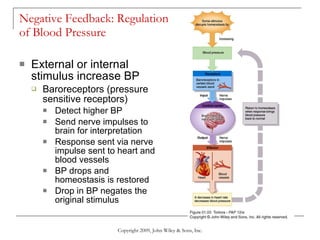
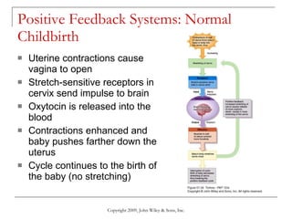
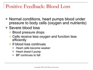
- It explains homeostasis as the maintenance of stable internal conditions and the feedback systems that regulate homeostasis.
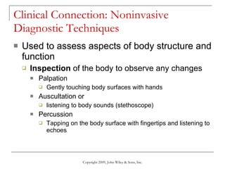
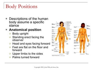
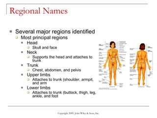
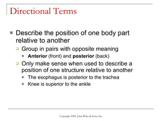
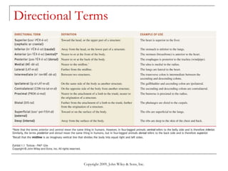
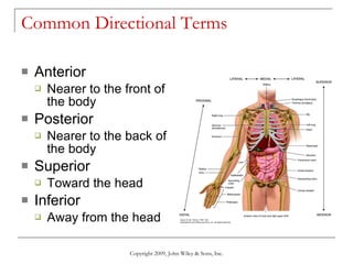
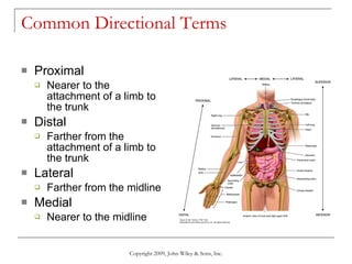
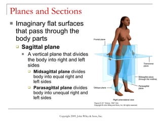
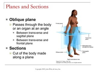
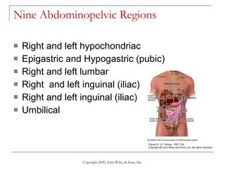
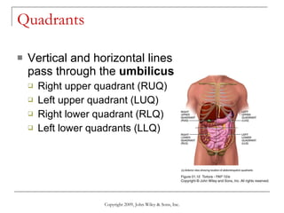
- It introduces anatomical terminology used to describe body positions, regions, directions, and planes.


![04 [chapter 4 the tissue level of organization][11e]](https://cdn.slidesharecdn.com/ss_thumbnails/04chapter4thetissueleveloforganization11e-170828035609-thumbnail.jpg?width=640&height=640&fit=bounds)


![03 [chapter 3 the cellular level of organization]](https://cdn.slidesharecdn.com/ss_thumbnails/03chapter3thecellularleveloforganization-170828035521-thumbnail.jpg?width=640&height=640&fit=bounds)
![20 [chapter 20 the cardiovascular system the heart]](https://cdn.slidesharecdn.com/ss_thumbnails/20chapter20thecardiovascularsystem-theheart-170828133506-thumbnail.jpg?width=640&height=640&fit=bounds)

![02 [chapter 2 the chemical level of organization]](https://cdn.slidesharecdn.com/ss_thumbnails/02chapter2thechemicalleveloforganization-170828035601-thumbnail.jpg?width=640&height=640&fit=bounds)


![22 [chapter 22 the lymphatic system and immunity]](https://cdn.slidesharecdn.com/ss_thumbnails/22chapter22thelymphaticsystemandimmunity-170828153258-thumbnail.jpg?width=640&height=640&fit=bounds)
![29 [chapter 29 development and inheritance]](https://cdn.slidesharecdn.com/ss_thumbnails/29chapter29developmentandinheritance-170828044352-thumbnail.jpg?width=640&height=640&fit=bounds)


