Downloaded 171 times
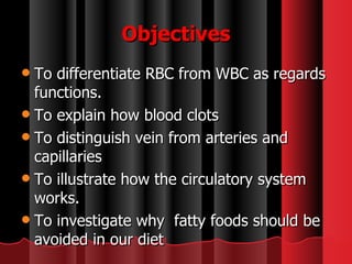
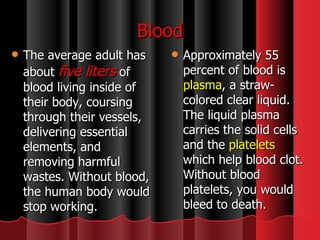
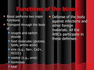
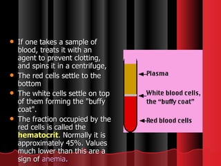
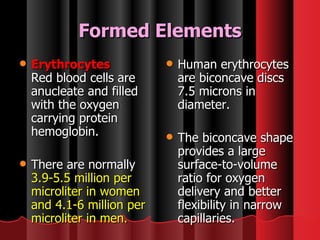
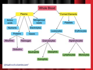
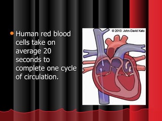
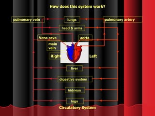
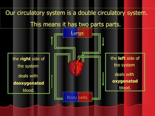
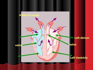
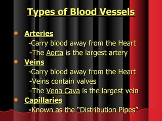
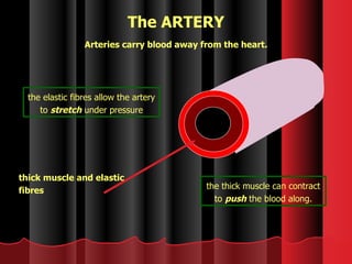
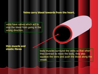

The document summarizes key aspects of blood and the circulatory system in 3 paragraphs or less: Blood contains red blood cells, white blood cells, platelets, and plasma. Red blood cells carry oxygen and carbon dioxide, while white blood cells help fight infection. Platelets help the blood clot to stop bleeding from cuts or wounds. The circulatory system is made up of the heart, arteries, veins, and capillaries. Oxygenated blood is pumped from the heart through arteries, then into capillaries to supply cells before returning to the heart as deoxygenated blood via veins. The double circulatory system separates oxygenated and deoxygenated blood flows. When blood vessels


