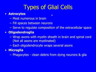
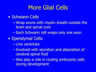
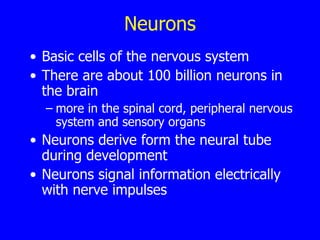
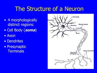
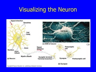
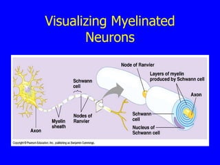
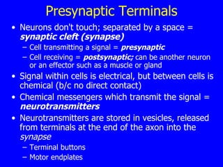
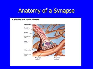
Cell Structure in the Nervous System
Cells & the Brain All info comes from sensory receptors This information is transformed in the brain into perceptions & commands for movement. The complexity of the brain results from the number, not the variety, of cells Brain contains ~ 100 billion nerve cells
Types of Cells 2 classes of cells in nervous system Neurons  or nerve cells Glial cells (glia) Neurons not initially recognized as single cells because they differ from other cells: much larger (axon can be 0.1 mm to 2 m) different in shape don't touch each other directly Golgi stain Glia surround neurons – from the Greek for ‘glue’
Glial Cells There are about 1 trillion glial cells in the brain 10 - 50 x  more than there are neurons Glial cells have perform important functions including: surround  & support neurons separate & insulate groups of neurons some produce myelin some are scavengers maintain concentration of K +  in extracellular space some take up & remove chemical transmitters some guide migration of neurons during development some help form an impermeable lining in capillaries & venules, creating the  blood-brain barrier
Types of Glial Cells Astrocytes  Most numerous in brain Fill spaces between neurons Serve to regulate composition of the extracellular space   Oligodendroglia Wrap axons with myelin sheath in brain and spinal cord (Not all axons are myelinated) Each oligodendrocyte wraps several axons Microglia Phagocytes - clean debris from dying neurons & glia
More Glial Cells Schwann Cells Wrap axons with myelin sheath outside the brain and spinal cord Each Schwann cell wraps only one axon Ependymal Cells Line ventricles Involved with secretion and absorption of cerebral spinal fluid Also play a role in routing embryonic cells during development 
Neurons Basic cells of the nervous system There are about 100 billion neurons in the brain more in the spinal cord, peripheral nervous system and sensory organs   Neurons derive form the neural tube during development Neurons signal information electrically with nerve impulses
The Neuron Doctrine Golgi stain  – A  reduced silver method using silver chromate that stains a few, isolated neurons in their entirety Invented by  Camillo Golgi Santiago Ramon y Cajal used it to survey the variety of structures in vertebrate nervous systems Led Cajal to propose the  neuron doctrine:   nerve cells are the structural and functional basis of the nervous system and they must communicate with one another  confirmed by electron microscopy 50 years later   Golgi and Cajal shared a Nobel Prize
The Structure of a Neuron 4 morphologically distinct regions: •  Cell Body ( soma ) •  Axon •  Dendrites •  Presynaptic Terminals
The Cell Body Also called  soma  ( somata ) - perikaryon   Metabolic center of the cell Assembly of new membrane Gives rise to dendrites & axon Contains the nucleus  Site of DNA in chromosomes  Contains rough & smooth endoplasmic reticulum Rough ER:  Stacks of flattened membrane compartments with ribosomes  attached Smooth Endoplasmic Reticulum: connects RER with Golgi apparatus and serves to further process membrane proteins; serves to sequester and release calcium to control cytoplasmic composition
Other Elements of the Cell Body Site of protein synthesis  (ribosomes)  Free ribosomes and polyribosomes loose in cytoplasm Contains the Golgi apparatus  Stacks of membrane compartments Packaging of secretory products (neurotransmitter peptides) Mitochondria  Site of conversion of energy from chemical bonds in food to ATP, the common energy currency of the cell
The Cytoskeleton Superstructure of the cell – 3 components: Microtubules  Largest - 20 nm thick-walled tubes Spirals of alpha and beta tubulin molecules Railroad tracks for movement of organelles, etc. Neurofilaments  10 nm twisted cables Tend to be the most static structures of the three Microfilaments  5 nm double helix of actin Especially common in neurites along with microtubules Also found in thick meshes associated with some regions of the cell membrane
The Neuronal Membrane A continuous sheet covering the neuron Separates the cytoplasm from the extra-cellular fluid Lipid bilayer with many embedded proteins The embedded proteins are channels critical to neuronal function
Dendrites Neurites which receive  signals from other nerve cells (message  IN )  Postsynaptic membrane in dendrites have molecular receptors for neurotransmitters (chemical messengers)  May have little bulges or pegs called dendritic spines where incoming synapses connect Carry impulses from other neurons or receptors toward the cell body Most neurons have several dendrites Have polyribosomes suggesting some local protein synthesis in dendrites
The Axon Main conducting unit of the neuron  Each neuron has only one axon Axon collaterals  – many axons do have branches Carries impulses away from the cell body to other neurons or to effectors (message  OUT )  Efferent - axon going away from reference neuron Afferent - axon coming toward reference neuro Axon hillock  = the conical region at the beginning of an axon where it joins the cell body Axon terminal  ( terminal bouton ) – the end of the axon
Visualizing the Neuron
Myelin Large axons have an insulating sheath -  myelin   Supports, insulates & nourishes the axon and helps maintain chemical balance.  This sheath is made up of  oligodendrocytes  in the CNS &  Schwann cells  in the PNS.  Nodes of Ranvier  =  Gaps in the sheath which allow passage of the electrical signal through ion channels  Saltatory conduction Transmission very fast .
Visualizing Myelinated Neurons
Presynaptic Terminals Neurons don't touch; separated by a space =  synaptic cleft (synapse) Cell transmitting a signal =  presynaptic Cell receiving =  postsynaptic;  can be another neuron or an effector such as a muscle or gland Signal within cells is electrical, but between cells is chemical (b/c no direct contact) Chemical messengers which transmit the signal =  neurotransmitters Neurotransmitters are stored in vesicles, released from terminals at the end of the axon into the  synapse Terminal buttons Motor endplates
Anatomy of a Synapse
Diversity of Neurons Neurons can be classified in several ways: Based on neurites  Based on dendrites  Based on connections/ function Based on axon length Based on neurotransmitter
Number of Neurite Processes Neurons classed in 3  (4) groups by number of processes: Unipolar the simplest neuron a single primary process with many branches no dendrites from the cell body common in invertebrates Bipolar oval shaped soma 2 processes: dendrite (info in) & axon (info out) most sensory neurons are bipolar
Number of Processes (Cont.) Pseudounipolar Sensory cells of touch, pressure, pain are special type of bipolar 1st develop as bipolar; the 2 processes fuse to form a single axon axon splits at the cell body one goes to spinal cord, other to periphery (skin, joints, muscles) Multipolar most common type in vertebrates one axon and one or more dendrite  vary in size and shape
Visualizing Cell Types
Classification Based on Dendrites Pyramidal cells/stellate cells Spiny cells/aspinous cells
Classification Based on Function 3 groups: Sensory  ( afferent )  neurons  receive stimuli & transmit them to the central nervous system Motor   neurons   carry impulses away from the central nervous  system to muscles or glands Interneurons   link sensory to motor neurons
Visualizing Cell Types - 2
Classification Based  on Neurotransmitter Cholinergic  neurons –  use acetylcholine Gabanergic   neurons –  use gamma aminobutyric acid Dopaminergic  neurons –  use dopamine
How Neurons Carry the Message Only neurons are involved in transmission of electrical signals. Within a nerve cell, message is an electrical signal =  action potential Cascading membrane depolarization creates the movement of the action potential as a nerve impulse rapid, all or none impulses in myelinated neurons, gaps at regular intervals allow regeneration of the action potential Between nerve cells the message is carried chemically
Principles of Impulse Transmission PRINCIPLE #1: Information conveyed by an action potential is determined not by the form of the signal, but by the pathway the signal travels. action potential for sound looks like action potential for odor PRINCIPLE #2: Principle of Dynamic Polarization :  electrical signals flow in a consistent direction PRINCIPLE #3:   Principle of Connectional Specificity: Contact is not random - cells communicate with certain targets and not others
Types of Signals Each sensory & motor nerve cell generates 4 types of signals: an input signal an integration signal (trigger) a conducting signal an output signal
Functional Regions Almost all neurons have 4 corresponding functional regions: receptive - local input integrative - trigger conductile - signaling secretory - output

Cells of Nervous System

  • 1.
    Cell Structure inthe Nervous System
  • 2.
    Cells & theBrain All info comes from sensory receptors This information is transformed in the brain into perceptions & commands for movement. The complexity of the brain results from the number, not the variety, of cells Brain contains ~ 100 billion nerve cells
  • 3.
    Types of Cells2 classes of cells in nervous system Neurons or nerve cells Glial cells (glia) Neurons not initially recognized as single cells because they differ from other cells: much larger (axon can be 0.1 mm to 2 m) different in shape don't touch each other directly Golgi stain Glia surround neurons – from the Greek for ‘glue’
  • 4.
    Glial Cells Thereare about 1 trillion glial cells in the brain 10 - 50 x more than there are neurons Glial cells have perform important functions including: surround & support neurons separate & insulate groups of neurons some produce myelin some are scavengers maintain concentration of K + in extracellular space some take up & remove chemical transmitters some guide migration of neurons during development some help form an impermeable lining in capillaries & venules, creating the blood-brain barrier
  • 5.
    Types of GlialCells Astrocytes Most numerous in brain Fill spaces between neurons Serve to regulate composition of the extracellular space Oligodendroglia Wrap axons with myelin sheath in brain and spinal cord (Not all axons are myelinated) Each oligodendrocyte wraps several axons Microglia Phagocytes - clean debris from dying neurons & glia
  • 6.
    More Glial CellsSchwann Cells Wrap axons with myelin sheath outside the brain and spinal cord Each Schwann cell wraps only one axon Ependymal Cells Line ventricles Involved with secretion and absorption of cerebral spinal fluid Also play a role in routing embryonic cells during development 
  • 7.
    Neurons Basic cellsof the nervous system There are about 100 billion neurons in the brain more in the spinal cord, peripheral nervous system and sensory organs Neurons derive form the neural tube during development Neurons signal information electrically with nerve impulses
  • 8.
    The Neuron DoctrineGolgi stain – A reduced silver method using silver chromate that stains a few, isolated neurons in their entirety Invented by Camillo Golgi Santiago Ramon y Cajal used it to survey the variety of structures in vertebrate nervous systems Led Cajal to propose the neuron doctrine: nerve cells are the structural and functional basis of the nervous system and they must communicate with one another confirmed by electron microscopy 50 years later Golgi and Cajal shared a Nobel Prize
  • 9.
    The Structure ofa Neuron 4 morphologically distinct regions: • Cell Body ( soma ) • Axon • Dendrites • Presynaptic Terminals
  • 10.
    The Cell BodyAlso called soma ( somata ) - perikaryon Metabolic center of the cell Assembly of new membrane Gives rise to dendrites & axon Contains the nucleus Site of DNA in chromosomes Contains rough & smooth endoplasmic reticulum Rough ER: Stacks of flattened membrane compartments with ribosomes attached Smooth Endoplasmic Reticulum: connects RER with Golgi apparatus and serves to further process membrane proteins; serves to sequester and release calcium to control cytoplasmic composition
  • 11.
    Other Elements ofthe Cell Body Site of protein synthesis (ribosomes) Free ribosomes and polyribosomes loose in cytoplasm Contains the Golgi apparatus Stacks of membrane compartments Packaging of secretory products (neurotransmitter peptides) Mitochondria Site of conversion of energy from chemical bonds in food to ATP, the common energy currency of the cell
  • 12.
    The Cytoskeleton Superstructureof the cell – 3 components: Microtubules Largest - 20 nm thick-walled tubes Spirals of alpha and beta tubulin molecules Railroad tracks for movement of organelles, etc. Neurofilaments 10 nm twisted cables Tend to be the most static structures of the three Microfilaments 5 nm double helix of actin Especially common in neurites along with microtubules Also found in thick meshes associated with some regions of the cell membrane
  • 13.
    The Neuronal MembraneA continuous sheet covering the neuron Separates the cytoplasm from the extra-cellular fluid Lipid bilayer with many embedded proteins The embedded proteins are channels critical to neuronal function
  • 14.
    Dendrites Neurites whichreceive signals from other nerve cells (message IN ) Postsynaptic membrane in dendrites have molecular receptors for neurotransmitters (chemical messengers) May have little bulges or pegs called dendritic spines where incoming synapses connect Carry impulses from other neurons or receptors toward the cell body Most neurons have several dendrites Have polyribosomes suggesting some local protein synthesis in dendrites
  • 15.
    The Axon Mainconducting unit of the neuron Each neuron has only one axon Axon collaterals – many axons do have branches Carries impulses away from the cell body to other neurons or to effectors (message OUT ) Efferent - axon going away from reference neuron Afferent - axon coming toward reference neuro Axon hillock = the conical region at the beginning of an axon where it joins the cell body Axon terminal ( terminal bouton ) – the end of the axon
  • 16.
  • 17.
    Myelin Large axonshave an insulating sheath - myelin Supports, insulates & nourishes the axon and helps maintain chemical balance. This sheath is made up of oligodendrocytes in the CNS & Schwann cells in the PNS. Nodes of Ranvier = Gaps in the sheath which allow passage of the electrical signal through ion channels Saltatory conduction Transmission very fast .
  • 18.
  • 19.
    Presynaptic Terminals Neuronsdon't touch; separated by a space = synaptic cleft (synapse) Cell transmitting a signal = presynaptic Cell receiving = postsynaptic; can be another neuron or an effector such as a muscle or gland Signal within cells is electrical, but between cells is chemical (b/c no direct contact) Chemical messengers which transmit the signal = neurotransmitters Neurotransmitters are stored in vesicles, released from terminals at the end of the axon into the synapse Terminal buttons Motor endplates
  • 20.
    Anatomy of aSynapse
  • 21.
    Diversity of NeuronsNeurons can be classified in several ways: Based on neurites Based on dendrites Based on connections/ function Based on axon length Based on neurotransmitter
  • 22.
    Number of NeuriteProcesses Neurons classed in 3 (4) groups by number of processes: Unipolar the simplest neuron a single primary process with many branches no dendrites from the cell body common in invertebrates Bipolar oval shaped soma 2 processes: dendrite (info in) & axon (info out) most sensory neurons are bipolar
  • 23.
    Number of Processes(Cont.) Pseudounipolar Sensory cells of touch, pressure, pain are special type of bipolar 1st develop as bipolar; the 2 processes fuse to form a single axon axon splits at the cell body one goes to spinal cord, other to periphery (skin, joints, muscles) Multipolar most common type in vertebrates one axon and one or more dendrite vary in size and shape
  • 24.
  • 25.
    Classification Based onDendrites Pyramidal cells/stellate cells Spiny cells/aspinous cells
  • 26.
    Classification Based onFunction 3 groups: Sensory ( afferent ) neurons receive stimuli & transmit them to the central nervous system Motor neurons carry impulses away from the central nervous system to muscles or glands Interneurons link sensory to motor neurons
  • 27.
  • 28.
    Classification Based on Neurotransmitter Cholinergic neurons – use acetylcholine Gabanergic neurons – use gamma aminobutyric acid Dopaminergic neurons – use dopamine
  • 29.
    How Neurons Carrythe Message Only neurons are involved in transmission of electrical signals. Within a nerve cell, message is an electrical signal = action potential Cascading membrane depolarization creates the movement of the action potential as a nerve impulse rapid, all or none impulses in myelinated neurons, gaps at regular intervals allow regeneration of the action potential Between nerve cells the message is carried chemically
  • 30.
    Principles of ImpulseTransmission PRINCIPLE #1: Information conveyed by an action potential is determined not by the form of the signal, but by the pathway the signal travels. action potential for sound looks like action potential for odor PRINCIPLE #2: Principle of Dynamic Polarization : electrical signals flow in a consistent direction PRINCIPLE #3: Principle of Connectional Specificity: Contact is not random - cells communicate with certain targets and not others
  • 31.
    Types of SignalsEach sensory & motor nerve cell generates 4 types of signals: an input signal an integration signal (trigger) a conducting signal an output signal
  • 32.
    Functional Regions Almostall neurons have 4 corresponding functional regions: receptive - local input integrative - trigger conductile - signaling secretory - output