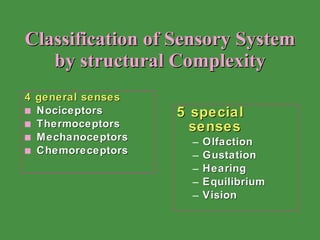
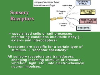
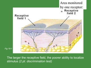
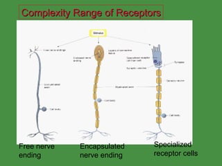
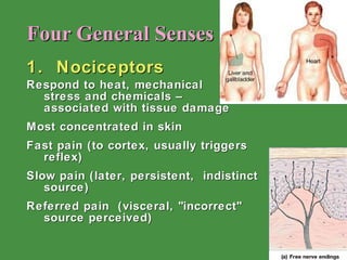
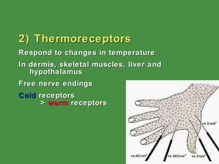
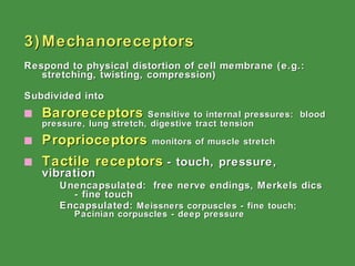
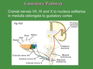
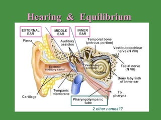
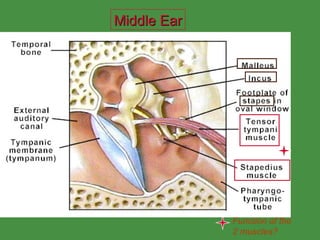
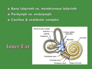
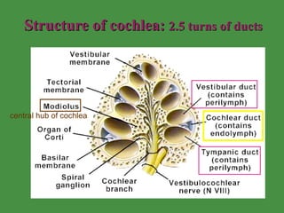
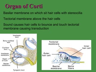
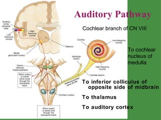
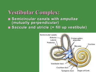
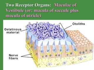
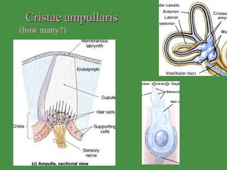
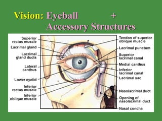
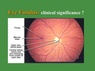
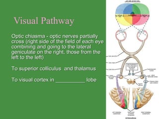
General & Special Senses Distinguish between general and specific senses Classify receptors according to stimulus detected, body location, and histological structure  Describe the structures of the ear and eye Explain the pathways of sound in the ear and light in the eye  Identify, describe, and discuss the receptors and neural pathways involved in each of the five special senses  Chapter objectives:
Classification of Sensory System by structural Complexity 4 general senses Nociceptors Thermoceptors Mechanoceptors Chemoreceptors 5 special senses Olfaction Gustation Hearing  Equilibrium Vision
Sensory  Receptors = specialized cells or cell processes monitoring conditions in/outside body (-> extero- and interoceptors) Receptors are specific for a certain type of stimulus -> “receptor specificity” All sensory receptors are transducers, changing incoming stimulus of pressure, vibration, light, etc., into electro-chemical neuron impulses. simplest receptor type: free nerve endings
The larger the receptive field, the poorer ability to localize stimulus  (2 pt. discrimination test) Fig 18-1 Area monitored by one receptor: =
Complexity Range of Receptors Free nerve ending Encapsulated nerve ending Specialized receptor cells
Four General Senses 1.  Nociceptors Respond to heat, mechanical  stress and chemicals –  associated with tissue damage Most concentrated in skin Fast pain (to cortex, usually triggers reflex)  Slow pain (later, persistent,  indistinct source) Referred pain  (visceral, "incorrect" source perceived)
2) Thermoreceptors Respond to changes in temperature  In dermis, skeletal muscles, liver and hypothalamus Free nerve endings Cold  receptors  >  warm  receptors
3) Mechanoreceptors Respond to physical distortion of cell membrane (e.g.: stretching, twisting, compression) Subdivided into Baroreceptors   Sensitive to internal pressures:  blood pressure, lung stretch, digestive tract tension   Proprioceptors   monitors of muscle stretch Tactile receptors   - touch, pressure, vibration   Unencapsulated:  free nerve endings, Merkels dics - fine touch Encapsulated:  Meissners corpuscles - fine touch; Pacinian corpuscles - deep pressure
4) Chemoreceptors Respond to small concentration changes of specific molecules (chemicals) Internal chemoreceptors monitor blood composition (e.g. Na + , pH, pCO 2  ) Found within aortic and carotid bodies Very important for homeostasis Fig 18-5
Special Senses Olfaction Taste Vision Hearing Equilibrium Organ responsible ??
Olfaction: Paired Olfactory Organs Olfactory epithelium (10-20 Mio receptors /  5 cm 2) Responds to molecules dissolved in mucus or lipids Easy to recognize – hard to categorize (Only) neuron that can be replaced in adult Through cribriform plate of ethmoid to olfactory bulb Type of receptor??
Olfactory Pathways Receptor neurons pass into cranium through cribiform plate and synapse in olfactory bulbs. Olfactory neurons are the only neurons known to routinely replace themselves  to reach the cerebrum without synapsing in the thalamus  Olfactory discrimination - Although difficult to describe, the number of different odors recognizable is immense.
Gustation 3 types of papillae 1) Filiform - thin, thread like projections  2) Fungiform  - shaped like mushrooms.  3) Circumvallate - large target-shaped bumps near the back of the tongue   Papillae contain taste buds Taste buds contain group of receptor & support cells How many 1 o  taste sensations? Fig 18.7
Gustatory Pathway  Cranial nerves VII, IX and X to nucleus solitarius in medulla oblongata to gustatory cortex  Fig 18.8
Hearing  &  Equilibrium 2 other names??
Middle Ear Function of the 2 muscles?
Inner Ear Bony labyrinth vs. membranous labyrinth Perilymph vs. endolymph Cochlea & vestibular complex
Structure of cochlea:  2.5 turns of ducts central hub of cochlea
Organ of Corti Basilar membrane on which sit hair cells with stereocilia Tectorial membrane above the hair cells Sound causes hair cells to bounce and touch tectorial membrane causing transduction
Auditory Pathway  To inferior colliculus of opposite side of midbrain To thalamus To auditory cortex  Cochlear branch of CN VIII To cochlear nucleus of medulla
Vestibular Complex: Semicircular canals with ampullae (mutually perpendicular) Saccule and utricle (= fill up vestibule)
Two Receptor Organs:   Maculae of Vestibule (or: macula of saccule plus macula of utricle)
Cristae ampullaris  (how many?)
Vision:  Eyeball  +  Accessory Structures
Palpebrae = Eyelid Continuation of skin Eyelashes Meibomian glands (on inner margin of lid) lipid rich product, fu? bacterial infection chalazion Conjunctiva (= mucous membrane) over cornea very thin (5-7 cells thick)
Lacrimal Apparatus Lacrimal gland  with several ducts - superior and lateral to eye Lacrimal puncta (superior and inferior) - holes near nose to drain tears Lacrimal canaliculi -  drain    tears to Nasolacrimal duct -    empties to nasal cavity Secretion contains  lysozyme Compare to fig 18.18
Extrinsic Eyemuscles  (see p.272) 4 recti 2 oblique Innervation?
The Three Tunics: 1) Fibrous Tunic (tough outer layer) sclera  -  white part of fibrous tunic cornea  -  transparent avascular anterior part limbus  - boundary between the above 2) Vascular Tunic (= Uvea) choroid  - heavily vascular iris  with pupil  hole - inner sphincter  and outer radial muscles ciliary body  - muscle attached to  suspensory ligaments,  regulates focus of lens
Lens and Chambers of the Eye Ciliary body Suspensory ligaments Anterior and posterior chambers (= anterior cavity) with aqueous humor Posterior cavity with vitreous humor Cataract   Glaucoma=? See fig 18.21
3) Nervous Tunic: Retina Outer layer pigmented - inner layer  photoreceptors a) rods - black/white vision, dim light b) cones - color vision, intense light Bipolar cells - synapse with rods and cones Ganglion cells - synapse with bipolar cells Ora serrata - anterior edge of retina Macula lutea – fovea centralis - all cones, best vision Optic disc – blind spot, where optic nerve exits eye Optic nerve See Fig 18.22
fu?
Eye Fundus:  clinical significance ?
Visual Pathway Optic chiasma - optic nerves partially cross (right side of the field of each eye combining and going to the lateral geniculate on the right, those from the left to the left)  To superior colliculus  and thalamus To visual cortex in __________ lobe

General & Special Senses

  • 1.
    General & SpecialSenses Distinguish between general and specific senses Classify receptors according to stimulus detected, body location, and histological structure Describe the structures of the ear and eye Explain the pathways of sound in the ear and light in the eye Identify, describe, and discuss the receptors and neural pathways involved in each of the five special senses Chapter objectives:
  • 2.
    Classification of SensorySystem by structural Complexity 4 general senses Nociceptors Thermoceptors Mechanoceptors Chemoreceptors 5 special senses Olfaction Gustation Hearing Equilibrium Vision
  • 3.
    Sensory Receptors= specialized cells or cell processes monitoring conditions in/outside body (-> extero- and interoceptors) Receptors are specific for a certain type of stimulus -> “receptor specificity” All sensory receptors are transducers, changing incoming stimulus of pressure, vibration, light, etc., into electro-chemical neuron impulses. simplest receptor type: free nerve endings
  • 4.
    The larger thereceptive field, the poorer ability to localize stimulus (2 pt. discrimination test) Fig 18-1 Area monitored by one receptor: =
  • 5.
    Complexity Range ofReceptors Free nerve ending Encapsulated nerve ending Specialized receptor cells
  • 6.
    Four General Senses1. Nociceptors Respond to heat, mechanical stress and chemicals – associated with tissue damage Most concentrated in skin Fast pain (to cortex, usually triggers reflex) Slow pain (later, persistent, indistinct source) Referred pain (visceral, "incorrect" source perceived)
  • 7.
    2) Thermoreceptors Respondto changes in temperature In dermis, skeletal muscles, liver and hypothalamus Free nerve endings Cold receptors > warm receptors
  • 8.
    3) Mechanoreceptors Respondto physical distortion of cell membrane (e.g.: stretching, twisting, compression) Subdivided into Baroreceptors Sensitive to internal pressures: blood pressure, lung stretch, digestive tract tension Proprioceptors monitors of muscle stretch Tactile receptors - touch, pressure, vibration Unencapsulated: free nerve endings, Merkels dics - fine touch Encapsulated: Meissners corpuscles - fine touch; Pacinian corpuscles - deep pressure
  • 9.
    4) Chemoreceptors Respondto small concentration changes of specific molecules (chemicals) Internal chemoreceptors monitor blood composition (e.g. Na + , pH, pCO 2 ) Found within aortic and carotid bodies Very important for homeostasis Fig 18-5
  • 10.
    Special Senses OlfactionTaste Vision Hearing Equilibrium Organ responsible ??
  • 11.
    Olfaction: Paired OlfactoryOrgans Olfactory epithelium (10-20 Mio receptors / 5 cm 2) Responds to molecules dissolved in mucus or lipids Easy to recognize – hard to categorize (Only) neuron that can be replaced in adult Through cribriform plate of ethmoid to olfactory bulb Type of receptor??
  • 12.
    Olfactory Pathways Receptorneurons pass into cranium through cribiform plate and synapse in olfactory bulbs. Olfactory neurons are the only neurons known to routinely replace themselves to reach the cerebrum without synapsing in the thalamus Olfactory discrimination - Although difficult to describe, the number of different odors recognizable is immense.
  • 13.
    Gustation 3 typesof papillae 1) Filiform - thin, thread like projections 2) Fungiform - shaped like mushrooms. 3) Circumvallate - large target-shaped bumps near the back of the tongue Papillae contain taste buds Taste buds contain group of receptor & support cells How many 1 o taste sensations? Fig 18.7
  • 14.
    Gustatory Pathway Cranial nerves VII, IX and X to nucleus solitarius in medulla oblongata to gustatory cortex Fig 18.8
  • 15.
    Hearing & Equilibrium 2 other names??
  • 16.
    Middle Ear Functionof the 2 muscles?
  • 17.
    Inner Ear Bonylabyrinth vs. membranous labyrinth Perilymph vs. endolymph Cochlea & vestibular complex
  • 18.
    Structure of cochlea: 2.5 turns of ducts central hub of cochlea
  • 19.
    Organ of CortiBasilar membrane on which sit hair cells with stereocilia Tectorial membrane above the hair cells Sound causes hair cells to bounce and touch tectorial membrane causing transduction
  • 20.
    Auditory Pathway To inferior colliculus of opposite side of midbrain To thalamus To auditory cortex Cochlear branch of CN VIII To cochlear nucleus of medulla
  • 21.
    Vestibular Complex: Semicircularcanals with ampullae (mutually perpendicular) Saccule and utricle (= fill up vestibule)
  • 22.
    Two Receptor Organs: Maculae of Vestibule (or: macula of saccule plus macula of utricle)
  • 23.
  • 24.
    Vision: Eyeball + Accessory Structures
  • 25.
    Palpebrae = EyelidContinuation of skin Eyelashes Meibomian glands (on inner margin of lid) lipid rich product, fu? bacterial infection chalazion Conjunctiva (= mucous membrane) over cornea very thin (5-7 cells thick)
  • 26.
    Lacrimal Apparatus Lacrimalgland with several ducts - superior and lateral to eye Lacrimal puncta (superior and inferior) - holes near nose to drain tears Lacrimal canaliculi - drain tears to Nasolacrimal duct - empties to nasal cavity Secretion contains lysozyme Compare to fig 18.18
  • 27.
    Extrinsic Eyemuscles (see p.272) 4 recti 2 oblique Innervation?
  • 28.
    The Three Tunics:1) Fibrous Tunic (tough outer layer) sclera - white part of fibrous tunic cornea - transparent avascular anterior part limbus - boundary between the above 2) Vascular Tunic (= Uvea) choroid - heavily vascular iris with pupil hole - inner sphincter and outer radial muscles ciliary body - muscle attached to suspensory ligaments, regulates focus of lens
  • 29.
    Lens and Chambersof the Eye Ciliary body Suspensory ligaments Anterior and posterior chambers (= anterior cavity) with aqueous humor Posterior cavity with vitreous humor Cataract Glaucoma=? See fig 18.21
  • 30.
    3) Nervous Tunic:Retina Outer layer pigmented - inner layer photoreceptors a) rods - black/white vision, dim light b) cones - color vision, intense light Bipolar cells - synapse with rods and cones Ganglion cells - synapse with bipolar cells Ora serrata - anterior edge of retina Macula lutea – fovea centralis - all cones, best vision Optic disc – blind spot, where optic nerve exits eye Optic nerve See Fig 18.22
  • 31.
  • 32.
    Eye Fundus: clinical significance ?
  • 33.
    Visual Pathway Opticchiasma - optic nerves partially cross (right side of the field of each eye combining and going to the lateral geniculate on the right, those from the left to the left) To superior colliculus and thalamus To visual cortex in __________ lobe