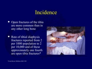
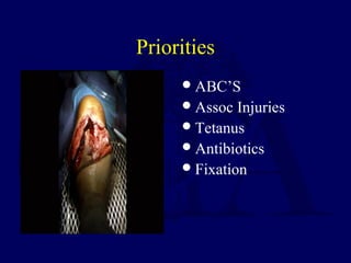
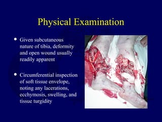
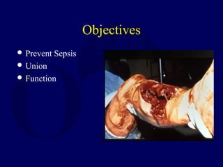
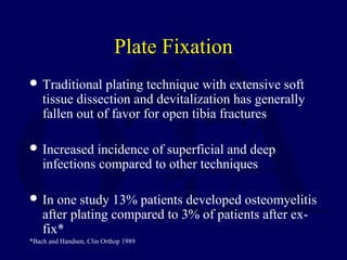
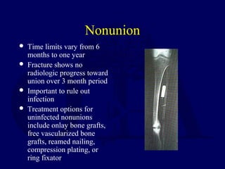
Open Fractures of the
Tibial Diaphysis
Robert V. Cantu, MD
David Templeman, MD
Incidence
 Open fractures of the tibia
are more common than in
any other long bone
 Rate of tibial diaphysis
fractures reported from 2
per 1000 population to 2
per 10,000 and of these
approximately one fourth
are open tibia fractures*
*Court-Brown; McBirnie JBJS 1995
Mechanism of Injury
Can occur in lower energy, torsional type
injury (eg, skiing)
More common with higher energy direct
force (eg car bumper)
Priorities
ABC’S
Assoc Injuries
Tetanus
Antibiotics
Fixation
Physical Examination
 Given subcutaneous
nature of tibia, deformity
and open wound usually
readily apparent
 Circumferential inspection
of soft tissue envelope,
noting any lacerations,
ecchymosis, swelling, and
tissue turgidity
Physical Exam
Neurologic and vascular exam of extremity
including ABI’s if indicated
Wounds should be assessed once in ER,
then covered with sterile gauze dressing
until treated in OR
True grading of wound best done at time of
surgical debridement
Radiographic Evaluation
 Full length AP and lateral
views from knee to ankle
required for all tibia
fractures
 Ankle views suggested to
examine mortise
 Arteriography indicated if
vascular compromise
present after reduction
Associated Injuries
 Approximately 30% of
patients have multiple
injuries
 Fibula commonly
fractured and its degree of
comminution correlates
with severity of injury
 Proximal or distal tib-fib
joints may be disrupted
 Ligamentous knee injury
and/or ipsilateral femur
(‘floating knee’) more
common in high energy
fractures
Associated Injuries
 Neurovascular structures
require repeated
assessment
 Foot fractures also
common
 Compartment syndrome
must be looked for
Classification Tibia Fractures
 Johner and Wruh’s
describes fracture
pattern as simple(A),
butterfly(B), or
comminuted(C)
Classification of Open Tibia
Fractures
 Gustilo and Anderson open fracture classification
first published in 1976 and later modified in 1984
 In one study interobserver agreement on
classification only 60%
 Grading of open injury likely best done in the OR
Gustilo and Anderson
Classification
 Grade 1- skin opening of 1cm or less, minimal
muscle contusion, usually inside out mechanism
 Grade 2- skin laceration 1-10cm, extensive soft
tissue damage
 Grade 3a- extensive soft tissue laceration(10cm)
but adequate bone coverage
 Grade 3b- extensive soft tissue injury with
periosteal stripping requiring flap advancement or
free flap
 Grade 3c- vascular injury requiring repair
Objectives
 Prevent Sepsis
 Union
 Function
Treament of Soft Tissue Injury
 After initial evaluation
wound covered with
sterile dressing and leg
splinted
 Appropriate tetanus
prophylaxis and
antibiotics begun
 Thorough debridement
and irrigation undertaken
in OR within 6 hours if
possible
Treatment of Soft Tissue Injury
Careful planning of skin incisions
Essential to fully explore wound as even
grade 1 fractures can pull dirt/debris back
into wound and on fracture ends
All foreign material, necrotic muscle,
unattached bone fragments, exposed fat and
fascia are debrided
Treatment of Soft Tissue Injury
 After debridement thorough irrigation with
Ringer’s lactate or normal saline
 Fasciotomies performed if indicated
 After I+D new gowns, gloves, drapes and sterile
instruments used for fracture fixation
 Large bone defects can be filled with antibiotic
cement beads until delayed bone grafting
performed
Bead Pouch: TOBRA 1.2 gm.
PMMA
Infection
 NO BEADS
– 16%
– + BEADS
– 4%
KEATING
Type III Open
 Timentin 3.1 gm.
 PCN barnyard
Large Fragments
Retained - 21%
Removed - 9%
 EDWARDS CORR
Soft Tissue Coverage
 Definitive coverage should be performed within 7
days if possible
 Most type 1 wounds will heal by secondary intent
or can be closed primarily after repeat I+D
 Delayed primary closure usually feasible for type
2 and type 3a fractures
Soft Tissue Coverage
 Type 3b fractures require
either local advancement
or rotation flap, split-
thickness skin graft, or
free flap
 STSG suitable for
coverage of large defects
with underlying viable
muscle
Soft Tissue Coverage
 Proximal third tibia
fractures can be covered
with gastrocnemius
rotation flap
 Middle third tibia
fractures can be covered
with soleus rotation flap
 Distal third fractures
usually require free flap
for coverage
Stabilization of Open Tibia
Fractures
Multiple options depending on fracture
pattern and soft tissue injury:
IM nail- reamed vs. unreamed
External fixation
ORIF
IM Nail
 Excellent results with
type 1 open fractures
Unreamed IM Nail
 Time to union with
unreamed nails can be
prolonged- in one study of
143 open tibia fractures
53% were united at 6
months
 Vast majority of fractures
united, but 11% required
at least one secondary
procedure to achieve
union*
*Tornetta and McConnell 16th
annual OTA 2000
Reamed Tibial Nailing
 In one study of type 2 and
type 3a fractures good
results- average time to
union 24 and 27 weeks
respectively; deep
infection rate 3.5%*
 Complications increased
with type 3b fractures-
average time to union was
50 weeks and infection
rate 23%*
*Court-Brown JBJS 1991
External Fixation
 Compared to IM nails,
increased rate of malunion
and need for secondary
procedures
 Most common
complication with ex-fix
is pin track infection (21%
in one study)*
*Tornetta JBJS 1994
Plate Fixation
 Traditional plating technique with extensive soft
tissue dissection and devitalization has generally
fallen out of favor for open tibia fractures
 Increased incidence of superficial and deep
infections compared to other techniques
 In one study 13% patients developed osteomyelitis
after plating compared to 3% of patients after ex-
fix*
*Bach and Handsen, Clin Orthop 1989
Percutaneous Plate Fixation
 Newer percutaneous
plating techniques
using indirect
reduction may be a
more beneficial
alternative
 Large prospective
studies yet to be
evaluated
Gunshot Wounds
 Tibia fractures due to low
energy missiles rarely
require debridement and
can often be treated like
closed injuries
 Fractures due to high
energy missiles (eg assault
rifle or close range shot
gun) treated as standard
open injuries
Amputation
 In general amputation
performed when limb
salvage poses significant
risk to patient survival,
when functional result
would be better with a
prosthesis, and when
duration and course of
treatment would cause
intolerable psychological
disturbance
Amputation
Lange proposed two absolute indications
for amputation of tibia fractures with
arterial injury: crush injury with warm
ischemia greater than 6 hours, and anatomic
division of the tibial nerve*
*Lange et al. J Trauma 1985
Mangled Extremity Severity
Score
 An attempt to help
guide between primary
amputation vs. limb
salvage
 In one study a score of
7 or higher was
predictive of
amputation*
*Johansen et al. J Trauma 1991
NISSA Score
Modification of Mess score adding a
component of nerve injury
Nerve, Ischemia, Soft tissue injury, Shock,
and Age Score
LEAP Study
 Plantar sensation not prognostic
 Scoring systems do not work
 10/55 Early Salvage
Complications
Nonunion
Malunion
Infection- deep and superficial
Compartment syndrome
Fatigue fractures
Hardware failure
Nonunion
 Time limits vary from 6
months to one year
 Fracture shows no
radiologic progress toward
union over 3 month period
 Important to rule out
infection
 Treatment options for
uninfected nonunions
include onlay bone grafts,
free vascularized bone
grafts, reamed nailing,
compression plating, or
ring fixator
Malunion
 In general varus malunion
more of a problem than
valgus
 In one study deformity up
to 15 degrees did not
produce ankle
complications*
 For symptomatic patients
with significant deformity
treatment is osteotomy
*Kristensen et al. Acta Orthop Scand 1989
Deep Infection
 Often presents with
increasing pain, wound
drainage, or sinus
formation
 Treatment involves
debridement, stabilization
(often with ex-fix),
coverage with healthy
tissue including muscle
flap if needed, IV
antibiotics, delayed bone
graft of defect if needed
Deep Infection
Not the Implant but the Management of the
Soft Tissues
Court-Brown
Deep Infection
If IM nail already in place, reamed
exchange nail with appropriate antibiotics
may prove adequate treatment
Superficial Infection
 Most superficial infections respond to elevation of
extremity and appropriate antibiotics (typically
gram + cocci coverage)
 If uncertain whether infection extends deeper
and/or it fails to respond to antibiotic treatment ,
then surgical debridement with tissue cultures
necessary
Compartment Sydrome
 Diagnosis same as in
closed tibial fractures
 Common with high
energy tibia fractures
 Treatment is 4
compartment
fasciotomies
Associated Fatigue Fractures
 Sometimes seen during rehab after prolonged non-
weight bearing
 Can present with localized tenderness in
metatarsal, calcaneus, or distal fibula
 Bone scan or MRI may be required to make
diagnosis as plain radiographs often normal
 Treatment is temporary reduction in weight
bearing
Hardware Failure
 Usually due to delayed
union or nonunion
 Important to rule out
infection as cause of
delayed healing
 Treatment depends on
type of failure- plate or
nail breakage requires
revision, whereas
breakage of locking screw
in nail may not require
operative intervention
Reamed Vs. Nonreamed
Nail Size
0
5
10
15
20
25
30
8 9 10 11 12 13 14
Nail Diameter
No. Used
Reamed
Unreamed
Outcomes
 Outcome most affected by severity of soft tissue
and neurovascular injury
 Most studies show major change in results
between type 3a and 3b/c fractures
 In one study of reamed nailing, the deep infection
rate was 3.5% for type 2 and 3a fractures, but 23%
for type 3b fractures*
*Court-Brown JBJS 1991
Outcomes
 For type 3b and 3c fractures early soft tissue
coverage gives best results
 In one study of 84 type 3b and 3c fractures, results
with single stage procedure involving fixation
with immediate flap coverage better than when
coverage delayed more than 72 hours (deep
infection 3% vs. 19%)*
*Gopal et al. JBJS[Br] 2000
Return to
Lower Extremity
Index

L09 open tibia

  • 1.
    Open Fractures ofthe Tibial Diaphysis Robert V. Cantu, MD David Templeman, MD
  • 2.
    Incidence  Open fracturesof the tibia are more common than in any other long bone  Rate of tibial diaphysis fractures reported from 2 per 1000 population to 2 per 10,000 and of these approximately one fourth are open tibia fractures* *Court-Brown; McBirnie JBJS 1995
  • 3.
    Mechanism of Injury Canoccur in lower energy, torsional type injury (eg, skiing) More common with higher energy direct force (eg car bumper)
  • 4.
  • 5.
    Physical Examination  Givensubcutaneous nature of tibia, deformity and open wound usually readily apparent  Circumferential inspection of soft tissue envelope, noting any lacerations, ecchymosis, swelling, and tissue turgidity
  • 6.
    Physical Exam Neurologic andvascular exam of extremity including ABI’s if indicated Wounds should be assessed once in ER, then covered with sterile gauze dressing until treated in OR True grading of wound best done at time of surgical debridement
  • 7.
    Radiographic Evaluation  Fulllength AP and lateral views from knee to ankle required for all tibia fractures  Ankle views suggested to examine mortise  Arteriography indicated if vascular compromise present after reduction
  • 8.
    Associated Injuries  Approximately30% of patients have multiple injuries  Fibula commonly fractured and its degree of comminution correlates with severity of injury  Proximal or distal tib-fib joints may be disrupted  Ligamentous knee injury and/or ipsilateral femur (‘floating knee’) more common in high energy fractures
  • 9.
    Associated Injuries  Neurovascularstructures require repeated assessment  Foot fractures also common  Compartment syndrome must be looked for
  • 10.
    Classification Tibia Fractures Johner and Wruh’s describes fracture pattern as simple(A), butterfly(B), or comminuted(C)
  • 11.
    Classification of OpenTibia Fractures  Gustilo and Anderson open fracture classification first published in 1976 and later modified in 1984  In one study interobserver agreement on classification only 60%  Grading of open injury likely best done in the OR
  • 12.
    Gustilo and Anderson Classification Grade 1- skin opening of 1cm or less, minimal muscle contusion, usually inside out mechanism  Grade 2- skin laceration 1-10cm, extensive soft tissue damage  Grade 3a- extensive soft tissue laceration(10cm) but adequate bone coverage  Grade 3b- extensive soft tissue injury with periosteal stripping requiring flap advancement or free flap  Grade 3c- vascular injury requiring repair
  • 13.
  • 14.
    Treament of SoftTissue Injury  After initial evaluation wound covered with sterile dressing and leg splinted  Appropriate tetanus prophylaxis and antibiotics begun  Thorough debridement and irrigation undertaken in OR within 6 hours if possible
  • 15.
    Treatment of SoftTissue Injury Careful planning of skin incisions Essential to fully explore wound as even grade 1 fractures can pull dirt/debris back into wound and on fracture ends All foreign material, necrotic muscle, unattached bone fragments, exposed fat and fascia are debrided
  • 16.
    Treatment of SoftTissue Injury  After debridement thorough irrigation with Ringer’s lactate or normal saline  Fasciotomies performed if indicated  After I+D new gowns, gloves, drapes and sterile instruments used for fracture fixation  Large bone defects can be filled with antibiotic cement beads until delayed bone grafting performed
  • 17.
    Bead Pouch: TOBRA1.2 gm. PMMA
  • 18.
    Infection  NO BEADS –16% – + BEADS – 4% KEATING
  • 19.
    Type III Open Timentin 3.1 gm.  PCN barnyard
  • 20.
    Large Fragments Retained -21% Removed - 9%  EDWARDS CORR
  • 21.
    Soft Tissue Coverage Definitive coverage should be performed within 7 days if possible  Most type 1 wounds will heal by secondary intent or can be closed primarily after repeat I+D  Delayed primary closure usually feasible for type 2 and type 3a fractures
  • 22.
    Soft Tissue Coverage Type 3b fractures require either local advancement or rotation flap, split- thickness skin graft, or free flap  STSG suitable for coverage of large defects with underlying viable muscle
  • 23.
    Soft Tissue Coverage Proximal third tibia fractures can be covered with gastrocnemius rotation flap  Middle third tibia fractures can be covered with soleus rotation flap  Distal third fractures usually require free flap for coverage
  • 24.
    Stabilization of OpenTibia Fractures Multiple options depending on fracture pattern and soft tissue injury: IM nail- reamed vs. unreamed External fixation ORIF
  • 25.
    IM Nail  Excellentresults with type 1 open fractures
  • 26.
    Unreamed IM Nail Time to union with unreamed nails can be prolonged- in one study of 143 open tibia fractures 53% were united at 6 months  Vast majority of fractures united, but 11% required at least one secondary procedure to achieve union* *Tornetta and McConnell 16th annual OTA 2000
  • 27.
    Reamed Tibial Nailing In one study of type 2 and type 3a fractures good results- average time to union 24 and 27 weeks respectively; deep infection rate 3.5%*  Complications increased with type 3b fractures- average time to union was 50 weeks and infection rate 23%* *Court-Brown JBJS 1991
  • 28.
    External Fixation  Comparedto IM nails, increased rate of malunion and need for secondary procedures  Most common complication with ex-fix is pin track infection (21% in one study)* *Tornetta JBJS 1994
  • 29.
    Plate Fixation  Traditionalplating technique with extensive soft tissue dissection and devitalization has generally fallen out of favor for open tibia fractures  Increased incidence of superficial and deep infections compared to other techniques  In one study 13% patients developed osteomyelitis after plating compared to 3% of patients after ex- fix* *Bach and Handsen, Clin Orthop 1989
  • 30.
    Percutaneous Plate Fixation Newer percutaneous plating techniques using indirect reduction may be a more beneficial alternative  Large prospective studies yet to be evaluated
  • 31.
    Gunshot Wounds  Tibiafractures due to low energy missiles rarely require debridement and can often be treated like closed injuries  Fractures due to high energy missiles (eg assault rifle or close range shot gun) treated as standard open injuries
  • 32.
    Amputation  In generalamputation performed when limb salvage poses significant risk to patient survival, when functional result would be better with a prosthesis, and when duration and course of treatment would cause intolerable psychological disturbance
  • 33.
    Amputation Lange proposed twoabsolute indications for amputation of tibia fractures with arterial injury: crush injury with warm ischemia greater than 6 hours, and anatomic division of the tibial nerve* *Lange et al. J Trauma 1985
  • 34.
    Mangled Extremity Severity Score An attempt to help guide between primary amputation vs. limb salvage  In one study a score of 7 or higher was predictive of amputation* *Johansen et al. J Trauma 1991
  • 35.
    NISSA Score Modification ofMess score adding a component of nerve injury Nerve, Ischemia, Soft tissue injury, Shock, and Age Score
  • 36.
    LEAP Study  Plantarsensation not prognostic  Scoring systems do not work  10/55 Early Salvage
  • 37.
    Complications Nonunion Malunion Infection- deep andsuperficial Compartment syndrome Fatigue fractures Hardware failure
  • 38.
    Nonunion  Time limitsvary from 6 months to one year  Fracture shows no radiologic progress toward union over 3 month period  Important to rule out infection  Treatment options for uninfected nonunions include onlay bone grafts, free vascularized bone grafts, reamed nailing, compression plating, or ring fixator
  • 39.
    Malunion  In generalvarus malunion more of a problem than valgus  In one study deformity up to 15 degrees did not produce ankle complications*  For symptomatic patients with significant deformity treatment is osteotomy *Kristensen et al. Acta Orthop Scand 1989
  • 40.
    Deep Infection  Oftenpresents with increasing pain, wound drainage, or sinus formation  Treatment involves debridement, stabilization (often with ex-fix), coverage with healthy tissue including muscle flap if needed, IV antibiotics, delayed bone graft of defect if needed
  • 41.
    Deep Infection Not theImplant but the Management of the Soft Tissues Court-Brown
  • 42.
    Deep Infection If IMnail already in place, reamed exchange nail with appropriate antibiotics may prove adequate treatment
  • 43.
    Superficial Infection  Mostsuperficial infections respond to elevation of extremity and appropriate antibiotics (typically gram + cocci coverage)  If uncertain whether infection extends deeper and/or it fails to respond to antibiotic treatment , then surgical debridement with tissue cultures necessary
  • 44.
    Compartment Sydrome  Diagnosissame as in closed tibial fractures  Common with high energy tibia fractures  Treatment is 4 compartment fasciotomies
  • 45.
    Associated Fatigue Fractures Sometimes seen during rehab after prolonged non- weight bearing  Can present with localized tenderness in metatarsal, calcaneus, or distal fibula  Bone scan or MRI may be required to make diagnosis as plain radiographs often normal  Treatment is temporary reduction in weight bearing
  • 46.
    Hardware Failure  Usuallydue to delayed union or nonunion  Important to rule out infection as cause of delayed healing  Treatment depends on type of failure- plate or nail breakage requires revision, whereas breakage of locking screw in nail may not require operative intervention
  • 47.
    Reamed Vs. Nonreamed NailSize 0 5 10 15 20 25 30 8 9 10 11 12 13 14 Nail Diameter No. Used Reamed Unreamed
  • 48.
    Outcomes  Outcome mostaffected by severity of soft tissue and neurovascular injury  Most studies show major change in results between type 3a and 3b/c fractures  In one study of reamed nailing, the deep infection rate was 3.5% for type 2 and 3a fractures, but 23% for type 3b fractures* *Court-Brown JBJS 1991
  • 49.
    Outcomes  For type3b and 3c fractures early soft tissue coverage gives best results  In one study of 84 type 3b and 3c fractures, results with single stage procedure involving fixation with immediate flap coverage better than when coverage delayed more than 72 hours (deep infection 3% vs. 19%)* *Gopal et al. JBJS[Br] 2000 Return to Lower Extremity Index