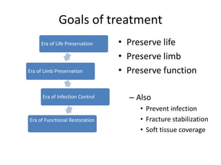
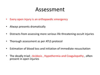
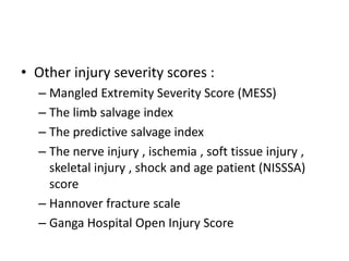
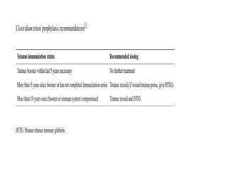
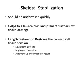
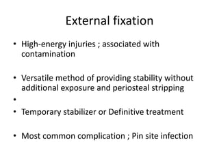
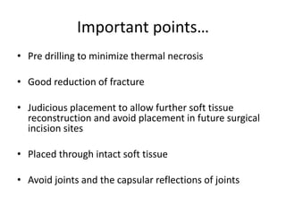
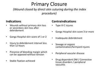
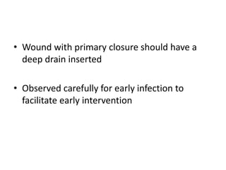
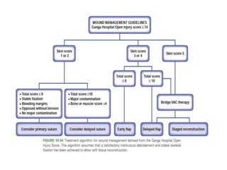
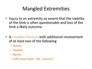
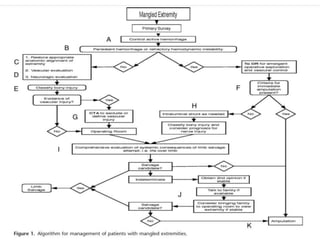
Open Fractures & Mangled
Extremities
Dr . Pranesh Chapagain
1st Year Resident , Orthopaedics
Nobel Medical College and Teaching Hospital
Introduction
• Defined as an injury where the fracture and fracture hematoma
communicate with the external environment through a traumatic
defect in the surrounding soft tissue and overlying skin
• Skin defect may lie at a distant site
• Any fracture associated with a wound in same region must be
considered an open injury unless proven otherwise by surgical
exploration
• Open tibial diaphyseal fracture are commonest open long bone
fracture
• The size and nature of external wound may not
reflect the damage to deeper structures
• Frequently small lacerated wounds are associated
with extensive degloving with severe soft tissue
damage and bone contamination
• Extent of injury to the soft tissues and bone may
not be fully exposed on day 1 and actual “zone of
injury “ may be revealed only over next few days
• Presence of open wound does not preclude
occurrence of compartment syndrome
• Open injury may damage one or more
compartment but compartment syndrome
may occur in other intact compartment of
same limb
Goals of treatment
• Preserve life
• Preserve limb
• Preserve function
– Also
• Prevent infection
• Fracture stabilization
• Soft tissue coverage
Era of Life Preservation
Era of Limb Preservation
Era of Infection Control
Era of Functional Restoration
Assessment
• Every open injury is an orthopaedic emergency
• Always presents dramatically
• Distracts from assessing more serious life threatening occult injuries
• Thorough assessment as per ATLS protocol
• Estimation of blood loss and initiation of immediate resuscitation
• The deadly triad ; Acidosis , Hypothermia and Coagulopathy , often
present in open injuries
• Proper history
• Thorough Physical Examination
• Proper written and photographic documentation
• Radiographic imaging and other diagnostic
studies
Culture in Emergency
• The practice of obtaining routine cultures
from wound either pre or post debridement is
no longer advocated
Lavage and Debridement
• “ SOLUTION OF POLLUTION IS DILUTION “
• Used before and after debridement
• Clears debris and hematoma
• Reduces contamination and bacterial count
• Volume
– Grade I 3 L
– Grade II 6 L
– Grade III 9 L
Wound debridement
• Must be performed by an experienced team
and as early as possible
• Orthoplastic approach with involvement of
plastic surgeons even at the time of index
surgery
• Pre-debridement photographs taken in
different angles
• Use of tourniquets
– Improves the thoroughness of debridement
and prevents unnecessary blood loss
– Viable muscle appear pale under torniquet
and blush immediately on release
– Avascular muscle appear dark red even
under torniquet with no change on release
– Debridement of skin without torniquet
Skin and Fascia
• Assess
– Orientation of wound
– Margins
– Quality of skin around the wound
– Presence of any flaps or closed degloving
• Wound longitudinally extended to provide adequate visualization of
deeper structures
• Margins trimmed to bleeding dermis
• Distally based skin flap have less vascularity , flaps with large base often
have sufficient vascularity
• Viable skin flaps retained; can be helpful in covering exposed bones
• Detatched , shredded or even doubtfully nonviable fascia excised
Muscle
• All muscles in compartment must be
evaluated for viability
• 4 C’s
– Color
– Consistency
– Contractility
– Capacity to bleed
• Debridement of non viable and doubtful
muscle
Bone
• Retained avascular bone is a rich source of infection
• Pieces with less than 50% soft tissue attachment considered to
have poor viability
• Diaphyseal fragments, regardless of size, devoid of soft tissue
attachments must be removed
• Metaphyseal bones have higher capacity for revascularization and
integration ; preserved if not grossly contaminated
• Cancellous bone involving articular surface usually retained so that
reconstruction of joint surface is possible
Classification
• Other injury severity scores :
– Mangled Extremity Severity Score (MESS)
– The limb salvage index
– The predictive salvage index
– The nerve injury , ischemia , soft tissue injury ,
skeletal injury , shock and age patient (NISSSA)
score
– Hannover fracture scale
– Ganga Hospital Open Injury Score
• Scores
– 14 or below : Salvage
– 17 and above : Amputation
– 15 and 16 : Gray zone, decision made on
individual basis
Antibiotics
• Intravenous antibiotics at the earliest, preferably in the emergency
room
• Ideally within 3 hours
• Decreases infection by 6 folds
• Type I and Type II , propensity for Gram Positive organisms
• Type III often have gram negative contamination
• In case of soil-contaminated wounds, additional coverage for
anaerobic bacteria
Skeletal Stabilization
• Should be undertaken quickly
• Helps to alleviate pain and prevent further soft
tissue damage
• Length restoration Restores the correct soft
tissue tension
• Decreases swelling
• Improves circulation
• Aids venous and lymphatic return
External fixation
• High-energy injuries ; associated with
contamination
• Versatile method of providing stability without
additional exposure and periosteal stripping
•
• Temporary stabilizer or Definitive treatment
• Most common complication ; Pin site infection
Important points…
• Pre drilling to minimize thermal necrosis
• Good reduction of fracture
• Judicious placement to allow further soft tissue
reconstruction and avoid placement in future surgical
incision sites
• Placed through intact soft tissue
• Avoid joints and the capsular reflections of joints
Primary Internal Fixation
• Was considered unacceptable even two decades ago
• With refinement techniques of debridement,
Interlocking nails and plate fixations have good results
• Upper limb : Plate fixation
• Lower Limb : Depends on Morphology, Availability and
Capability
Plate fixation
• Increased soft tissue exposure and periosteal stripping
• Method of choice :
– Most Open upper limb fracture
– Femoral fractures involving periarticular and articular
regions
– All intra-articular and Juxta articular fractures
– Open injuries with vascular involvement
• Wound coverage within 3 days , critical factor
for maximizing chance of success
• Locking plates provide greater stability
Intramedullary Nails
• Provides superior biomechanical conditions
and maintain length and rotation
• Ideally suited for Gustilo Type I and II injuries
and even in Type III injuries with minimal
contamination and effective debridement
Intramedullary Nails
• Provides superior biomechanical conditions and
maintain length and rotation
• Ideally suited for Gustilo Type I and II injuries and
even in Type III injuries with minimal
contamination and effective debridement
• A study showed >88% surgeons use IM Nail for
Type I and II tibial shaft fractures, 68% for IIIA and
48% for IIIB
Reamed vs Unreamed
• Studies in 70s and 80s reported unacceptably high infection rates
(13.6% to 33 % ) in small series of open tibial fractues treated with
reamed nailing
• Studies of fracture treated with unreamed Ender pins and Lottes
nails during same time period reported infection rates of 6 to 7%
• Led to conclusion that medullary reaming is contraindicated in open
tibial fractures , especially GA II and III
• Problems with delayed union and implant failure with smaller
implants have led investigators to return to use of reamed nailing
• Unreamed nails
– Less devascularization
– Quicker to perform
– Lower incidence of fat embolism and thermal necrosis
– Increased rate of implant failure with screw and nail
breakages
– Fracture disruption during surgery
– Higher rate of non union and mal union
• Reamed nails
– Reamed products stimulate osteogenesis at
fracture site
– Shorter union time with fewer non unions
– Allows insertion of larger nails with increased
stability
• There has been considerable debate in
literature regarding reamed and nonreamed
nails
• To answer this question, one of largest studies
in orthopedic trauma surgery was done
• The Study to Prospectively evaluate Reamed
Intramedullary Nails in Tibial Fractues (SPRINT)
• More than 1300 patients enrolled, 400 open
fractures
• 27% risk of revision in open fractures regardless
of treatment used
• Although not statistically significant , a trend was
noted toward need for revision surgery (P = 0.16 )
when reamed nails were used
• The severity of soft tissue injury and adequacy
of debridement and soft tissue coverage are
more important in the prevention of infection
than is the type of implant used
Wound Cover
• Primary closure
• Immediate cover
• Soft tissue cover performed within 48 hours
• Early cover
• Soft tissue cover performed within 1 week
• Delayed cover
• Soft tissue cover performed within 3 weeks
• Staged reconstruction
• Soft tissue reconstruction done after 3 weeks
Primary Closure
(Wound closed by direct skin suturing during the index
procedure)
Indications
• Wounds without primary skin loss
or secondary skin loss after
debridement
• Ganga Hospital skin score of 1 or 2
• Injury to debridement interval less
than 12 hours
• Presence of bleeding margin which
can be apposed without tension
• Stable fixation achieved
Contraindications
• Type III C injures
• Ganga Hospital skin score 3 or more
• Inadequate debridement
• Sewage or organic
contamination/farmyard injures
• Peripheral vascular disease
• Drug dependent DM / Connective
tissue disorders / peripheral
vasculitis
• Wound with primary closure should have a
deep drain inserted
• Observed carefully for early infection to
facilitate early intervention
Mangled Extremities
• Injury to an extremity so severe that the viability
of the limb is often questionable and loss of the
limb a likely outcome
• A complex fracture with additional involvement
of at least two of the following
– Artery
– Tendon
– Nerve
– Soft tissue (skin , fat , muscle )
Potential Scenarios
• Immediate Amputation
• Attempted salvage with early amputation
• Successful salvage
• Unsuccessful salvage with late amputation
Criteria for Immediate Amputation
• Life threatening injury to the extremity
• Hemodynamic instability
• Prolonged Limb ischemia ( >6 hr :lower extremity ,>8 hr :upper extremity )
• Severe soft tissue loss without option for free flap reconstruction
• Nonreconstructable bone injury
• Muscle loss affecting more than two lower leg compartments
• Bone loss involving more than 1/3rd length of tibia
A score of > 7 has been reported to predict
amputation acccurately in both prospective and
retrospective studies
Others…
• Mangled Extremity Syndrome Index (MESI)
• Predictive Salvage Index (PSI)
• Limb Salvage Index (LSI)
• Hanover Fracture Scale 98 (HFS 98 )
Used for documentation and guides in clinical
decision making ; Not absolute indicators for salvage
or amputation
References
• Rockwood and Green’s Fractures in Adults , 8th
edition
• Campbell’s Operative Orthopaedics , 14th edition
• Treatment principles in the management of open
fractures, William W Cross and Marc F Swiontkowski
• Western trauma association critical decisions in trauma :
Management of the mangled extremity
Open Fracture.pptx

Open Fracture.pptx

  • 1.
    Open Fractures &Mangled Extremities Dr . Pranesh Chapagain 1st Year Resident , Orthopaedics Nobel Medical College and Teaching Hospital
  • 2.
    Introduction • Defined asan injury where the fracture and fracture hematoma communicate with the external environment through a traumatic defect in the surrounding soft tissue and overlying skin • Skin defect may lie at a distant site • Any fracture associated with a wound in same region must be considered an open injury unless proven otherwise by surgical exploration • Open tibial diaphyseal fracture are commonest open long bone fracture
  • 3.
    • The sizeand nature of external wound may not reflect the damage to deeper structures • Frequently small lacerated wounds are associated with extensive degloving with severe soft tissue damage and bone contamination • Extent of injury to the soft tissues and bone may not be fully exposed on day 1 and actual “zone of injury “ may be revealed only over next few days
  • 4.
    • Presence ofopen wound does not preclude occurrence of compartment syndrome • Open injury may damage one or more compartment but compartment syndrome may occur in other intact compartment of same limb
  • 5.
    Goals of treatment •Preserve life • Preserve limb • Preserve function – Also • Prevent infection • Fracture stabilization • Soft tissue coverage Era of Life Preservation Era of Limb Preservation Era of Infection Control Era of Functional Restoration
  • 6.
    Assessment • Every openinjury is an orthopaedic emergency • Always presents dramatically • Distracts from assessing more serious life threatening occult injuries • Thorough assessment as per ATLS protocol • Estimation of blood loss and initiation of immediate resuscitation • The deadly triad ; Acidosis , Hypothermia and Coagulopathy , often present in open injuries
  • 7.
    • Proper history •Thorough Physical Examination • Proper written and photographic documentation • Radiographic imaging and other diagnostic studies
  • 8.
    Culture in Emergency •The practice of obtaining routine cultures from wound either pre or post debridement is no longer advocated
  • 9.
    Lavage and Debridement •“ SOLUTION OF POLLUTION IS DILUTION “ • Used before and after debridement • Clears debris and hematoma • Reduces contamination and bacterial count • Volume – Grade I 3 L – Grade II 6 L – Grade III 9 L
  • 11.
  • 12.
    • Must beperformed by an experienced team and as early as possible • Orthoplastic approach with involvement of plastic surgeons even at the time of index surgery • Pre-debridement photographs taken in different angles
  • 13.
    • Use oftourniquets – Improves the thoroughness of debridement and prevents unnecessary blood loss – Viable muscle appear pale under torniquet and blush immediately on release – Avascular muscle appear dark red even under torniquet with no change on release – Debridement of skin without torniquet
  • 14.
    Skin and Fascia •Assess – Orientation of wound – Margins – Quality of skin around the wound – Presence of any flaps or closed degloving • Wound longitudinally extended to provide adequate visualization of deeper structures • Margins trimmed to bleeding dermis • Distally based skin flap have less vascularity , flaps with large base often have sufficient vascularity • Viable skin flaps retained; can be helpful in covering exposed bones • Detatched , shredded or even doubtfully nonviable fascia excised
  • 15.
    Muscle • All musclesin compartment must be evaluated for viability • 4 C’s – Color – Consistency – Contractility – Capacity to bleed • Debridement of non viable and doubtful muscle
  • 16.
    Bone • Retained avascularbone is a rich source of infection • Pieces with less than 50% soft tissue attachment considered to have poor viability • Diaphyseal fragments, regardless of size, devoid of soft tissue attachments must be removed • Metaphyseal bones have higher capacity for revascularization and integration ; preserved if not grossly contaminated • Cancellous bone involving articular surface usually retained so that reconstruction of joint surface is possible
  • 17.
  • 21.
    • Other injuryseverity scores : – Mangled Extremity Severity Score (MESS) – The limb salvage index – The predictive salvage index – The nerve injury , ischemia , soft tissue injury , skeletal injury , shock and age patient (NISSSA) score – Hannover fracture scale – Ganga Hospital Open Injury Score
  • 23.
    • Scores – 14or below : Salvage – 17 and above : Amputation – 15 and 16 : Gray zone, decision made on individual basis
  • 24.
    Antibiotics • Intravenous antibioticsat the earliest, preferably in the emergency room • Ideally within 3 hours • Decreases infection by 6 folds • Type I and Type II , propensity for Gram Positive organisms • Type III often have gram negative contamination • In case of soil-contaminated wounds, additional coverage for anaerobic bacteria
  • 27.
    Skeletal Stabilization • Shouldbe undertaken quickly • Helps to alleviate pain and prevent further soft tissue damage • Length restoration Restores the correct soft tissue tension • Decreases swelling • Improves circulation • Aids venous and lymphatic return
  • 28.
    External fixation • High-energyinjuries ; associated with contamination • Versatile method of providing stability without additional exposure and periosteal stripping • • Temporary stabilizer or Definitive treatment • Most common complication ; Pin site infection
  • 29.
    Important points… • Predrilling to minimize thermal necrosis • Good reduction of fracture • Judicious placement to allow further soft tissue reconstruction and avoid placement in future surgical incision sites • Placed through intact soft tissue • Avoid joints and the capsular reflections of joints
  • 30.
    Primary Internal Fixation •Was considered unacceptable even two decades ago • With refinement techniques of debridement, Interlocking nails and plate fixations have good results • Upper limb : Plate fixation • Lower Limb : Depends on Morphology, Availability and Capability
  • 31.
    Plate fixation • Increasedsoft tissue exposure and periosteal stripping • Method of choice : – Most Open upper limb fracture – Femoral fractures involving periarticular and articular regions – All intra-articular and Juxta articular fractures – Open injuries with vascular involvement
  • 32.
    • Wound coveragewithin 3 days , critical factor for maximizing chance of success • Locking plates provide greater stability
  • 33.
    Intramedullary Nails • Providessuperior biomechanical conditions and maintain length and rotation • Ideally suited for Gustilo Type I and II injuries and even in Type III injuries with minimal contamination and effective debridement
  • 34.
    Intramedullary Nails • Providessuperior biomechanical conditions and maintain length and rotation • Ideally suited for Gustilo Type I and II injuries and even in Type III injuries with minimal contamination and effective debridement • A study showed >88% surgeons use IM Nail for Type I and II tibial shaft fractures, 68% for IIIA and 48% for IIIB
  • 35.
    Reamed vs Unreamed •Studies in 70s and 80s reported unacceptably high infection rates (13.6% to 33 % ) in small series of open tibial fractues treated with reamed nailing • Studies of fracture treated with unreamed Ender pins and Lottes nails during same time period reported infection rates of 6 to 7% • Led to conclusion that medullary reaming is contraindicated in open tibial fractures , especially GA II and III • Problems with delayed union and implant failure with smaller implants have led investigators to return to use of reamed nailing
  • 36.
    • Unreamed nails –Less devascularization – Quicker to perform – Lower incidence of fat embolism and thermal necrosis – Increased rate of implant failure with screw and nail breakages – Fracture disruption during surgery – Higher rate of non union and mal union
  • 37.
    • Reamed nails –Reamed products stimulate osteogenesis at fracture site – Shorter union time with fewer non unions – Allows insertion of larger nails with increased stability
  • 38.
    • There hasbeen considerable debate in literature regarding reamed and nonreamed nails • To answer this question, one of largest studies in orthopedic trauma surgery was done
  • 39.
    • The Studyto Prospectively evaluate Reamed Intramedullary Nails in Tibial Fractues (SPRINT) • More than 1300 patients enrolled, 400 open fractures • 27% risk of revision in open fractures regardless of treatment used • Although not statistically significant , a trend was noted toward need for revision surgery (P = 0.16 ) when reamed nails were used
  • 40.
    • The severityof soft tissue injury and adequacy of debridement and soft tissue coverage are more important in the prevention of infection than is the type of implant used
  • 41.
    Wound Cover • Primaryclosure • Immediate cover • Soft tissue cover performed within 48 hours • Early cover • Soft tissue cover performed within 1 week • Delayed cover • Soft tissue cover performed within 3 weeks • Staged reconstruction • Soft tissue reconstruction done after 3 weeks
  • 42.
    Primary Closure (Wound closedby direct skin suturing during the index procedure) Indications • Wounds without primary skin loss or secondary skin loss after debridement • Ganga Hospital skin score of 1 or 2 • Injury to debridement interval less than 12 hours • Presence of bleeding margin which can be apposed without tension • Stable fixation achieved Contraindications • Type III C injures • Ganga Hospital skin score 3 or more • Inadequate debridement • Sewage or organic contamination/farmyard injures • Peripheral vascular disease • Drug dependent DM / Connective tissue disorders / peripheral vasculitis
  • 43.
    • Wound withprimary closure should have a deep drain inserted • Observed carefully for early infection to facilitate early intervention
  • 45.
    Mangled Extremities • Injuryto an extremity so severe that the viability of the limb is often questionable and loss of the limb a likely outcome • A complex fracture with additional involvement of at least two of the following – Artery – Tendon – Nerve – Soft tissue (skin , fat , muscle )
  • 46.
    Potential Scenarios • ImmediateAmputation • Attempted salvage with early amputation • Successful salvage • Unsuccessful salvage with late amputation
  • 47.
    Criteria for ImmediateAmputation • Life threatening injury to the extremity • Hemodynamic instability • Prolonged Limb ischemia ( >6 hr :lower extremity ,>8 hr :upper extremity ) • Severe soft tissue loss without option for free flap reconstruction • Nonreconstructable bone injury • Muscle loss affecting more than two lower leg compartments • Bone loss involving more than 1/3rd length of tibia
  • 48.
    A score of> 7 has been reported to predict amputation acccurately in both prospective and retrospective studies
  • 49.
    Others… • Mangled ExtremitySyndrome Index (MESI) • Predictive Salvage Index (PSI) • Limb Salvage Index (LSI) • Hanover Fracture Scale 98 (HFS 98 ) Used for documentation and guides in clinical decision making ; Not absolute indicators for salvage or amputation
  • 52.
    References • Rockwood andGreen’s Fractures in Adults , 8th edition • Campbell’s Operative Orthopaedics , 14th edition • Treatment principles in the management of open fractures, William W Cross and Marc F Swiontkowski • Western trauma association critical decisions in trauma : Management of the mangled extremity