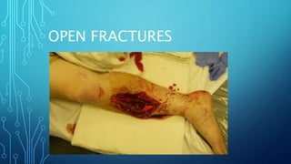
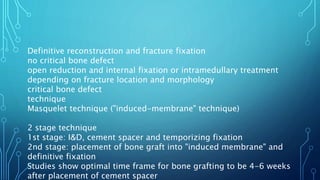
OPEN FRACTURES
EPIDEMIOLOGY
Incidence
common
30.7 per 100,000 persons per year
Demographics
average age is 45 years old
Anatomic location
tibia and finger phalanx are most common
ETIOLOGY
Pathophysiology
mechanism of injury
high-energy trauma
"inside-out" open fractures
Associated conditions
often associated with additional injuries
(30%)
compartment syndrome
the presence of an open wound does not
preclude the occurrence of compartment
syndrome in the injured limb
CLASSIFICATION
Gustilo classification
Tscherne classification
SHORTER VERSION
Type I
wound ≤1 cm, minimal contamination or muscle damage
Type II
wound 1-10 cm, moderate soft tissue injury
Type IIIA
wound usually >10 cm, high energy, extensive soft-tissue damage,
contaminated
adequate tissue for flap coverage
farm injuries are automatically at least Gustillo IIIA
Type IIIB
extensive periosteal stripping, wound requires soft tissue coverage
(rotational or free flap)
Type IIIC
vascular injury requiring vascular repair, regardless of degree of soft
tissue injury
Most accurate way to grade open fratures is by intra-operative
examination
PRESENTATION
History
obtain information regarding mechanism, location, and timing
of injury
Physical exam
inspection
assess soft-tissue damage
the size and nature of the external wound may not reflect the
damage to the deeper structures
neurovascular
if concern for vascular insult, ankle brachial index (ABI) should
be obtained
normal ratio is > 0.9
vascular surgery consult and angiogram is warranted if ABI <
0.9
IMAGING
RADIOGRAPHS
INDICATIONS
OBTAIN RADIOGRAPHS INCLUDING
JOINT ABOVE AND BELOW FRACTURE
CT
INDICATIONS
PERI-ARTICULAR INJURIES
EVALUATION FOR TRAUMATIC
ARTHROTOMY OF THE KNEE
TREATMENT
Nonoperative
urgent IV antibiotics, tetanus prophylaxis, and
extremity stabilization and dressing
indications
initial treatment for all open fractures
a soft tissue wound in proximity to a fracture
should be treated as an open fracture until
proven otherwise
mutlidisciplinary training of open fracture
management has been associated with
decreased timing to antibiotic administration
antibiotic type indicated by injury pattern and
location
Operative
I&D, temporary fracture stabilization, local antibiotic
administration and soft tissue coverage
indications
consider I&D as soon as possible
ideal time of soft tissue coverage controversial, but
most centers perform within 5-7 days
outcomes
infection rates of open fracture depend on zone of
injury, periosteal stripping and delay in treatment
incidence of fracture-related infection range from <1%
in type I open fractures to 30% in type III fractures
definitive reconstruction and fracture
fixation
indications
once soft tissue coverage is obtained
and an adequate sterility is achieved
outcomes
definitive treatment with internal
fixation leads to significantly
decreased time to union, improved
functional outcomes, and decreased
time in the hospital compared to
TECHNIQUE
Urgent IV antibiotics, tetanus prophylaxis, extremity
stabilization and dressings
Antibiotics
timing
initiate as soon as possible
studies show increased infection rate when antibiotics are
delayed for more than 3 hours from time of injury
continue for 24 hours after initial injury if wound is able to
be closed primarily
continue for 24 hours after final closure if wound is not
closed during initial surgical debridement (48 hours for
type III wounds)
types
Gustilo type I and II
1st generation cephalosporin
clindamycin or vancomycin can also be used if allergies exist
Gustilo type III
1st generation cephalosporin + aminoglycoside
some institutions use vancomycin + cefepime
farm injuries, heavy contamination, or possible bowel
contamination
add high dose penicillin for anaerobic coverage (clostridium)
special considerations
fresh water wounds
fluoroquinolones or 3rd or 4th generation cephalosporin
saltwater wounds
doxycycline + ceftazidime or a fluoroquinolone
Tetanus prophylaxis
timing
initiate in emergency room or trauma bay
two forms of prophylaxis
toxoid
0.5 mL, regardless of age
immunoglobulin
< 5 years old receive 75 U
5-10 years old receive 125 U
>10 years old receive 250 U
toxoid and immunoglobulin should be given
intramuscularly with two different syringes in two
different locations
guidelines for tetanus prophylaxis depend on 3
factors
complete or incomplete vaccination history (3
doses)
Extremity stabilization & dressing
stabilization
splint, brace, or traction for temporary stabilization
decreases pain, minimizes soft tissue trauma, and
prevents disruption of clots
dressing
remove gross debris from wound, do not remove any
bone fragments
place sterile saline-soaked dressing on wound
little evidence to support aggressive irrigation or
irrigation with antiseptic solution in the ED, as this can
push debris further into wound
I&D, temporary fracture stabilization, local
antibiotic administration and soft tissue
coverage
Irrigation and debridement
timing
recent meta-analysis (GOLIATH study) have
recommended debridement within 24 hours to
minimize risk of infection for type III fractures
within 12 hours for type IIIB open tibia
fractures
staged debridement and irrigation
perform every 24 to 48 hours as needed
temporary fracture stabilization
technique
performed at the time of initial debridement
external fixation is temporary initial treatment of choice
for majority of high energy open fractures of the lower
extremity
local antibiotic administration
indications
significantly contaminated wounds with large soft tissue
defects
large bony defects
technique
beads made by mixing methylmethacrylate with heat-
stable antibiotic powder
vancomycin and tobramycin most commonly used
soft tissue coverage
timing
early soft tissue coverage or wound closure is ideal
timing of flap coverage for open tibial fractures
remains controversial, < 7 days is desired
increased risk of infection beyond 7 days
odds of infection increase by 16% for each day
beyond day 7
early studies demonstrated increased infection with
delay beyond 72 hours, however recent studies do
not support this finding (LEAP study)
studies have not shown any statistical difference
between rate of infection when ORIF is performed
before fasciotomy closure, at fasciotomy closure, or
after fasciotomy closure
technique
can proceed with bone grafting after wound
is clean and closed
negative-pressure wound therapy may be
utilized during debridement until definitive
coverage can be achieved (increased risk of
infection if open >7 days)
Definitive reconstruction and fracture fixation
no critical bone defect
open reduction and internal fixation or intramedullary treatment
depending on fracture location and morphology
critical bone defect
technique
Masquelet technique ("induced-membrane" technique)
2 stage technique
1st stage: I&D, cement spacer and temporizing fixation
2nd stage: placement of bone graft into "induced membrane" and
definitive fixation
Studies show optimal time frame for bone grafting to be 4-6 weeks
after placement of cement spacer
COMPLICATIONS
Surgical site infection
incidence
fracture-related infection ranges from <1% in type I open fractures to
30% in type III fractures
Osteomyelitis
incidence
ranges between 1.8% to 27% depending on the bone involved and
fracture characteristics.
the tibia is the most common site of post-surgical osteomyelitis
following surgical treatment of open fractures
risk factors include:
blast mechanism of injury
acute surgical amputation
delay in defintive soft tissue coverage greater than 7 days
more severe Gustillo-Anderson classification.
Neurovascular injury
Compartment syndrome
PROGNOSIS
To minimize risk of infection, debridement
recommended to be performed within 24 hours for all
type III fractures and within 12 hours for type IIIB open
tibia fractures
Contamination with dirt and debris and devitalization of
the soft tissues increase the risk of infection and other
complications
Infection rates higher in open injuries due to blunt
trauma compared to penetrating trauma
THE END

Open Fractures.pptx

  • 1.
  • 2.
    EPIDEMIOLOGY Incidence common 30.7 per 100,000persons per year Demographics average age is 45 years old Anatomic location tibia and finger phalanx are most common
  • 3.
    ETIOLOGY Pathophysiology mechanism of injury high-energytrauma "inside-out" open fractures Associated conditions often associated with additional injuries (30%) compartment syndrome the presence of an open wound does not preclude the occurrence of compartment syndrome in the injured limb
  • 4.
  • 5.
    SHORTER VERSION Type I wound≤1 cm, minimal contamination or muscle damage Type II wound 1-10 cm, moderate soft tissue injury Type IIIA wound usually >10 cm, high energy, extensive soft-tissue damage, contaminated adequate tissue for flap coverage farm injuries are automatically at least Gustillo IIIA Type IIIB extensive periosteal stripping, wound requires soft tissue coverage (rotational or free flap) Type IIIC vascular injury requiring vascular repair, regardless of degree of soft tissue injury Most accurate way to grade open fratures is by intra-operative examination
  • 8.
    PRESENTATION History obtain information regardingmechanism, location, and timing of injury Physical exam inspection assess soft-tissue damage the size and nature of the external wound may not reflect the damage to the deeper structures neurovascular if concern for vascular insult, ankle brachial index (ABI) should be obtained normal ratio is > 0.9 vascular surgery consult and angiogram is warranted if ABI < 0.9
  • 9.
    IMAGING RADIOGRAPHS INDICATIONS OBTAIN RADIOGRAPHS INCLUDING JOINTABOVE AND BELOW FRACTURE CT INDICATIONS PERI-ARTICULAR INJURIES EVALUATION FOR TRAUMATIC ARTHROTOMY OF THE KNEE
  • 10.
    TREATMENT Nonoperative urgent IV antibiotics,tetanus prophylaxis, and extremity stabilization and dressing indications initial treatment for all open fractures a soft tissue wound in proximity to a fracture should be treated as an open fracture until proven otherwise mutlidisciplinary training of open fracture management has been associated with decreased timing to antibiotic administration antibiotic type indicated by injury pattern and location
  • 11.
    Operative I&D, temporary fracturestabilization, local antibiotic administration and soft tissue coverage indications consider I&D as soon as possible ideal time of soft tissue coverage controversial, but most centers perform within 5-7 days outcomes infection rates of open fracture depend on zone of injury, periosteal stripping and delay in treatment incidence of fracture-related infection range from <1% in type I open fractures to 30% in type III fractures
  • 12.
    definitive reconstruction andfracture fixation indications once soft tissue coverage is obtained and an adequate sterility is achieved outcomes definitive treatment with internal fixation leads to significantly decreased time to union, improved functional outcomes, and decreased time in the hospital compared to
  • 13.
    TECHNIQUE Urgent IV antibiotics,tetanus prophylaxis, extremity stabilization and dressings Antibiotics timing initiate as soon as possible studies show increased infection rate when antibiotics are delayed for more than 3 hours from time of injury continue for 24 hours after initial injury if wound is able to be closed primarily continue for 24 hours after final closure if wound is not closed during initial surgical debridement (48 hours for type III wounds)
  • 14.
    types Gustilo type Iand II 1st generation cephalosporin clindamycin or vancomycin can also be used if allergies exist Gustilo type III 1st generation cephalosporin + aminoglycoside some institutions use vancomycin + cefepime farm injuries, heavy contamination, or possible bowel contamination add high dose penicillin for anaerobic coverage (clostridium) special considerations fresh water wounds fluoroquinolones or 3rd or 4th generation cephalosporin saltwater wounds doxycycline + ceftazidime or a fluoroquinolone
  • 15.
    Tetanus prophylaxis timing initiate inemergency room or trauma bay two forms of prophylaxis toxoid 0.5 mL, regardless of age immunoglobulin < 5 years old receive 75 U 5-10 years old receive 125 U >10 years old receive 250 U toxoid and immunoglobulin should be given intramuscularly with two different syringes in two different locations guidelines for tetanus prophylaxis depend on 3 factors complete or incomplete vaccination history (3 doses)
  • 16.
    Extremity stabilization &dressing stabilization splint, brace, or traction for temporary stabilization decreases pain, minimizes soft tissue trauma, and prevents disruption of clots dressing remove gross debris from wound, do not remove any bone fragments place sterile saline-soaked dressing on wound little evidence to support aggressive irrigation or irrigation with antiseptic solution in the ED, as this can push debris further into wound
  • 17.
    I&D, temporary fracturestabilization, local antibiotic administration and soft tissue coverage Irrigation and debridement timing recent meta-analysis (GOLIATH study) have recommended debridement within 24 hours to minimize risk of infection for type III fractures within 12 hours for type IIIB open tibia fractures staged debridement and irrigation perform every 24 to 48 hours as needed
  • 18.
    temporary fracture stabilization technique performedat the time of initial debridement external fixation is temporary initial treatment of choice for majority of high energy open fractures of the lower extremity local antibiotic administration indications significantly contaminated wounds with large soft tissue defects large bony defects technique beads made by mixing methylmethacrylate with heat- stable antibiotic powder vancomycin and tobramycin most commonly used
  • 19.
    soft tissue coverage timing earlysoft tissue coverage or wound closure is ideal timing of flap coverage for open tibial fractures remains controversial, < 7 days is desired increased risk of infection beyond 7 days odds of infection increase by 16% for each day beyond day 7 early studies demonstrated increased infection with delay beyond 72 hours, however recent studies do not support this finding (LEAP study) studies have not shown any statistical difference between rate of infection when ORIF is performed before fasciotomy closure, at fasciotomy closure, or after fasciotomy closure
  • 20.
    technique can proceed withbone grafting after wound is clean and closed negative-pressure wound therapy may be utilized during debridement until definitive coverage can be achieved (increased risk of infection if open >7 days)
  • 21.
    Definitive reconstruction andfracture fixation no critical bone defect open reduction and internal fixation or intramedullary treatment depending on fracture location and morphology critical bone defect technique Masquelet technique ("induced-membrane" technique) 2 stage technique 1st stage: I&D, cement spacer and temporizing fixation 2nd stage: placement of bone graft into "induced membrane" and definitive fixation Studies show optimal time frame for bone grafting to be 4-6 weeks after placement of cement spacer
  • 22.
    COMPLICATIONS Surgical site infection incidence fracture-relatedinfection ranges from <1% in type I open fractures to 30% in type III fractures Osteomyelitis incidence ranges between 1.8% to 27% depending on the bone involved and fracture characteristics. the tibia is the most common site of post-surgical osteomyelitis following surgical treatment of open fractures risk factors include: blast mechanism of injury acute surgical amputation delay in defintive soft tissue coverage greater than 7 days more severe Gustillo-Anderson classification. Neurovascular injury Compartment syndrome
  • 23.
    PROGNOSIS To minimize riskof infection, debridement recommended to be performed within 24 hours for all type III fractures and within 12 hours for type IIIB open tibia fractures Contamination with dirt and debris and devitalization of the soft tissues increase the risk of infection and other complications Infection rates higher in open injuries due to blunt trauma compared to penetrating trauma
  • 24.