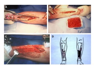
2. • The reconstruction of wide long bone
diaphyseal defects is often the major
challenge in limb salvage whatever the
etiology of bone loss.
• The most common and widely accepted
procedures are the autologous bone transfer
and the Ilizarov bone transport method.
• The critical size for non-vascularized bone-
grafting is 6.0 - 7.0 cm
3. PROBLEM WITH AUTOLOGOUS BONE
GRAFT
• Bone autograft is not advocated when the
defect is over 4 to 5 cm. When diaphyseal
defects larger than 6 cm are reconstructed
with autologous bone graft, healing is
incomplete because of graft resorption even
in a good vascularized muscular envelope*.
*Hertel, L., Gerber, A., Schlegel, U. et al. Cancellous bone graft for skeletal
reconstruction muscular versus periosteal bed. Preliminaryresults. Injury. 1994; 25:
59–70
4. • Vascularized bonegrafting is technically demanding and
require micro vascular surgical skills. The technique is
reliable but the donor sites are limited.
• The Ilizarov technique is the one commonly used to
address intermediate and large bone defects. The
technique is very demanding and patient’s cooperation
is critical.
• There is no single current technique that is reliably
successful in the management of large bone defects.
• The Masquelet technique does offer an alternative and
a viable management strategy for large bone defects.
5. • The technique was developed in 1986 to
address defects larger than 15cm. It was later
established that it can successfully address
bone defects as large as 25cm.
• It can be safely used in irradiated or infected
areas provided the membrane is formed
around the defect to protect and vascularize
the bone graft.
6. PRINCIPLE OF TECHNIQUE
• The first stage is comprised of a radical debridement, a
soft tissue repair by flaps when needed, and the
insertion of a polymethyl methacrylate (PMMA)
cement spacer mixed with or without antibiotic into
the bone defect
• The second stage is performed 6 to 8 weeks later, when
the definitive healing of soft tissue is acquired. The
spacer is removed, but the membrane that is induced
by the cement is left in place. The cavity is filled up by
morcellized cancellous bone autograft harvested from
the iliac crests
7.
8. FIRST STAGE
• 1. Radical debridement :- the first stage involves radical
debridement of all infected or non-viable bone and
interposed fibrous tissue.
• devitalized tissue serves as a nidus for recurrent
infection and predisposes to increased risk of
postoperative complications such as delayed union,
nonunion, and vascular thrombosis.
• The margins of debridement should extend until viable
bony edges are encountered, determined
intraoperatively using the “paprika sign” (punctate
bleeding upon drilling with a 2.5 mm drill bit)
9. • 2. Limb Stabilization :- Following debridement, stabilization
must be achieved to maintain length, alignment and
rotation prior to insertion of the cement spacer. Choice of
stabilizer depends on the site of the defect.
• For bone loss in the mid-diaphysis, an IM nail offers stable
fixation that allows early weight bearing.
• For defects close to an articular surface, external fixation is
preferred.
• Ring-fixators offer stable fixation and the ability to modify
the bony alignment postoperatively.
• When placing external fixation, care must be taken to keep
pins away from the site of definitive fixation so that the
external fixator can be left in place until healing is achieved
10. CHOICE OF FIXATION DEVICE
• Since most cases treated by this method are
of septic non unions requiring serial
debridements and excisions, as well as wound
dressings and flap surgeries, external fixators
were preferred choice of fixation initially.
11. DISADVANTAGES OF EXTERNAL
FIXATOR
• external fixator doesn’t provide rigid fixation
which favors the action of growth factors as
well as fusion of bone grafts
• With bone defect >20cm, it is difficult to
maintain axis of the limb with the EF alone
forcing the surgeon to do osteotomies at later
stages
12. • Locked nails or plates are now preferred because
of more stable fixation and reduced rate of
infection post debridement due to better
antibiotics and wound care.
• They also reduce the graft requirement and also
prevent the central core of densely packed graft
(unlikely to revascularize)
• To avoid biofilm formation and to create a
complete induced membrane,it is adviced that
the implant should be completely covered with
the antibiotic cement
13. • 3. Placement of antibiotic impregnated Cement Spacer :- The
role of the cement spacer is to avoid the collapse of the soft
tissue into the bone defect and to maintain the dead space for
the bone reconstruction.
• For optimal outcome, the cement spacer should fill the
intramedullary canal and edges of surrounding viable bone to
allow detaching small pieces of the ends of the bone when
cement is removed and lifting it with a bit of the induced
membrane at the second stage
14. • The biological function of cement i.e. induction of
foreign-body surrounding membrane was realized
when masquelet noticed that after thorough
debridement and using cement spacer in all post
traumatic septic non unions showed absence of
recurrent infection after 2 months.
• Beads leave an irregular membrane that is less than
ideal for containment of the graft.
• Since cement itself act as a foreign body, absence of
infection after 2 months shows the adequateness of
the debridement and readiness of the site for bone
grafting.
15. • 4. Soft-tissue coverage and wound healing :-
the least technically demanding strategy that
enables successful soft-tissue coverage should
be chosen ranging from wet-to-dry dressings
to a flap procedure to provide adequate soft-
tissue coverage
16. After STAGE 1
• Weight-bearing is determined based upon the stability of
the defect size, location and implant. Patients with small
and medium diaphyseal defects treated by IM nailing can
bear weight as tolerated.
• The patient is then placed on a prolonged systemic
antibiotic regimen for a period of 6-8 weeks. This is done in
order to allow adequate time for a number of processes to
occur: (1) epithelialization of free or pedicled muscle flaps
to prevent surgical site contamination by bacterial skin
flora, (2) revascularization of marginally viable tissue
surrounding the bony defect, (3) formation of the self-
induced periosteal membrane, and (4) treatment of any
residual infection by systemic and/or local antibiotics
17. FOREIGN-BODY INDUCED MEMBRANE
• Experimental study was done at the AO
Development Institute of Davos and it was
proved that the membrane avoided the
resorption of the cancellous bone and had a
positive effect upon the healing of the
autograft*
*Klaue, K., Anton, C., Knothe, U. et al. Biological implementation of “in situ”
induced autologous foreign body membranes in consolidation of massive
cancellous bone grafts. J Bone Joint Surg. 1993; 79B: 236
18. • They created segmental femoral defect in 30
sheep, 3 cm in length, filled it up with a PMMA
cement spacer, and stabilized with a plate. One
month later, four groups were constituted after
removing the spacer:
• Group A: the membrane was maintained and filled up with
cancellous bone chips.
• Group B: the membrane was excised and the defect was filled up
with cancellous bone chips.
• Group C: the membrane alone was maintained without filling.
• Group D: the membrane was excised and the defect was not filled
up.
19. • No bone formation was noted in groups C and
D. In group B, bone healing was partially
obtained with an important resorption in all
cases. In group A, bone healing was acquired
without reduction of the volume of the initial
graft.
20. HISTOLOGIC AND
BIOCHEMICAL CHARACTERISTICS
• The membrane is richly vascularized in all its
layers.
• The inner part (face to the cement) is a
synovial like epithelium and the outer part is
made of fibroblasts, myofibroblasts, and
collagen.
21. • The membrane secretes growth factors: high
concentration of VEGF and TGF Beta 1 were
observed as early as the second week. BMP2
is at its highest level at the fourth week.
• Membrane extracts has been shown to
stimulate bone marrow cell proliferation and
differentiation to osteoblastic lineage in vitro.
22. • the cancellous bone inside the membrane is
not submitted to resorption.
• Macroscopic examination of transverse
section of the healed bone graft exhibited
normal bone anatomy, and the junction
between the normal bone and the graft was
difficult to see by macroscopic examination of
longitudinal sections
23. ADVANTAGES
• Induced membrane acts as a biological chamber
keeping the cancellous graft together and prevents
its resorption
• The membrane promotes the vascularization and the
corticalization of the cancellous bone, even in bad
vascularized bed like irradiated tissue or in very
specific bone disease like congenital pseudarthrosis.
• Membrane is considered an in situ delivery system
for growth and osteoinductive factors( MC is BMP-2)
24. INDUCED MEMBRANE V/S
POLYLACTIDE MEMBRANE
• Polylactide membrane have not been proven
to secrete growth factors.
• Their mechanism is based on the exclusion of
fibrous tissue inside the defects.
• When cancellous bone graft is added within
the lumen, small perforation of the membrane
will allow the revascularization of the graft.
• They do not have the same biological
properties as that of foreign body membrane.
25. SECOND STAGE
• 1.Clearence of infection :- Complete eradication
of infection is a prerequisite to reconstruction of
bone defects due to osteomyelitis (Stage 2 of the
Masquelet technique)
• Levels of acute inflammatory markers, including
CRP and ESR, should be normal in patients lacking
comorbidities.
• If there remains any doubt as to the presence of
residual infection, then tissue specimens at the
site of the segmental defect can be harvested for
culture and pathology
26. • 2.Removal of the cement spacer and permanent fixation of
the fracture :- A single longitudinal incision is made
centrally through the self-induced periosteal membrane.
• The cement spacer should be removed in one piece or a
few small pieces created with a saw or osteotome.
• Injury to the induced periosteal membrane should be
avoided so that it remains a self-contained compartment.
• The ends of the resected bone margins should be
freshened with a drill bit or rasp to remove sclerotic bone
and facilitate bone graft integration. The medullary canal
should also be debrided to enable communicate with the
graft.
• Definitive fixation can be revised at this point if necessary.
27. • The initial reason for which Masquelet did not excise
the membrane was to prevent excessive bleeding.
• The best period to perform the graft is 1 month after
the set of the cement spacer*
*Klaue, K., Anton, C., Knothe, U. et al. Biological implementation of “in situ” induced
autologous foreign body membranes in consolidation of massive cancellous bone grafts. J
Bone Joint Surg. 1993; 79B: 236
• This is because of the secretion of highest level of
BMP-2 at 4 weeks by the induction membrane.
However, Soft-tissue status often results in delaying
of this graft-insertion time in some patients.
28. • 3.Harvest of autogenous bone graft :-Morcellized, fresh, cancellous
bone autograft is the gold standard and is harvested from the iliac
crests.
• The cancellous bone is capable of forming bone even without stress
to the bone. But the cancellous bone will resorb if the recipient bed
is poorly vascularized.
• In some cases with large bone defects, the bone substitute can be
added with a ratio of 1:3; with no difference on rate of complication
or time of healing when compared to the reconstruction performed
with cancellous bone alone.
• Autogenous bone graft can also be enhanced with synthetic bone
morphogenetic protein (BMP), bisphosphonates, or hydroxyapatite,
however increased autograft resorption has been observed in
patients receiving additional local injections of recombinant BMP-7
29. ROLE OF RIA
• Intramedullary canal bone reamings (ICBR)/(RIA) can be
used as a source of viable bone graft in relation to their
osteoblastic potential and living bone cells similar to bone
cells from the iliac crest.
• It provides graft with minimal incision, minimal donor site
pain and also higher graft yield ( 47cm3 > 35cm3)
• They provide graft which contain multipotent stem cells
with osteosynthesis capability and higher level of growth
factors like FGF, PDGF, BMP-2 when compared with graft
from iliac crest
• Requires cautious use in osteoporotic bones
• Graft harvested from RIA is becoming the new gold
standard
30. • Densely packing of the graft should be prevented
as it has been shown that tightly packed graft
does not consolidate well. On tight packing of the
graft, While the peripheral graft seems to
revascularize and ossify, the central core of graft
remains unhealed and non-vascular.
• After the bone graft is placed in the membrane,
the soft tissues including the membrane are
sutured close to the graft resulting in a
containment chamber
31. • ‘watertight’ induced membrane chamber can
be considered to be a complex bioreactor,
continuously providing osteogenesic factors,
cells as well as a blood supply for bone graft.
• Induced membrane technique must be
combined with stable fixation, as instability
may lead to deformity or nonunion.
32. CONCLUSION
• The Masquelet’s technique is a practical
alternative in the management of bone
defects. It has a relatively low complication
rate. It does not ‘burn bridges’: other complex
procedures may be done if the technique fails.