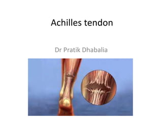
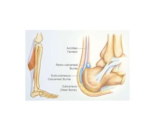
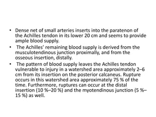
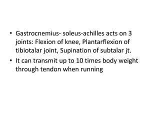
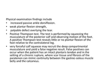
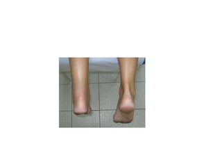
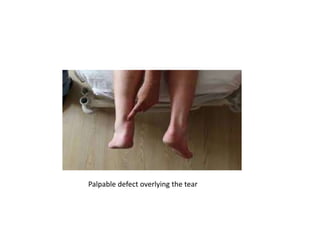
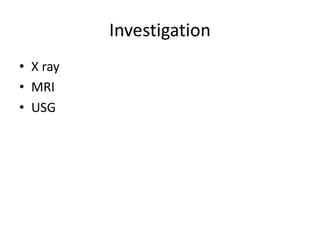
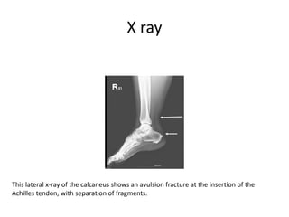
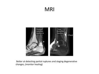
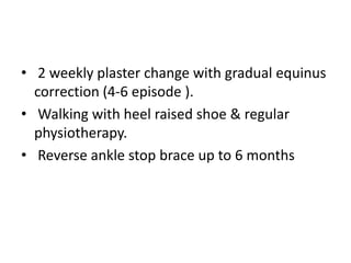
The Achilles tendon connects the calf muscles to the heel bone and is active during walking, running, and jumping. It is vulnerable to rupture, which commonly occurs in the watershed area 2-6cm above the heel in males ages 25-50 during sports. Diagnosis involves physical exam findings like inability to raise the toes and a gap or defect being felt. Treatment options include immobilization in a cast or surgical repair, with the goal of restoring proper tension and length. Post-operative care focuses on protecting the repair during healing over 6 months using casts, braces, and physical therapy.
2. INTRODUCTION
• The Achilles tendon is the largest and strongest
tendon in the human body.
• The incidence rises rapidly after 25 years of age
with males in their fourth or fifth decade
accounting for the overwhelming majority of
acute ruptures.
• Another peak is seen between the sixth and
eighth decade, which predominantly occurs from
a longstanding degenerative condition of the
tendon, with the incidence remaining higher in
men.
3. ANATOMY
• The tendon receives equal contributions from
both the gastrocnemius and soleus muscle
and tendinous fibers. These fibers converge
approximately 15 cm from the insertion point.
As the tendon courses inferiorly in the
posterior aspect of the leg, the fibers twist
approximatedly 120° internally before its
insertion point on the calcaneal tuberosity.
4. • The Achilles tendon lacks a true synovial
sheath. Rather, the tendon is enveloped
within a paratenon. The paratenon permits
gliding of the tendon between the skin and
surrounding posterior soft tissues of the leg.
• The paratenon is responsible for a significant
portion of the tendon’s blood supply through
a highly vascularized areolar tissue on its
anterior aspect.
5.
6. • Dense net of small arteries inserts into the paratenon of
the Achilles tendon in its lower 20 cm and seems to provide
ample blood supply.
• The Achilles’ remaining blood supply is derived from the
musculotendinous junction proximally, and from the
osseous insertion, distally.
• The pattern of blood supply leaves the Achilles tendon
vulnerable to injury in a watershed area approximately 2–6
cm from its insertion on the posterior calcaneus. Rupture
occurs in this watershed area approximately 75 % of the
time. Furthermore, ruptures can occur at the distal
insertion (10 %–20 %) and the myotendinous junction (5 %–
15 %) as well.
7. • Gastrocnemius- soleus-achilles acts on 3
joints: Flexion of knee, Plantarflexion of
tibiotalar joint, Supination of subtalar jt.
• It can transmit up to 10 times body weight
through tendon when running
8. Risk factors for rupture
Local corticosteroids
Systemic corticosteroids
Peritendinous injection
Flouroquinolones
Degenerative changes in tendon
Obesity
Diabetes mellitus
Vascular degeneration/ irregularitiees
Hyperthermia of tendon
Training error
Malalignment of foot and/or ankle
Chronic tendinopathy ( with haglund’s
deformity)
Previous tendon injury
9. Presentation, physical examination
and diagnosis
• Patient will often describe sudden onset of pain
in the posterior aspect of the foot and ankle,
usually during activity that calls for maximum
forceful plantar flexion.
• Patients will describe feeling a “pop” or a
sensation of being kicked in the back of the leg.
• They will often lose the ability to bear weight
and/or report weakness in plantar flexion of the
ankle. Interestingly enough, patients rarely
present with significant pain. Rather, they present
with bruising and a functional deficit.
10. Physical examination findings include
• increased passive ankle dorsiflexion.
• weak plantar flexion strength,
• palpable defect overlying the tear.
• Positive Thompson test. The test is performed by squeezing the
musculature of the posterior calf and observing motion of the foot.
A positive Thompson test reveals little or no plantar flexion of the
foot relative to the contralateral leg.
• very forceful calf squeeze may recruit the deep compartmental
musculature and yield a false negative result. False positives can
occur when the patient has an intact plantaris tendon and in the
setting of a chronic rupture, where scar tissue and fibrosis of the
paratenon can mimic continuity between the gastroc-soleus muscle
belly and the calcaneus.
15. X ray
This lateral x-ray of the calcaneus shows an avulsion fracture at the insertion of the
Achilles tendon, with separation of fragments.
16. MRI
Better at detecting partial ruptures and staging degenerative
changes, (monitor healing)
17. USG
Inexpensive, fast, reproducable, dynamic examination possible
Operator dependent
Best to measure thickness and gap
Good screening test for complete rupture
18. MANAGEMENT
• The fundamental goals of treatment of an
acute Achilles tendon rupture are to restore
length and tension of the tendon in order to
optimize a patient’s ability to return to their
desired level of activity.
• Treatment options include nonoperative
regimens, traditional open repair, and
percutaneous or mini-open repairs.
19. Conservative management
• Conservative treatment regimens vary greatly but
commonly involve immobilisation with rigid casting or
functional bracing. The foot is initially placed in full
equinus (30° namely full plantarflexion). The foot is
then brought into neutral sequentially over a period of
8-12 wk. Once ankle position permits it, weight bearing
is allowed.
• patients can be allowed to weight-bear early in an off-
the-shelf orthosis/CAM walker/Sheffield splint with no
detriment in any long term outcomes.
• Rerupture rate is high
20. Surgical management
Principles:
• Preserve anterior paratenon bl. supply
• Beware of sural nerve
• Debride and approximate tendon ends
• Use 2-4 stranded locked suture technique
• Close paratenon separately
21. A: Defects of 1 cm or less: Direct end to end
repair without augmentation
• Bunnell Suture
• Modified Kessler
• Many techniques available
B: Defects 1 - 2 cm Muscle mobilization
augmentation (plantaris) Can gain up to 2 cm
with mobilization
22. C: Defects 2 - 5 cm
• No consensus on best reconstruction technique
• Semi-T tendon transfer
• Flexor hallucis longus (FHL) tendon transfer loss
of great toe flexion(Not acceptable in Athletes)
• Others: FDL , Peroneus Brevis
• V-Y myotendinous lengthening FHL transfer
D: Defects > 5 cm: SemiT Transfer, V-Y
myotendinous lengthening
25. Post op care
• Assess strength of repair, tension and ROM
intra-op.
• Apply long leg cast with ankle in the least
amount of planterflexion(gravityequinus) &
knee 60 degree flexion with window at
operated site.
• Stitch removal after 2 wks.
• Short leg cast after 3 wks with partial equinus
correction
26. • 2 weekly plaster change with gradual equinus
correction (4-6 episode ).
• Walking with heel raised shoe & regular
physiotherapy.
• Reverse ankle stop brace up to 6 months