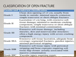
SEMINAR :-OPEN
FRACTURE ,EVALUATION
AND MANAGEMENT
DR. B.BORTHAKUR
PROFESSOR & HOD, DEPT. OF ORTHOPAEDICS,
SMCH
LEARNING OBJECTIVES
• INTRODUCTION
• ASSESMENT AND CLASSIFICATION
• PREVENTION OF INFECTION
• WOUND MANAGEMENT
• FRACTURE STABILISATION
• SUMMARY
 DEFINITION
Open fractures is defined as an injury
the fracture and fracture hematoma
communicate with the external
through a traumatic defect in the
surrounding soft tissues and overlying skin.
Assessment and Classification of
Open Fractures
• Patient is evaluated and resuscitated according
to ATLS protocols.
• Injured extremities then should be assessed for
neurovascular injury and compartment
• Photographic documentation of the wound
should ideally be undertaken.
• If immediate operative intervention is planned do
not irrigate , debride or probe the wound in ER.
• If surgical delay (>24hrs) is anticipated ,gentle
irrigation with normal saline can be done.
• Gross contamination should be removed.
• Wound is covered with saline soaked dressing
under impervious seal.
SPLINTAGE :-
• Any skeletal trauma must be splinted with
whatever best available before shifting the
patient for any investigation or OT
• After temporary splintage, stabilization with
external fixator or definitive internal fixation
is done.
ROLE OF CULTURES IN EMERGENCY ROOM
• There is disparity between the organism grown on the initial
wound swabs and organism grown subsequently after the
development of wound infection.
• The practice of obtaining routine cultures from the wound
either pre or post debridement is no longer advocated.
CLASSIFICATION OF OPEN FRACTURE
• For most of the open fracture
modified Gustilo and Anderson’s
classification is used.
• But for open tibia fracture Ganga
Hospital open injury score is used.
• Classification of open fracture is done
in OT not in emergency room.
DISADVANTAGES OF GUSTILO`S AND ANDERSON`S CLASSIFICATION
• Definition has undergone many modification and
does not have uniformity in application.
• Includes wide spectrum of injuries in type IIIB
• Mainly depends on size of the skin wound.
• Does not address the question of salvage
• Poor interobserver reliability.
PREVENTION OF INFECTION
• All open fracture wounds should be considered
contaminated.
• Infection is enhanced by the following:-
# bacterial contamination
# colonization of the wound
# presence of dead space with
devitalized tissues
# foreign material
ANTIBIOTICS COVERAGE FOR OPEN FRACTURE
TYPE I & II :- First generation cephalosporin
TYPE III:- add an Aminoglycoside.
Antibiotics should be started as soon as possible
• Tetanus prophylaxis should be given in
emergency department.
• Current dose of toxoid is 0.5ml regardless of
age.
• DOSE OF IMMUNOGLOBULIN :-
 < 5 years of age:- 75 IU
 5-10 years of age:- 125 IU
 > 10 years of age :- 250 IU
• Toxoid and immunoglobulin should be given
intramuscularly with two different syringes in two
different locations.
GUIDELINES FOR TETANUS PROPHYLAXIS:-
Depends on 3 factors:-
* complete or incomplete vaccination history ( 3 doses)
*date of most recent vaccination
*severity of wound.
Immunization history Clean, minor wound (type I
open fracture)
Type II & III open fracture
Unknown history or < 3 doses Give vaccine only *Give vaccine
*Give Ig
Vaccination complete (3 prior
doses)
*No prophylaxis if last dose
given within 10 years
*Give vaccine if > 10 years since
last dose
*No prophylaxis if last dose given within
years
*Give vaccine if > 5 years since last dose
WOUND
MANAGEMENT
Irrigation and Debridement
• Adequate Irrigation and debridement are the
important steps in open fracture treatment.
• The most commonly used irrigant is normal saline.
• High pressure irrigation removes more bacteria and
necrotic tissue than syringe.
• The current consensus seems to lean towards
high volume , low pressure lavage repeated
adequate number of times in pulsatile manner
to effect the best healing and prevention of
infection.
• Debridement is done by senior surgeon.
TECHNIQUE OF DEBRIDEMENT:-
Wash and drape the wound as for normal surgical procedure
Remove devitalizes skin untill bleeding is visible
Remove the subcutaneous tissue including all contaminated tissue
Remove devitalized fat beneath the flaps
Open the fascia to allow exposure of the muscle tendon and removal of all devitalized
muscle
Trim completely severed tendon back to viable tendon
Enlarge the wound for proper debridement and exposure of the fracture.
Irrigate the wound with NS after removal of all dead tissue
Close the surgically created wound first
Loosely close the remaining wound over a drain if necessary.
VOLUME OF FLUID REQUIRED:-
• FOR TYPE I OPEN FRACTURE :- 3 L
• FOR TYPE II OPEN FRACTURE :- 6L
• FOR TYPE III OPEN FRACTURE :- 9L
.
• Retained avascular bones and small fragments which
are completely devoid of soft tissues must be
removed.
FRACTURE STABILIZATION :-
For type I open fracture, any technique that is suitable
for closed fracture management is satisfactory.
Upto type IIIA closed internal fixation can be done
The method to reduce and stabilize the fracture
depends on following:-
#Bone that is involved
#Type of fracture
#The efficacy of the debridement
#patient`s general condition
#Surgeons choice.
OPTIONS AVAIALABLE FOR FRACTURE STABILIZATION :-
# EXTERNAL FIXATOR
# INTRAMEDULLARY NAILING
# OCCASIONALLY PLATE AND SCREW FIXATION
EXTERNAL SKELETAL FIXATION:-
• Tubular external fixators and dynamic external devices
LRS)
• Ring external fixators (Ilizarov, Taylors spatial frame)
• Hybrid external fixators.
• External fixators are mainly used as temporary
stabilizers.
• Can be used as a definitive treatment when a stable
fracture configuration with good reduction and
circumferential contact is achieved.
• Pin tract infection is the most frequent complication
With external fixation
• External fixation is preferred for metaphyseal -
diaphyseal fracture
PRIMARY INTERNAL FIXATION :-
• Done by interlocking nails and plate fixation.
• Open diaphyseal femoral and tibial fractures have been
treated successfully with nailing.
PLATE FIXATION :-
Disadvantage:-
• Need increased soft tissue exposure and periosteal
stripping
INDICATION:-
• Most open upper limb fracture
• Femoral fractures involving periarticular and articular regions
• All intra-articular and juxta articular fractures
INTRAMEDULLARY NAILS :-
• Often the first choice for lower limb diaphyseal fractures.
• Ideally suited for Type I & Type II & even in Type III injuries
where contamination is minimal.
.
UNREAMED NAILS:-
ADVANTAGE:-
• Causes less devascularization
• Shorter operating time.
• Lower incidence of fat embolism and thermal
necrosis.
DISADVANTAGE:-
• Increased rate of implant failure
• Fracture disruption during surgery
• Higher rate of nonunion and malunion
Wound Closure:-
Primary wound closure is controversial, but good results
are reported after primary closure.
INDICATIONS :-
• Type I & II & IIIA & B injuries of limb..
• Wounds without primary skin loss or secondary skin loss after
debridement.
• Injuries to debridement interval is less than 12hrs.
• Presence of bleeding wound margins which can be apposed
without tension.
• Stable fixation achieved by internal or external fixation.
SKIN GRAFTING PRINCIPLES:-
• Harvest skin from donor site to cover the
defect.
• Split thickness skin graft:- when a graft
includes only portion of dermis.
• Full thickness skin graft:- when a graft
contains entire dermis.
• Split thickness skin graft survive in
conditions with less vascularity but they
have likelihood of contracture.
FLAP COVERAGE :-
• Unlike grafts , flap maintains its own blood supply.
• Used for large wounds or to cover underlying bone and
tendons that may not be managed by graft alone..
NEGATIVE PRESSURE WOUND THERAPY (NPWT):-
Useful treatment in all injuries where soft tissue cover
is not immediately possible is vacuum assisted wound
closure (VAC) using NPWT.
BENEFICIAL EFFECTS OF VAC THERAPY:-
• Promotes wound contraction and increases the chance of
delayed primary closure.
• Removes excess oedematous fluid
• Causes reactive increase in blood flow and promotes
• Decreases bacterial burden
• Removes protein and electrolytes that are harmful for
healing.
REHABILITATION :-
Patients are advised for various active and
passive physiotherapy.
SUMMARY:-
• Assessment and classification of open fractures should be
done intra operatively based on the degree of bacterial
contamination, soft-tissue damage, and fracture
characteristics.
• To avoid the complication the wound should be thoroughly
irrigated and debrided.
• Tetanus toxoid and immun oglobulins should be given in
emergency department.
• Early, systemic, wide-spectrum antibiotic therapy is
to cover both gram-positive and gram negative organisms.
• In the presence of extensive soft-tissue loss and exposed
bone, coverage is accomplished with early transfer of local or
free muscle flaps.
• Stable fracture fixation is important; the method chosen
depends on the bone and soft-tissue characteristics.
• Early bone grafting is indicated for bone defects, unstable
fractures treated with external fixation, and delayed union.
THANK YOU

Open fractures

  • 1.
    SEMINAR :-OPEN FRACTURE ,EVALUATION ANDMANAGEMENT DR. B.BORTHAKUR PROFESSOR & HOD, DEPT. OF ORTHOPAEDICS, SMCH
  • 2.
    LEARNING OBJECTIVES • INTRODUCTION •ASSESMENT AND CLASSIFICATION • PREVENTION OF INFECTION • WOUND MANAGEMENT • FRACTURE STABILISATION • SUMMARY
  • 3.
     DEFINITION Open fracturesis defined as an injury the fracture and fracture hematoma communicate with the external through a traumatic defect in the surrounding soft tissues and overlying skin.
  • 4.
    Assessment and Classificationof Open Fractures • Patient is evaluated and resuscitated according to ATLS protocols. • Injured extremities then should be assessed for neurovascular injury and compartment • Photographic documentation of the wound should ideally be undertaken.
  • 5.
    • If immediateoperative intervention is planned do not irrigate , debride or probe the wound in ER. • If surgical delay (>24hrs) is anticipated ,gentle irrigation with normal saline can be done. • Gross contamination should be removed. • Wound is covered with saline soaked dressing under impervious seal.
  • 6.
    SPLINTAGE :- • Anyskeletal trauma must be splinted with whatever best available before shifting the patient for any investigation or OT • After temporary splintage, stabilization with external fixator or definitive internal fixation is done.
  • 7.
    ROLE OF CULTURESIN EMERGENCY ROOM • There is disparity between the organism grown on the initial wound swabs and organism grown subsequently after the development of wound infection. • The practice of obtaining routine cultures from the wound either pre or post debridement is no longer advocated.
  • 8.
  • 9.
    • For mostof the open fracture modified Gustilo and Anderson’s classification is used. • But for open tibia fracture Ganga Hospital open injury score is used. • Classification of open fracture is done in OT not in emergency room.
  • 11.
    DISADVANTAGES OF GUSTILO`SAND ANDERSON`S CLASSIFICATION • Definition has undergone many modification and does not have uniformity in application. • Includes wide spectrum of injuries in type IIIB • Mainly depends on size of the skin wound. • Does not address the question of salvage • Poor interobserver reliability.
  • 12.
    PREVENTION OF INFECTION •All open fracture wounds should be considered contaminated. • Infection is enhanced by the following:- # bacterial contamination # colonization of the wound # presence of dead space with devitalized tissues # foreign material
  • 13.
    ANTIBIOTICS COVERAGE FOROPEN FRACTURE TYPE I & II :- First generation cephalosporin TYPE III:- add an Aminoglycoside. Antibiotics should be started as soon as possible
  • 14.
    • Tetanus prophylaxisshould be given in emergency department. • Current dose of toxoid is 0.5ml regardless of age. • DOSE OF IMMUNOGLOBULIN :-  < 5 years of age:- 75 IU  5-10 years of age:- 125 IU  > 10 years of age :- 250 IU
  • 15.
    • Toxoid andimmunoglobulin should be given intramuscularly with two different syringes in two different locations. GUIDELINES FOR TETANUS PROPHYLAXIS:- Depends on 3 factors:- * complete or incomplete vaccination history ( 3 doses) *date of most recent vaccination *severity of wound.
  • 16.
    Immunization history Clean,minor wound (type I open fracture) Type II & III open fracture Unknown history or < 3 doses Give vaccine only *Give vaccine *Give Ig Vaccination complete (3 prior doses) *No prophylaxis if last dose given within 10 years *Give vaccine if > 10 years since last dose *No prophylaxis if last dose given within years *Give vaccine if > 5 years since last dose
  • 17.
  • 18.
    Irrigation and Debridement •Adequate Irrigation and debridement are the important steps in open fracture treatment. • The most commonly used irrigant is normal saline. • High pressure irrigation removes more bacteria and necrotic tissue than syringe.
  • 19.
    • The currentconsensus seems to lean towards high volume , low pressure lavage repeated adequate number of times in pulsatile manner to effect the best healing and prevention of infection. • Debridement is done by senior surgeon.
  • 20.
    TECHNIQUE OF DEBRIDEMENT:- Washand drape the wound as for normal surgical procedure Remove devitalizes skin untill bleeding is visible Remove the subcutaneous tissue including all contaminated tissue Remove devitalized fat beneath the flaps Open the fascia to allow exposure of the muscle tendon and removal of all devitalized muscle Trim completely severed tendon back to viable tendon Enlarge the wound for proper debridement and exposure of the fracture. Irrigate the wound with NS after removal of all dead tissue Close the surgically created wound first Loosely close the remaining wound over a drain if necessary.
  • 21.
    VOLUME OF FLUIDREQUIRED:- • FOR TYPE I OPEN FRACTURE :- 3 L • FOR TYPE II OPEN FRACTURE :- 6L • FOR TYPE III OPEN FRACTURE :- 9L .
  • 22.
    • Retained avascularbones and small fragments which are completely devoid of soft tissues must be removed.
  • 23.
    FRACTURE STABILIZATION :- Fortype I open fracture, any technique that is suitable for closed fracture management is satisfactory. Upto type IIIA closed internal fixation can be done
  • 24.
    The method toreduce and stabilize the fracture depends on following:- #Bone that is involved #Type of fracture #The efficacy of the debridement #patient`s general condition #Surgeons choice.
  • 25.
    OPTIONS AVAIALABLE FORFRACTURE STABILIZATION :- # EXTERNAL FIXATOR # INTRAMEDULLARY NAILING # OCCASIONALLY PLATE AND SCREW FIXATION
  • 26.
    EXTERNAL SKELETAL FIXATION:- •Tubular external fixators and dynamic external devices LRS) • Ring external fixators (Ilizarov, Taylors spatial frame) • Hybrid external fixators.
  • 27.
    • External fixatorsare mainly used as temporary stabilizers. • Can be used as a definitive treatment when a stable fracture configuration with good reduction and circumferential contact is achieved. • Pin tract infection is the most frequent complication With external fixation • External fixation is preferred for metaphyseal - diaphyseal fracture
  • 28.
    PRIMARY INTERNAL FIXATION:- • Done by interlocking nails and plate fixation. • Open diaphyseal femoral and tibial fractures have been treated successfully with nailing.
  • 29.
    PLATE FIXATION :- Disadvantage:- •Need increased soft tissue exposure and periosteal stripping INDICATION:- • Most open upper limb fracture • Femoral fractures involving periarticular and articular regions • All intra-articular and juxta articular fractures
  • 30.
    INTRAMEDULLARY NAILS :- •Often the first choice for lower limb diaphyseal fractures. • Ideally suited for Type I & Type II & even in Type III injuries where contamination is minimal. .
  • 31.
    UNREAMED NAILS:- ADVANTAGE:- • Causesless devascularization • Shorter operating time. • Lower incidence of fat embolism and thermal necrosis. DISADVANTAGE:- • Increased rate of implant failure • Fracture disruption during surgery • Higher rate of nonunion and malunion
  • 32.
    Wound Closure:- Primary woundclosure is controversial, but good results are reported after primary closure. INDICATIONS :- • Type I & II & IIIA & B injuries of limb.. • Wounds without primary skin loss or secondary skin loss after debridement. • Injuries to debridement interval is less than 12hrs. • Presence of bleeding wound margins which can be apposed without tension. • Stable fixation achieved by internal or external fixation.
  • 34.
    SKIN GRAFTING PRINCIPLES:- •Harvest skin from donor site to cover the defect. • Split thickness skin graft:- when a graft includes only portion of dermis. • Full thickness skin graft:- when a graft contains entire dermis. • Split thickness skin graft survive in conditions with less vascularity but they have likelihood of contracture.
  • 35.
    FLAP COVERAGE :- •Unlike grafts , flap maintains its own blood supply. • Used for large wounds or to cover underlying bone and tendons that may not be managed by graft alone..
  • 36.
    NEGATIVE PRESSURE WOUNDTHERAPY (NPWT):- Useful treatment in all injuries where soft tissue cover is not immediately possible is vacuum assisted wound closure (VAC) using NPWT.
  • 37.
    BENEFICIAL EFFECTS OFVAC THERAPY:- • Promotes wound contraction and increases the chance of delayed primary closure. • Removes excess oedematous fluid • Causes reactive increase in blood flow and promotes • Decreases bacterial burden • Removes protein and electrolytes that are harmful for healing.
  • 38.
    REHABILITATION :- Patients areadvised for various active and passive physiotherapy.
  • 39.
    SUMMARY:- • Assessment andclassification of open fractures should be done intra operatively based on the degree of bacterial contamination, soft-tissue damage, and fracture characteristics. • To avoid the complication the wound should be thoroughly irrigated and debrided. • Tetanus toxoid and immun oglobulins should be given in emergency department. • Early, systemic, wide-spectrum antibiotic therapy is to cover both gram-positive and gram negative organisms.
  • 40.
    • In thepresence of extensive soft-tissue loss and exposed bone, coverage is accomplished with early transfer of local or free muscle flaps. • Stable fracture fixation is important; the method chosen depends on the bone and soft-tissue characteristics. • Early bone grafting is indicated for bone defects, unstable fractures treated with external fixation, and delayed union.
  • 41.

Editor's Notes