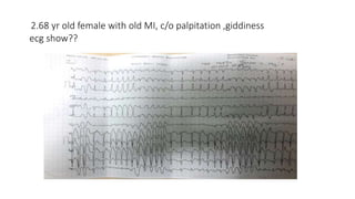
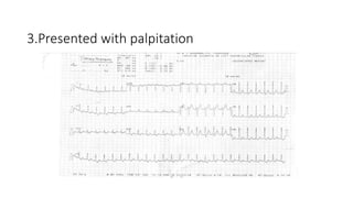
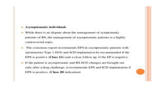
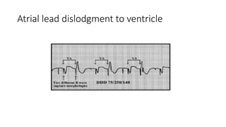
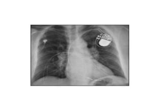
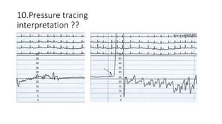
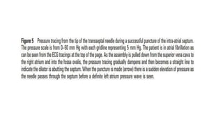
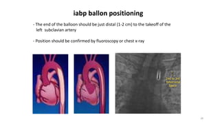
SPOTTERS
(ECG,CXR,ECHO)
DR. MAHENDRA
CARDIOLOGY,JIPMER
1.A 75 year old man with a history of COPD presents with fever and increased
sputum production. An ECG is taken in the emergency department. What does it
show?
• Diagnosis: polymorphic atrial tachycardia
• Ecg feature-
• Heart rate > 100 bpm (usually 100-150 bpm; may be as high as 250 bpm).
• Irregularly irregular rhythm with varying PP, PR and RR intervals.
• At least 3 distinct P-wave morphologies in the same lead.
• Isoelectric baseline between P-waves (i.e. no flutter waves).
• Absence of a single dominant atrial pacemaker (i.e. not just sinus rhythm with frequent
PACs).
• Some P waves may be nonconducted
• others may be aberrantly conducted to the ventricles.
2.68 yr old female with old MI, c/o palpitation ,giddiness
ecg show??
• Irregularly irregular rhythm
• Rate approx. 200/min
• Initially narrow complex change into wide complex tachycardia with changing
axis.
• p/o
• AF with aberrancy
• Preexcited AF
• VT
3.Presented with palpitation
After adenosine
• Regular narrow complex tachycardia
• Short RP
• Rate 150/min, normal axis
• Pseudo s and r present
• Respond with adenosine
• p/o AVNRT
4.18 year old man signs up to join the army. He is fit and well. This is his ECG taken at
his medical examination. Is it normal
• ECG is characteristic of Brugada Syndrome (Type 1)
•
5.Diagnosis??
• R on T phenomenon:
• sinus rhythm with frequent PVCs in a pattern of ventricular bigeminy.
• QT interval is markedly prolonged (at least 600ms), with each PVC falling on the
preceding T wave (= ‘R on T’ phenomenon).
• ECG is extremely high risk for TdP
6.diagnosis??
• HYPOKALEMIA
• Typical U wave seen
• Camel hump sign
• Prolonged repolarisation
7.Diagnosis ??
• Exercise stress test in a patient with CPVT.
• Progressively worsening ventricular arrhythmias are observed during exercise.
• Typical bidirectional VT develops after 1 minute of exercise
• Arrhythmias rapidly recede during recovery.
8.
Atrial lead dislodgment to ventricle
9.c/o recurrent palpitation diagnosis?
Typical atrial flutter
10.Pressure tracing
interpretation ??
11.6 yr old child with cyanosis
left side LV/RV tracing, rt side PA tracing
• RV and Ao pressures are equal
• PA pressures –Normal
• Diagnosis
• VSD,PS physiology
12.60 yr old male with cardiogenic shock
xray taken after cardiac intervention
28
- The end of the balloon should be just distal (1-2 cm) to the takeoff of the
left subclavian artery
- Position should be confirmed by fluoroscopy or chest x-ray
iabp ballon positioning
THANK YOU

interesting ECG,CXR,ECHO

  • 1.
  • 2.
    1.A 75 yearold man with a history of COPD presents with fever and increased sputum production. An ECG is taken in the emergency department. What does it show?
  • 3.
    • Diagnosis: polymorphicatrial tachycardia • Ecg feature- • Heart rate > 100 bpm (usually 100-150 bpm; may be as high as 250 bpm). • Irregularly irregular rhythm with varying PP, PR and RR intervals. • At least 3 distinct P-wave morphologies in the same lead. • Isoelectric baseline between P-waves (i.e. no flutter waves). • Absence of a single dominant atrial pacemaker (i.e. not just sinus rhythm with frequent PACs). • Some P waves may be nonconducted • others may be aberrantly conducted to the ventricles.
  • 4.
    2.68 yr oldfemale with old MI, c/o palpitation ,giddiness ecg show??
  • 5.
    • Irregularly irregularrhythm • Rate approx. 200/min • Initially narrow complex change into wide complex tachycardia with changing axis. • p/o • AF with aberrancy • Preexcited AF • VT
  • 6.
  • 7.
  • 8.
    • Regular narrowcomplex tachycardia • Short RP • Rate 150/min, normal axis • Pseudo s and r present • Respond with adenosine • p/o AVNRT
  • 9.
    4.18 year oldman signs up to join the army. He is fit and well. This is his ECG taken at his medical examination. Is it normal
  • 10.
    • ECG ischaracteristic of Brugada Syndrome (Type 1) •
  • 12.
  • 13.
    • R onT phenomenon: • sinus rhythm with frequent PVCs in a pattern of ventricular bigeminy. • QT interval is markedly prolonged (at least 600ms), with each PVC falling on the preceding T wave (= ‘R on T’ phenomenon). • ECG is extremely high risk for TdP
  • 14.
  • 15.
    • HYPOKALEMIA • TypicalU wave seen • Camel hump sign • Prolonged repolarisation
  • 16.
  • 17.
    • Exercise stresstest in a patient with CPVT. • Progressively worsening ventricular arrhythmias are observed during exercise. • Typical bidirectional VT develops after 1 minute of exercise • Arrhythmias rapidly recede during recovery.
  • 18.
  • 19.
  • 21.
  • 22.
  • 23.
  • 25.
    11.6 yr oldchild with cyanosis left side LV/RV tracing, rt side PA tracing
  • 26.
    • RV andAo pressures are equal • PA pressures –Normal • Diagnosis • VSD,PS physiology
  • 27.
    12.60 yr oldmale with cardiogenic shock xray taken after cardiac intervention
  • 28.
    28 - The endof the balloon should be just distal (1-2 cm) to the takeoff of the left subclavian artery - Position should be confirmed by fluoroscopy or chest x-ray iabp ballon positioning
  • 29.