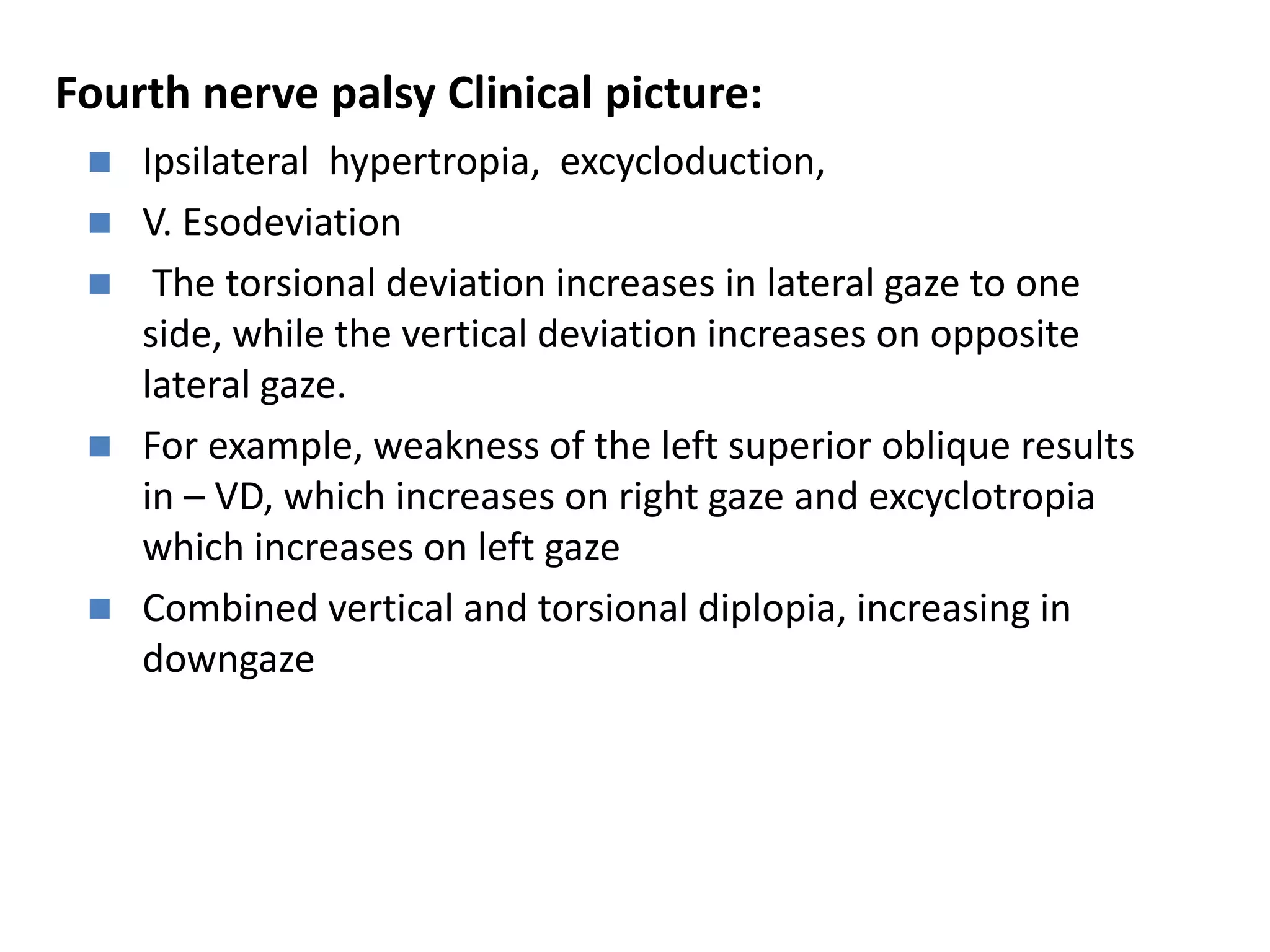
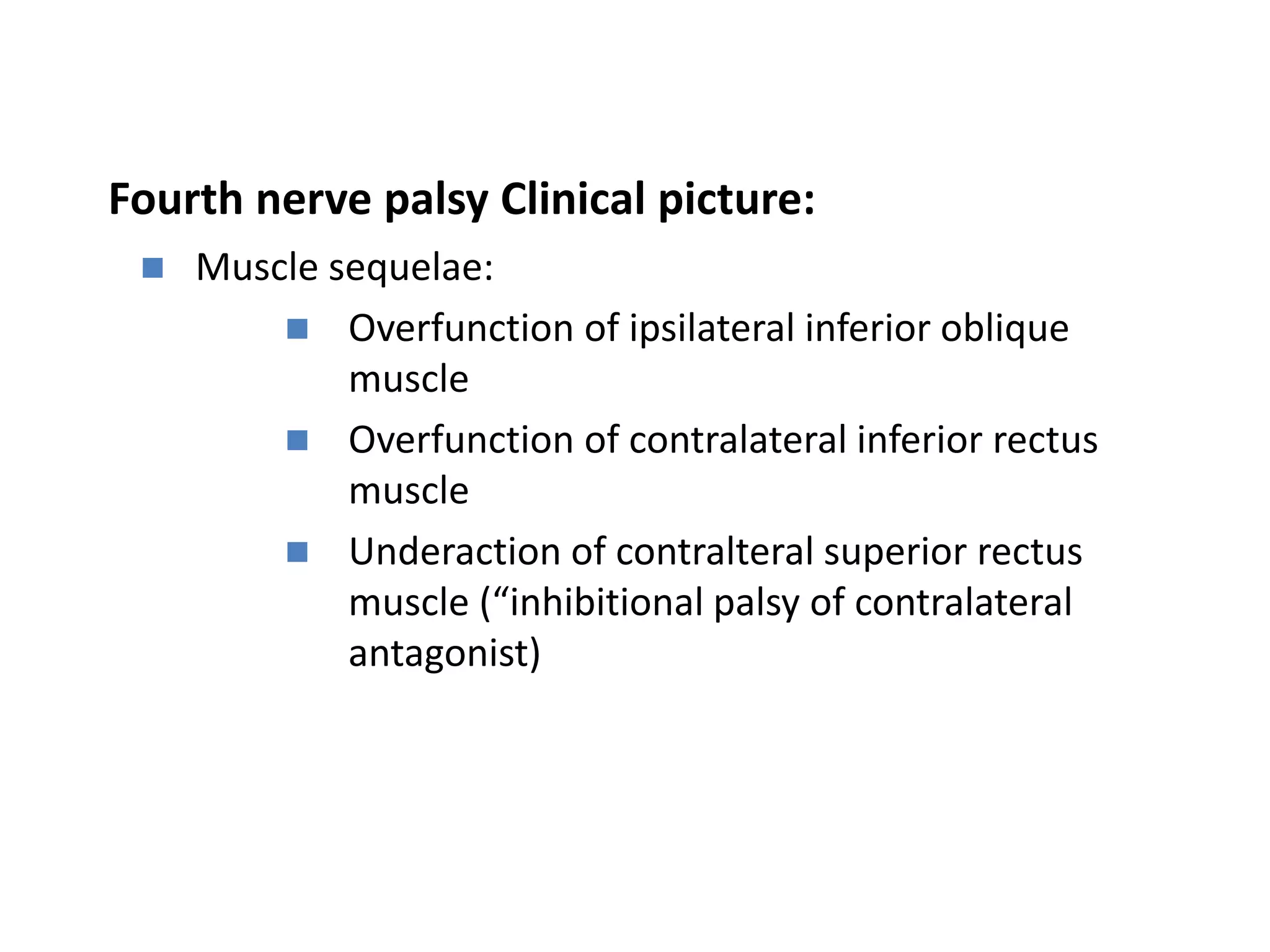
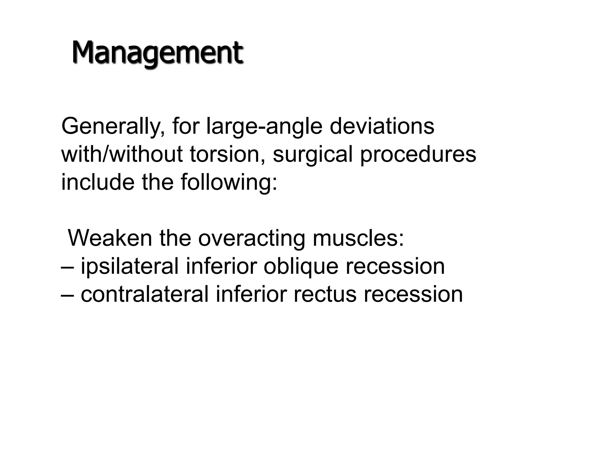
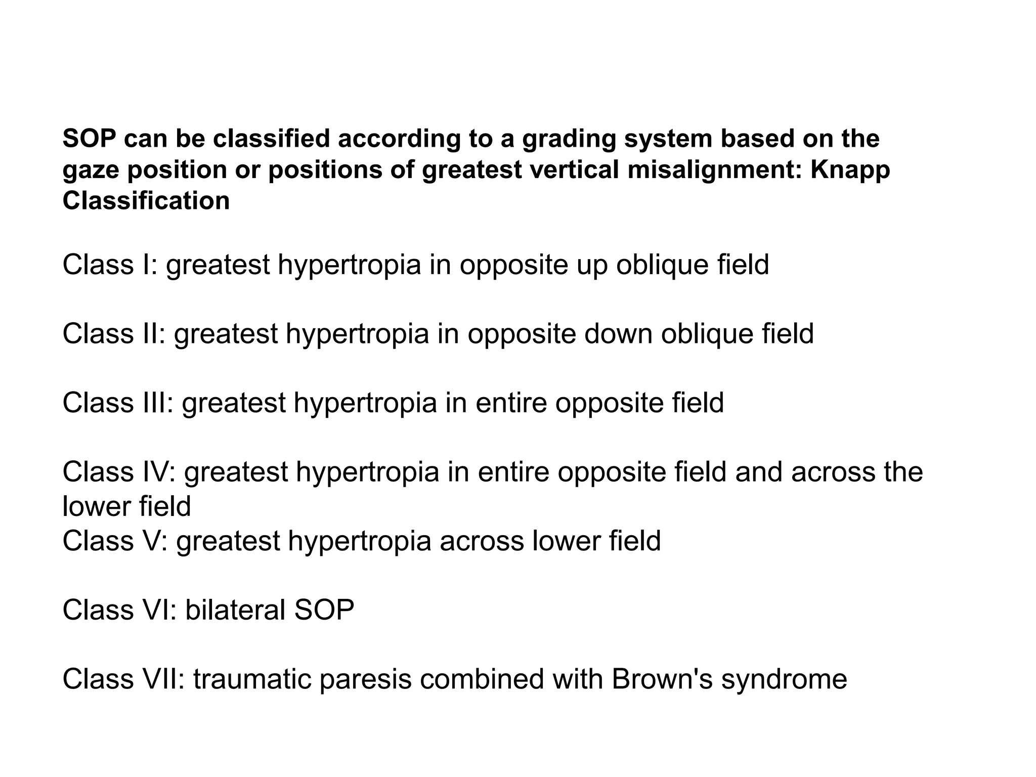
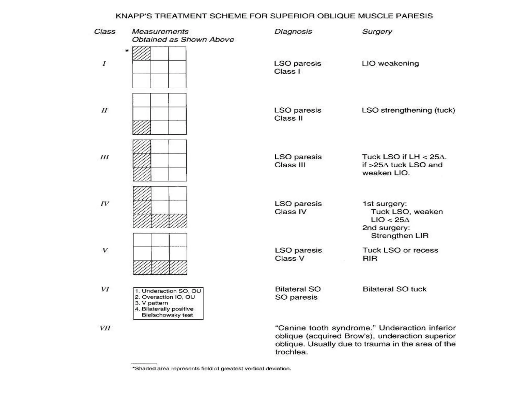
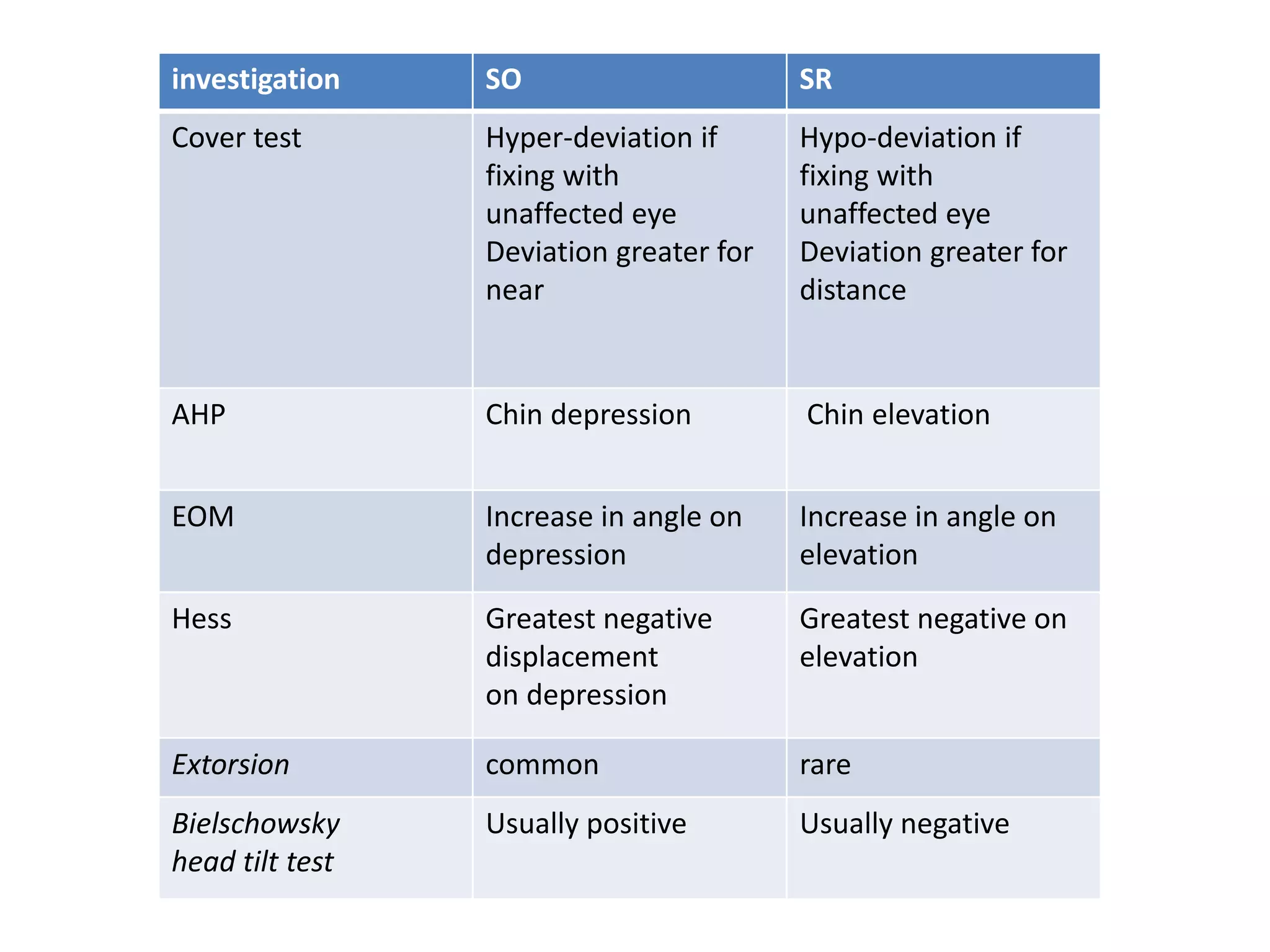
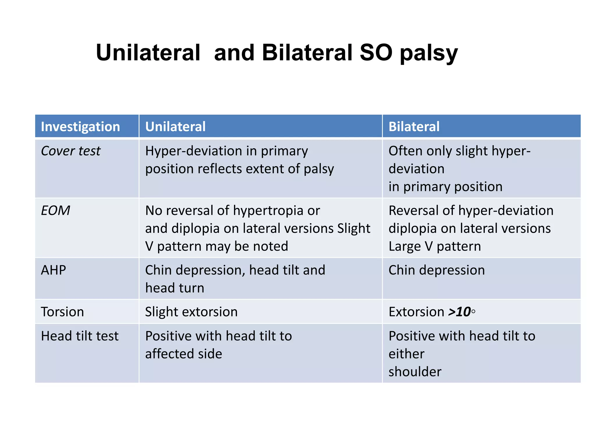
Fourth Nerve Palsy (SOP)
SIRAJ SAFI
Anatomy
• The fourth (trochlear) cranial nerve supplies only the
superior oblique muscle.
• Key features
• It is a very long and slender(slim) nerve, increasing
its vulnerability.
• It is the only cranial nerve to emerge from the dorsal
aspect of the brain.
• innervating the superior oblique muscle contralateral
to its nucleus.
Fourth nerve
Internal carotid artery
Posterior communicating
artery
Oculomotor nerve (III)
6th nerve
Fourth nerve palsy:
 The most common isolated cyclovertical muscle
palsy (congenital and acquired)
 Function of the superior oblique muscle:
 Incycloduction in abduction
 Depression in adduction
 Abduction
Trauma
Vascular, for example hypertension.
 Diabetes
 Space-occupying lesions
Other
Aetiology
Fourth nerve palsy Clinical picture:
 Ipsilateral hypertropia, excycloduction,
 V. Esodeviation
 The torsional deviation increases in lateral gaze to one
side, while the vertical deviation increases on opposite
lateral gaze.
 For example, weakness of the left superior oblique results
in – VD, which increases on right gaze and excyclotropia
which increases on left gaze
 Combined vertical and torsional diplopia, increasing in
downgaze
Fourth nerve palsy Clinical picture:
 AHP –
typical chin depression and head tilt and face
turn to the opposite shoulder
 Bielschowsky head tilt test positive hypertropia
increases when head tilted to ipsilateral shoulder
 EOM: limitation of depression in adduction
Fourth nerve palsy Clinical picture:
 Muscle sequelae:
 Overfunction of ipsilateral inferior oblique
muscle
 Overfunction of contralateral inferior rectus
muscle
 Underaction of contralteral superior rectus
muscle (“inhibitional palsy of contralateral
antagonist)
Management:
•In recent cases ,investigation and wait till
6 months if recovery occur
•Prism for small vertical deviations
•Surgery for large decompensating vertical
deviation
The fields of greatest deviation should be matched to
the muscles that exert their strongest
action in those fields.
For example, if the inferior oblique muscles overact,
this should be recessed.
If the inferior oblique does not overact and the
ipsilateral superior rectus is restricted, the superior
rectus should be recessed.
If the superior oblique is lax, it should be tucked.
Management
Management
 If the deviation is 15 prism pd. With an
overacting antagonistic inferior oblique
muscle, then weaken the inferior oblique.
Generally, for large-angle deviations
with/without torsion, surgical procedures
include the following:
Weaken the overacting muscles:
– ipsilateral inferior oblique recession
– contralateral inferior rectus recession
Management
Management
 If the deviation is greater than 35 prism
dpt. In primary position, consider 3
muscle surgery.
 This might include recession of the
overacting antagonistic inferior oblique
muscle and vertical rectus muscle surgery
as necessary
For torsion, a Harada–Ito (Fell’s
modification) can be performed (Harada&Ito
1964).
In this procedure, the anterior portion of the
affected superior oblique tendon is placed
further anteriorly and laterally
Management
(LSOP)
SOP can be classified according to a grading system based on the
gaze position or positions of greatest vertical misalignment: Knapp
Classification
Class I: greatest hypertropia in opposite up oblique field
Class II: greatest hypertropia in opposite down oblique field
Class III: greatest hypertropia in entire opposite field
Class IV: greatest hypertropia in entire opposite field and across the
lower field
Class V: greatest hypertropia across lower field
Class VI: bilateral SOP
Class VII: traumatic paresis combined with Brown's syndrome
investigation SO SR
Cover test Hyper-deviation if
fixing with
unaffected eye
Deviation greater for
near
Hypo-deviation if
fixing with
unaffected eye
Deviation greater for
distance
AHP Chin depression Chin elevation
EOM Increase in angle on
depression
Increase in angle on
elevation
Hess Greatest negative
displacement
on depression
Greatest negative on
elevation
Extorsion common rare
Bielschowsky
head tilt test
Usually positive Usually negative
Investigation Unilateral Bilateral
Cover test Hyper-deviation in primary
position reflects extent of palsy
Often only slight hyper-
deviation
in primary position
EOM No reversal of hypertropia or
and diplopia on lateral versions Slight
V pattern may be noted
Reversal of hyper-deviation
diplopia on lateral versions
Large V pattern
AHP Chin depression, head tilt and
head turn
Chin depression
Torsion Slight extorsion Extorsion >10◦
Head tilt test Positive with head tilt to
affected side
Positive with head tilt to
either
shoulder
Unilateral and Bilateral SO palsy
THANK YOU

Superior oblique palsy

  • 1.
    Fourth Nerve Palsy(SOP) SIRAJ SAFI
  • 2.
    Anatomy • The fourth(trochlear) cranial nerve supplies only the superior oblique muscle. • Key features • It is a very long and slender(slim) nerve, increasing its vulnerability. • It is the only cranial nerve to emerge from the dorsal aspect of the brain. • innervating the superior oblique muscle contralateral to its nucleus.
  • 3.
    Fourth nerve Internal carotidartery Posterior communicating artery Oculomotor nerve (III) 6th nerve
  • 4.
    Fourth nerve palsy: The most common isolated cyclovertical muscle palsy (congenital and acquired)  Function of the superior oblique muscle:  Incycloduction in abduction  Depression in adduction  Abduction
  • 5.
    Trauma Vascular, for examplehypertension.  Diabetes  Space-occupying lesions Other Aetiology
  • 6.
    Fourth nerve palsyClinical picture:  Ipsilateral hypertropia, excycloduction,  V. Esodeviation  The torsional deviation increases in lateral gaze to one side, while the vertical deviation increases on opposite lateral gaze.  For example, weakness of the left superior oblique results in – VD, which increases on right gaze and excyclotropia which increases on left gaze  Combined vertical and torsional diplopia, increasing in downgaze
  • 7.
    Fourth nerve palsyClinical picture:  AHP – typical chin depression and head tilt and face turn to the opposite shoulder  Bielschowsky head tilt test positive hypertropia increases when head tilted to ipsilateral shoulder  EOM: limitation of depression in adduction
  • 8.
    Fourth nerve palsyClinical picture:  Muscle sequelae:  Overfunction of ipsilateral inferior oblique muscle  Overfunction of contralateral inferior rectus muscle  Underaction of contralteral superior rectus muscle (“inhibitional palsy of contralateral antagonist)
  • 9.
    Management: •In recent cases,investigation and wait till 6 months if recovery occur •Prism for small vertical deviations •Surgery for large decompensating vertical deviation
  • 10.
    The fields ofgreatest deviation should be matched to the muscles that exert their strongest action in those fields. For example, if the inferior oblique muscles overact, this should be recessed. If the inferior oblique does not overact and the ipsilateral superior rectus is restricted, the superior rectus should be recessed. If the superior oblique is lax, it should be tucked. Management
  • 11.
    Management  If thedeviation is 15 prism pd. With an overacting antagonistic inferior oblique muscle, then weaken the inferior oblique.
  • 12.
    Generally, for large-angledeviations with/without torsion, surgical procedures include the following: Weaken the overacting muscles: – ipsilateral inferior oblique recession – contralateral inferior rectus recession Management
  • 13.
    Management  If thedeviation is greater than 35 prism dpt. In primary position, consider 3 muscle surgery.  This might include recession of the overacting antagonistic inferior oblique muscle and vertical rectus muscle surgery as necessary
  • 14.
    For torsion, aHarada–Ito (Fell’s modification) can be performed (Harada&Ito 1964). In this procedure, the anterior portion of the affected superior oblique tendon is placed further anteriorly and laterally Management
  • 16.
  • 17.
    SOP can beclassified according to a grading system based on the gaze position or positions of greatest vertical misalignment: Knapp Classification Class I: greatest hypertropia in opposite up oblique field Class II: greatest hypertropia in opposite down oblique field Class III: greatest hypertropia in entire opposite field Class IV: greatest hypertropia in entire opposite field and across the lower field Class V: greatest hypertropia across lower field Class VI: bilateral SOP Class VII: traumatic paresis combined with Brown's syndrome
  • 20.
    investigation SO SR Covertest Hyper-deviation if fixing with unaffected eye Deviation greater for near Hypo-deviation if fixing with unaffected eye Deviation greater for distance AHP Chin depression Chin elevation EOM Increase in angle on depression Increase in angle on elevation Hess Greatest negative displacement on depression Greatest negative on elevation Extorsion common rare Bielschowsky head tilt test Usually positive Usually negative
  • 21.
    Investigation Unilateral Bilateral Covertest Hyper-deviation in primary position reflects extent of palsy Often only slight hyper- deviation in primary position EOM No reversal of hypertropia or and diplopia on lateral versions Slight V pattern may be noted Reversal of hyper-deviation diplopia on lateral versions Large V pattern AHP Chin depression, head tilt and head turn Chin depression Torsion Slight extorsion Extorsion >10◦ Head tilt test Positive with head tilt to affected side Positive with head tilt to either shoulder Unilateral and Bilateral SO palsy
  • 22.