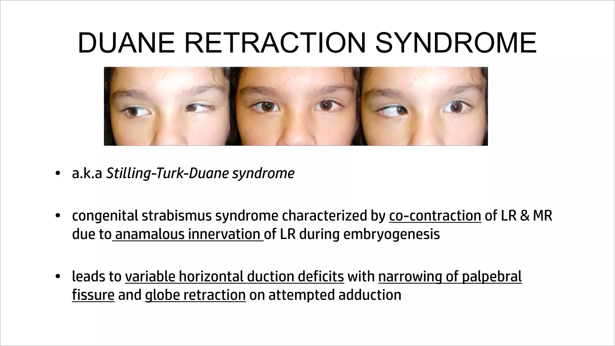
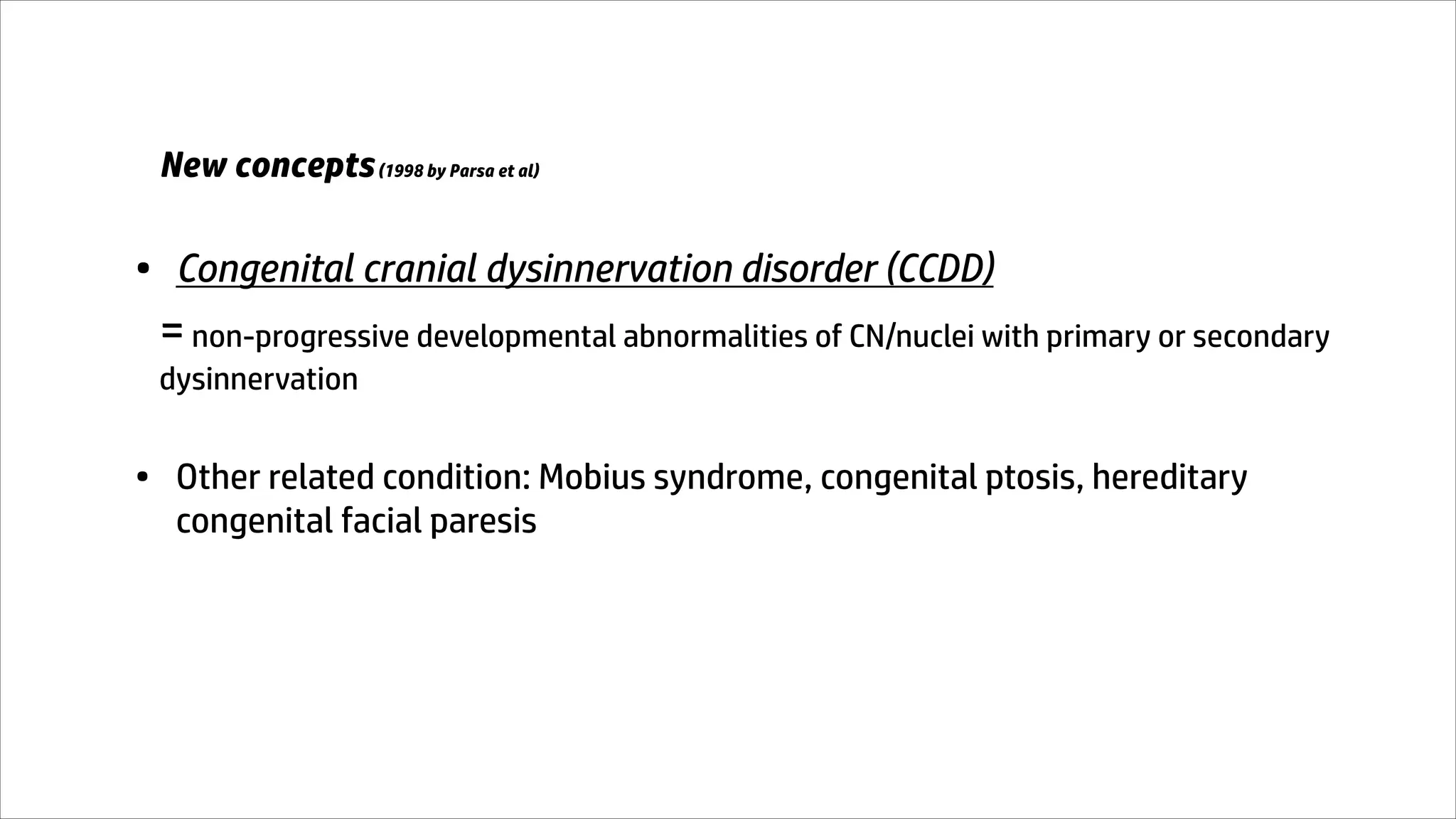
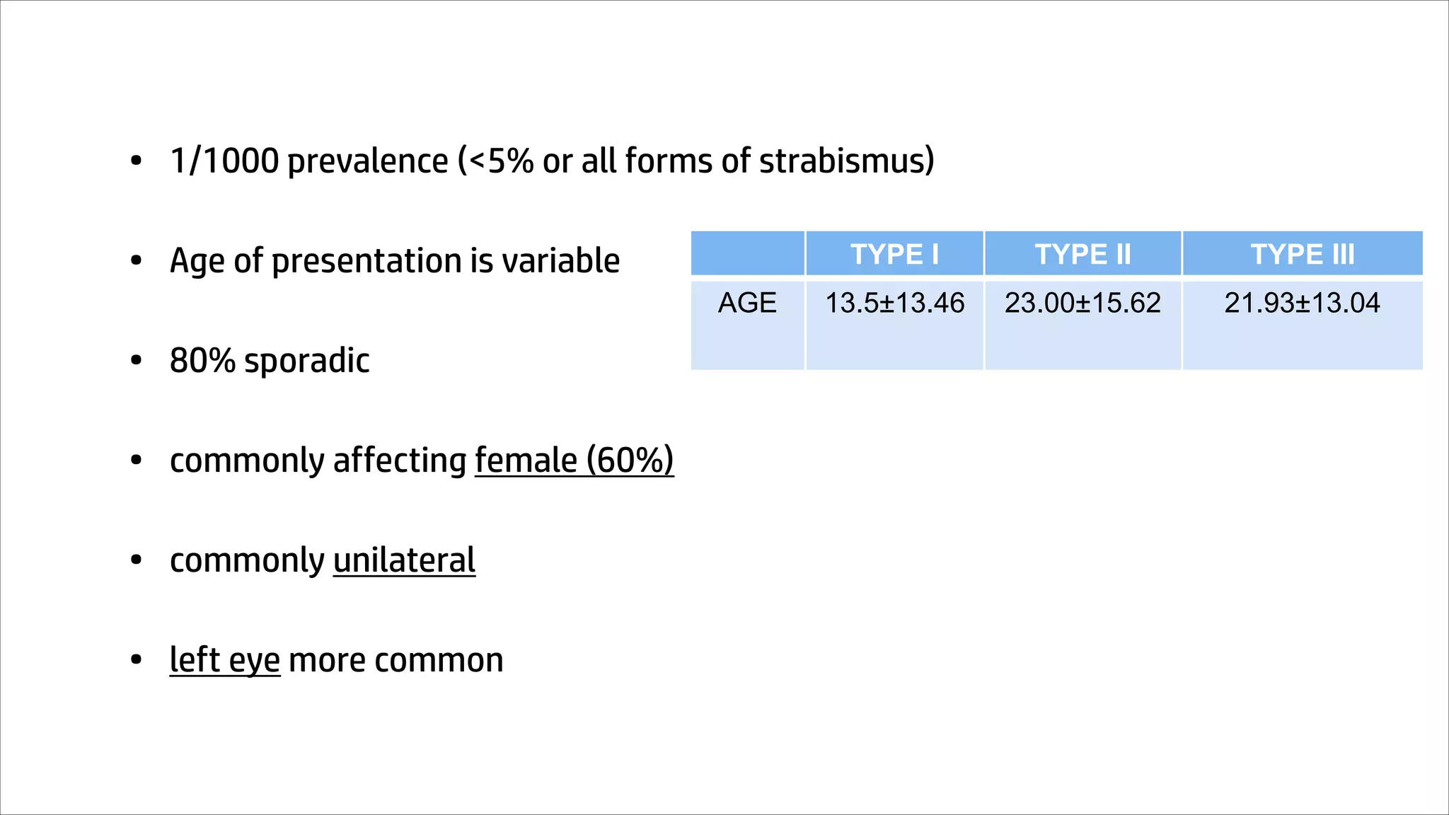
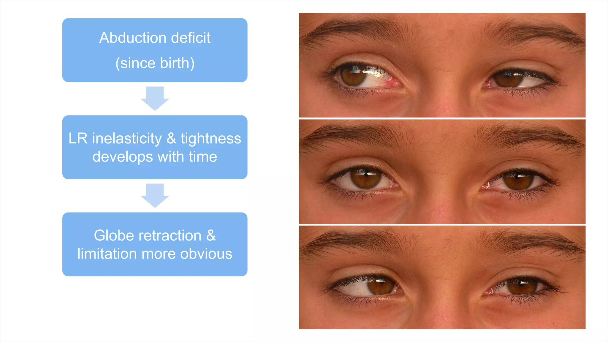
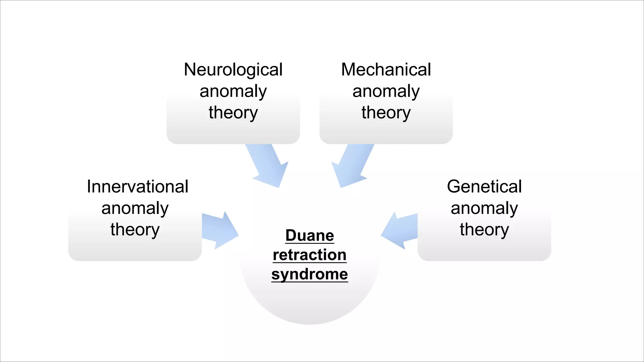
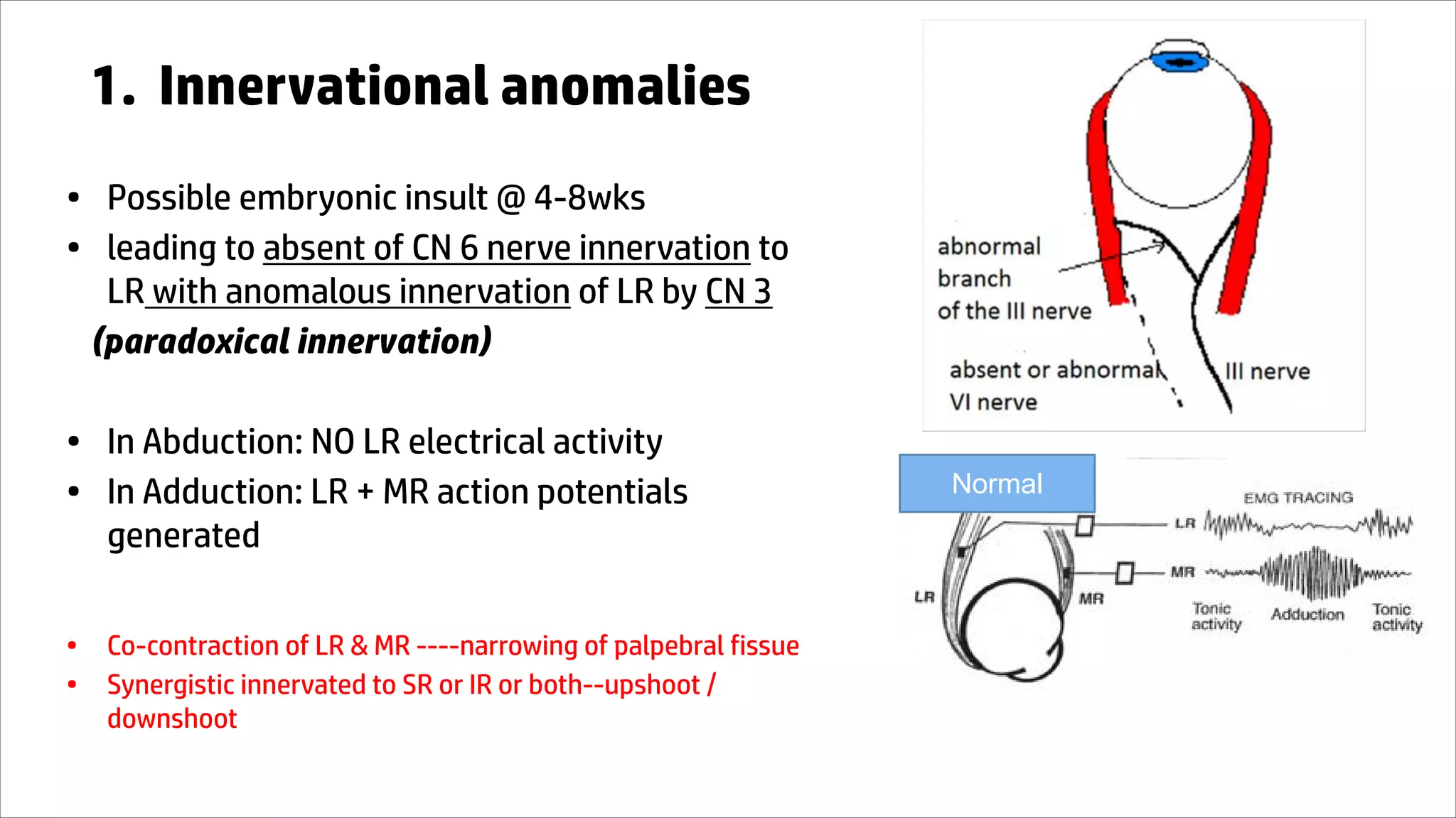
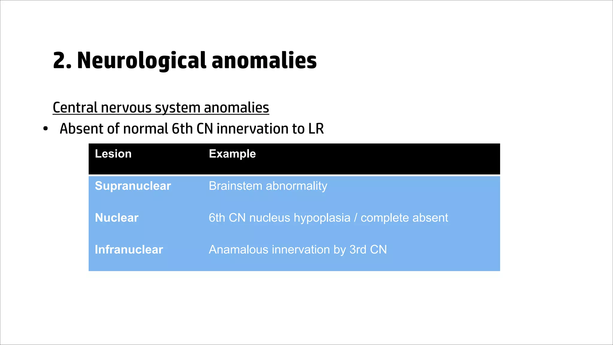
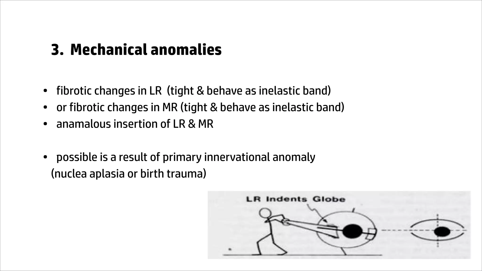
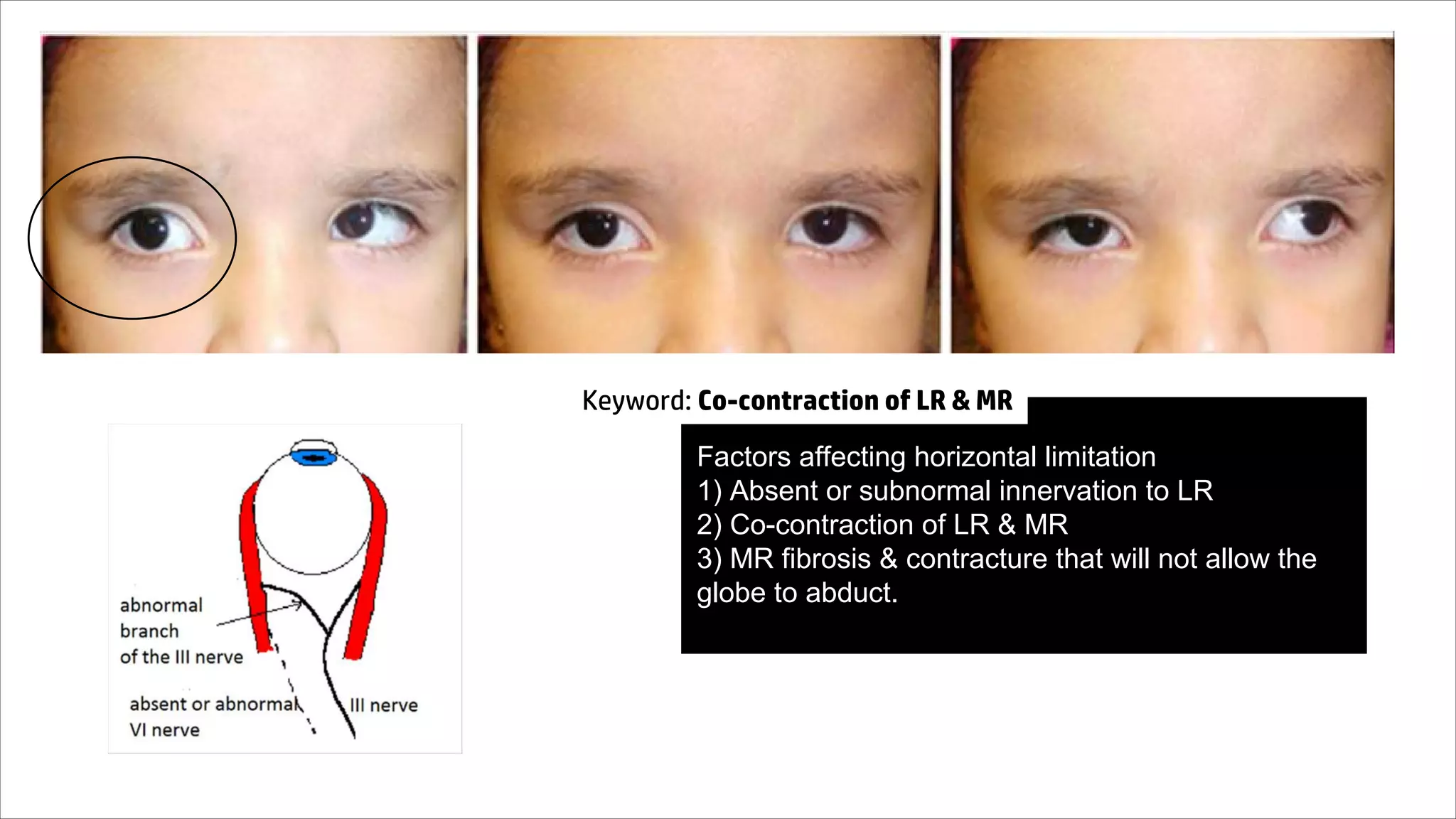
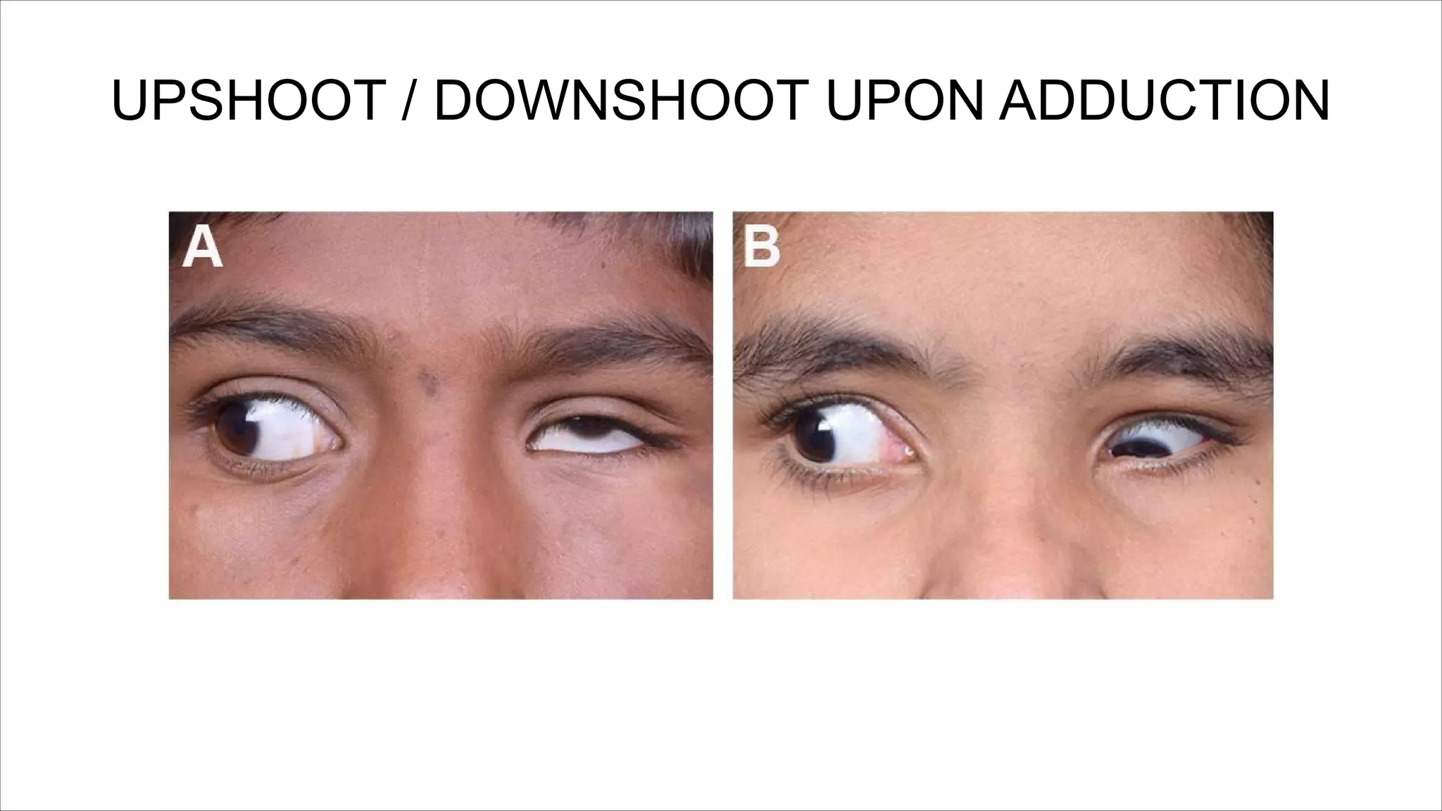
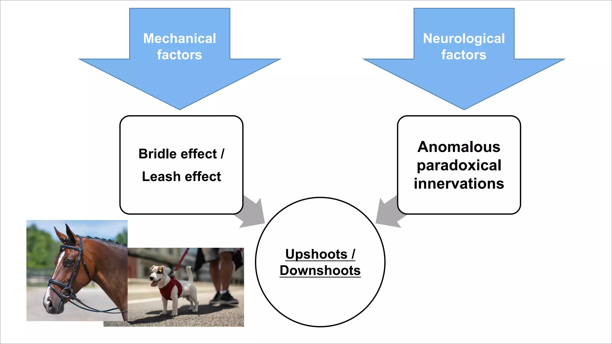
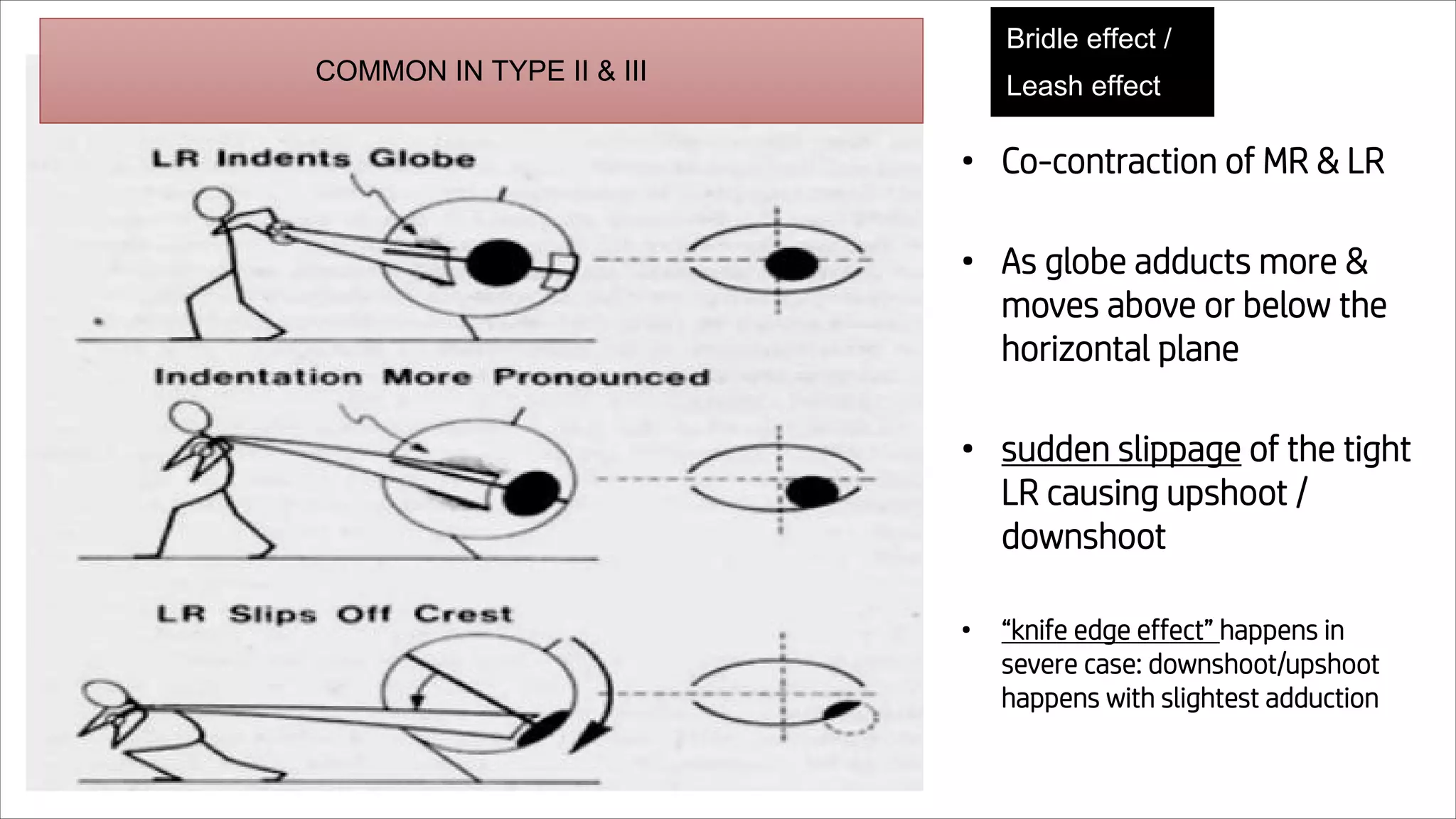
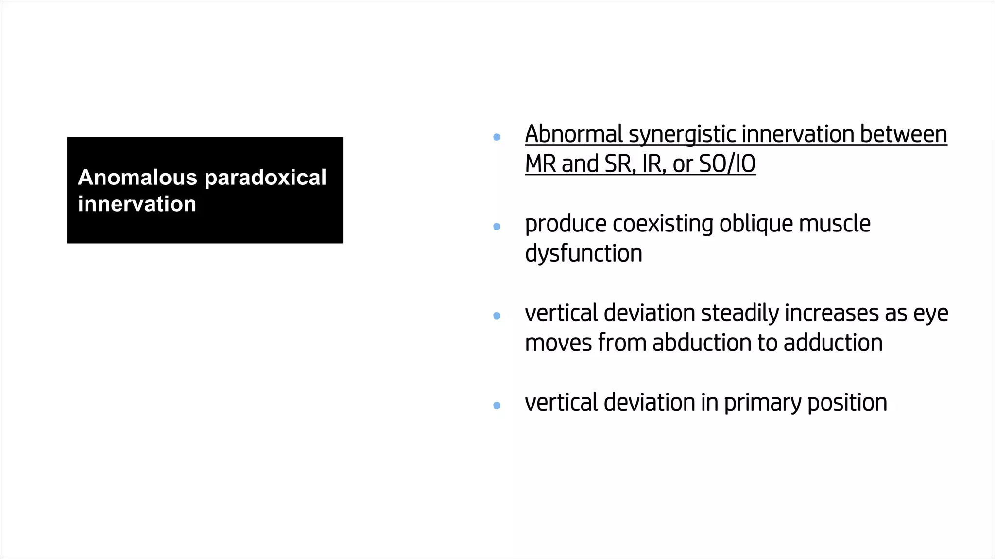
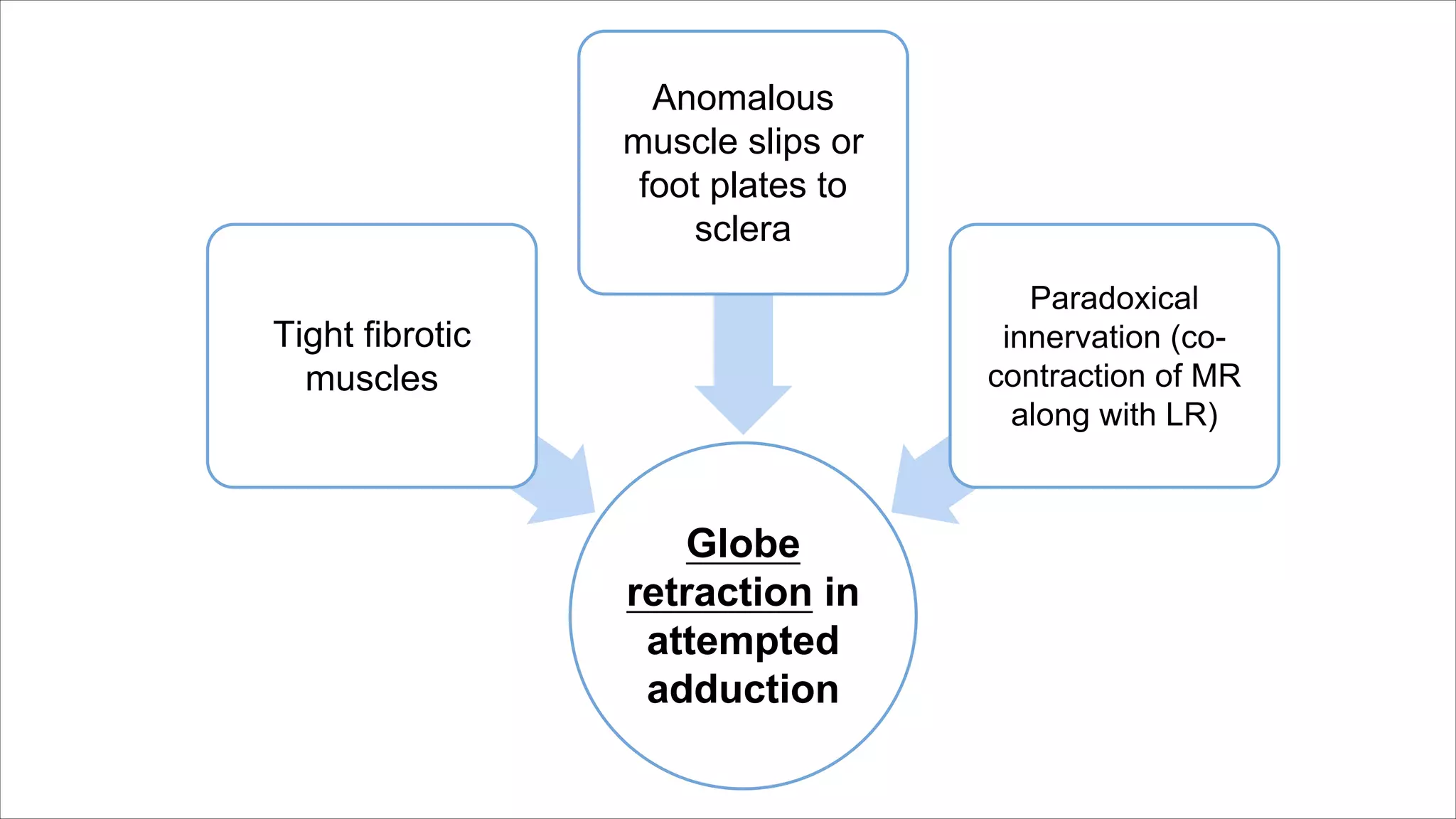
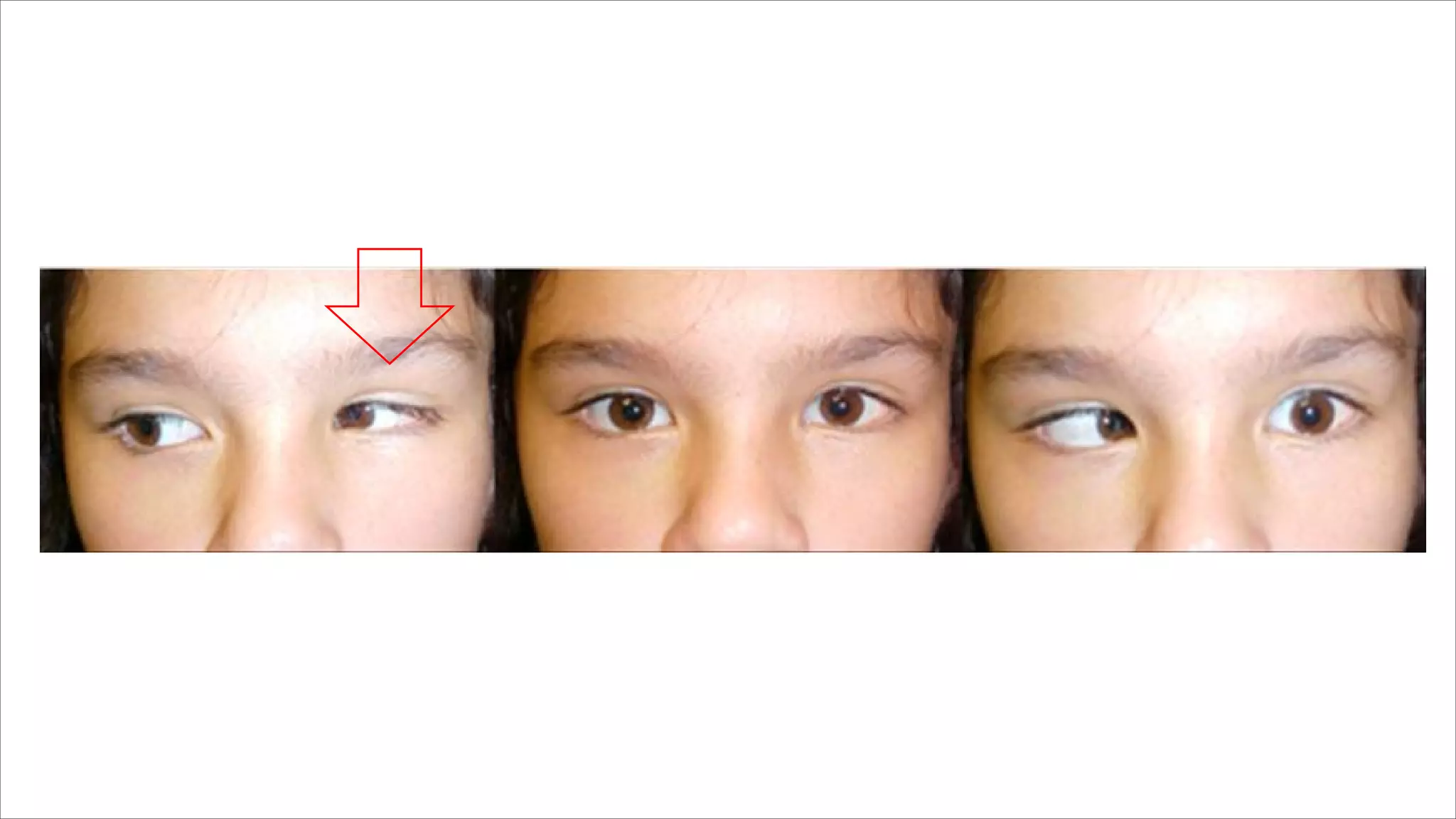
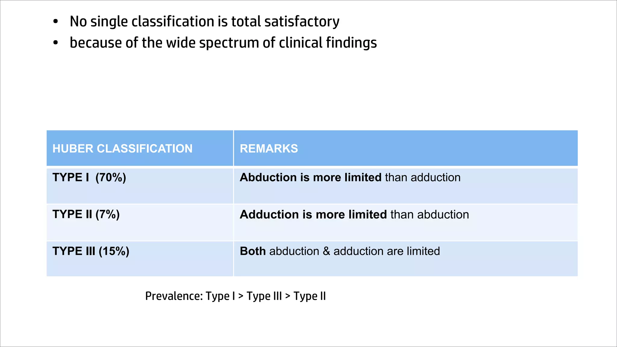
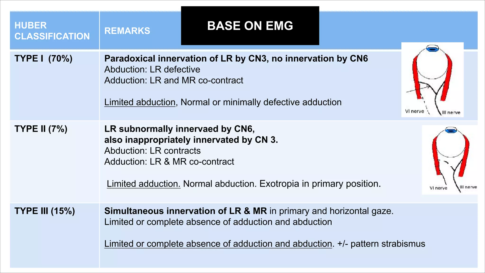
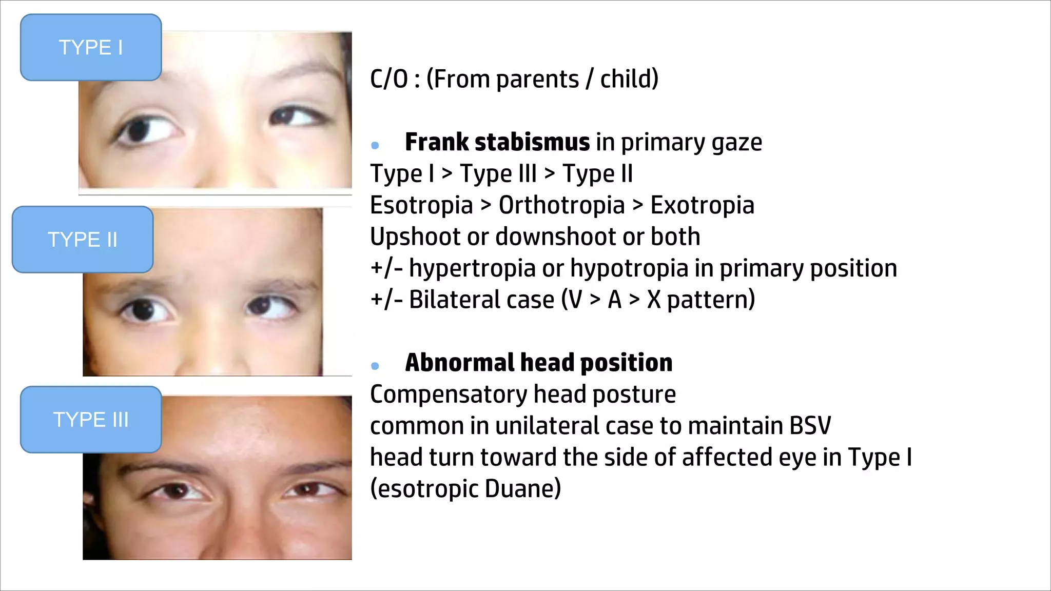
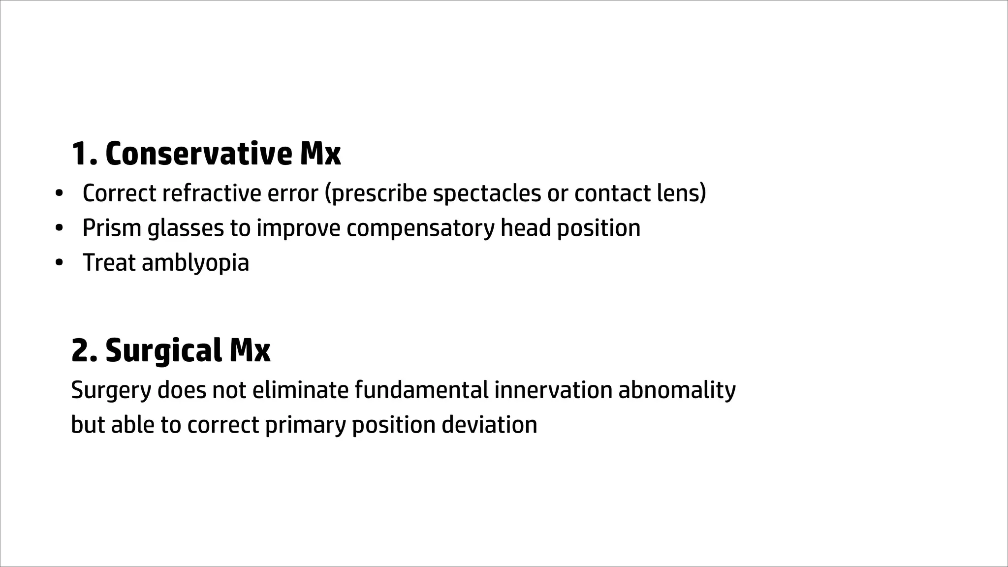
Duane Retraction
Syndrome
Prepared by
Dr Robin Goh Chon Han
Ophthalmology Post-graduate
Student (UM), MBBS (IMU)
General Paediatric Department
Hospital Kuala Lumpur, Malaysia
Index
• Introduction
• Etiology
• Pathophysiology
• Classification
• Clinical Presentation
• Diagnosis
• Differential Diagnosis
• Management
• Reference
INTRODUCTION
DUANE RETRACTION SYNDROME
• a.k.a Stilling-Turk-Duane syndrome
• congenital strabismus syndrome characterized by co-contraction of LR & MR
due to anamalous innervation of LR during embryogenesis
• leads to variable horizontal duction deficits with narrowing of palpebral
fissure and globe retraction on attempted adduction
New concepts(1998 by Parsa et al)
• Congenital cranial dysinnervation disorder (CCDD)
=non-progressive developmental abnormalities of CN/nuclei with primary or secondary
dysinnervation
• Other related condition: Mobius syndrome, congenital ptosis, hereditary
congenital facial paresis
• 1/1000 prevalence (<5% or all forms of strabismus)
• Age of presentation is variable
• 80% sporadic
• commonly affecting female (60%)
• commonly unilateral
• left eye more common
TYPE I TYPE II TYPE III
AGE 13.5±13.46 23.00±15.62 21.93±13.04
Abduction deficit
(since birth)
LR inelasticity & tightness
develops with time
Globe retraction &
limitation more obvious
ETIOLOGY
Various theories
Duane
retraction
syndrome
Innervational
anomaly
theory
Neurological
anomaly
theory
Mechanical
anomaly
theory
Genetical
anomaly
theory
1. Innervational
anomalies
1. Innervational anomalies
• Possible embryonic insult @ 4-8wks
• leading to absent of CN 6 nerve innervation to
LR with anomalous innervation of LR by CN 3
(paradoxical innervation)
• In Abduction: NO LR electrical activity
• In Adduction: LR + MR action potentials
generated
• Co-contraction of LR & MR ----narrowing of palpebral fissue
• Synergistic innervated to SR or IR or both--upshoot /
downshoot
Normal
2.
Neurological
anomalies
2. Neurological anomalies
Central nervous system anomalies
• Absent of normal 6th CN innervation to LR
Lesion Example
Supranuclear Brainstem abnormality
Nuclear 6th CN nucleus hypoplasia / complete absent
Infranuclear Anamalous innervation by 3rd CN
• Absent of normal 6th CN innervation to LR
Supportive evidence (Synkinesis)
• Marcus Gunn Jaw Winking phenomenon
• Crocodile tears
Lesion Example
Supranuclear Brainstem abnormality
Nuclear 6th nucleus hypoplasia / complete absent
Infranuclear anamalous innervation by 3rd CN
3.
Mechanical
anomalies
3. Mechanical anomalies
• fibrotic changes in LR (tight & behave as inelastic band)
• or fibrotic changes in MR (tight & behave as inelastic band)
• anamalous insertion of LR & MR
• possible is a result of primary innervational anomaly
(nuclea aplasia or birth trauma)
4.
Genetic
anomalies
4. Genetics
• Sporadic mutation (>80%)
• PHOX2A, HOXA1, ROB03
(growth & maturation of brainstem motor neurons & axons)
• DURS1 (chromosone 2q31)
• SALL 4 gene
• if familial (10%)suspect:
CHN1 gene (autosomal dominant)
CHN1
ü autosomal dominant
ü bilateral DRS
ü assocaited vertical
anomalies
ü significant family history
PATHOPHYSIOLOGY
• Why is there limited horizontal gaze?
Factors affecting horizontal limitation
1) Absent or subnormal innervation to LR
2) Co-contraction of LR & MR
3) MR fibrosis & contracture that will not allow the
globe to abduct.
Keyword: Co-contraction of LR & MR
• What causes downshoot / upshoot?
UPSHOOT / DOWNSHOOT UPON ADDUCTION
Upshoots /
Downshoots
Bridle effect /
Leash effect
Anomalous
paradoxical
innervations
Mechanical
factors
Neurological
factors
• Co-contraction of MR & LR
• As globe adducts more &
moves above or below the
horizontal plane
• sudden slippage of the tight
LR causing upshoot /
downshoot
• “knife edge effect” happens in
severe case: downshoot/upshoot
happens with slightest adduction
Bridle effect /
Leash effect
COMMON IN TYPE II & III
Anomalous paradoxical
innervation
● Abnormal synergistic innervation between
MR and SR, IR, or SO/IO
● produce coexisting oblique muscle
dysfunction
● vertical deviation steadily increases as eye
moves from abduction to adduction
● vertical deviation in primary position
• What is the mechanism of globe retraction?
Globe
retraction in
attempted
adduction
Tight fibrotic
muscles
Anomalous
muscle slips or
foot plates to
sclera
Paradoxical
innervation (co-
contraction of MR
along with LR)
CLASSIFICATION
• No single classification is total satisfactory
• because of the wide spectrum of clinical findings
HUBER CLASSIFICATION REMARKS
TYPE I (70%) Abduction is more limited than adduction
TYPE II (7%) Adduction is more limited than abduction
TYPE III (15%) Both abduction & adduction are limited
Prevalence: Type I > Type III > Type II
HUBER
CLASSIFICATION
REMARKS
TYPE I (70%) Paradoxical innervation of LR by CN3, no innervation by CN6
Abduction: LR defective
Adduction: LR and MR co-contract
Limited abduction, Normal or minimally defective adduction
TYPE II (7%) LR subnormally innervaed by CN6,
also inappropriately innervated by CN 3.
Abduction: LR contracts
Adduction: LR & MR co-contract
Limited adduction. Normal abduction. Exotropia in primary position.
TYPE III (15%) Simultaneous innervation of LR & MR in primary and horizontal gaze.
Limited or complete absence of adduction and abduction
Limited or complete absence of adduction and abduction. +/- pattern strabismus
BASE ON EMG
CLINICAL
PRESENTATION
TYPE I
TYPE II
TYPE III
C/O : (From parents / child)
● Frank stabismus in primary gaze
Type I > Type III > Type II
Esotropia > Orthotropia > Exotropia
Upshoot or downshoot or both
+/- hypertropia or hypotropia in primary position
+/- Bilateral case (V > A > X pattern)
● Abnormal head position
Compensatory head posture
common in unilateral case to maintain BSV
head turn toward the side of affected eye in Type I
(esotropic Duane)
• Induced ptosis on adduction
passive movement of eyelids over retracting globe + decreased levator muscle action
on adduction
• Unilateral involvement
Bilateral less frequent
Bilateral involvement usually low visual acuity, less likely abnormal head posture
usually with associated congenital anomalies
• Refractive error
mostly hypermetropia / hypermetropic astigmatism
+/- Anisometropia
+/- Amblyopia (anisometropic/ametropic/strabismic/stimulus deprivation)
OCULAR & SYSTEMIC ASSOCIATION
Summary of Evaluation
• History
• Family history
• Examination include VA, head position, EOM motility, aberrant
movement
• General physical examination (TRO associated syndrome)
• Further assessment include orthoptic assessment (Refraction, EOM
motility, BSV, Accommodation)
GRADING OF GLOBE
RETRACTION (ADDUCTION)
COMPARED TO PALPEBRAL FISSURE HEIGHT
OF NORMAL EYE
0 no narrowing
1 <25%
2 25-50%
3 50%-75%
4 >75%
GRADING OF
OVERSHOOT
(ADDUCTION)
* Imaginary straight line parallel to
the intermedial canthal line is drawn
from pupillary center of fellow eye
0 Line bisects pupil of involved eye
1 Line lies between pupillary center &
pupillary margin
2 Line lies between pupillary margin &
limbus
3 Line lies at the limbus or over sclera
4 Cornea disappears below lid (pumpkin
seed sign)
DIAGNOSIS
● Diagnosis is primarily clinical
Laboratory Test
Molecular genetic testing of CHN1 recommended only in familial case
Imaging
Not recommended for diagnostic purposes
Electromyography
DIFFERENTIAL
DIAGNOSIS
• 6th CN Palsy
• 3rd CN Palsy
• INO
• Acquired retraction syndrome (pseudoduane syndrome)
= limited abduction along with globe retraction in abduction
MANAGEMENT
• Mx depends on:
1. Primary position deviation
2. Degree of abnormal head posture
3. Severity of globe retraction and overshoots
4. Degree of limitation of ductions
1. Conservative Mx
• Correct refractive error (prescribe spectacles or contact lens)
• Prism glasses to improve compensatory head position
• Treat amblyopia
2. Surgical Mx
Surgery does not eliminate fundamental innervation abnomality
but able to correct primary position deviation
Goals of Surgical Mx:
• Improve anomalous head posture
• Correct any angle of strabismus in primary position
• Improve globe retraction
• Expand BSV
General rules (AAO)
To correct ocular deviation in primary position
A. Type I = MR recession (unilateral/bilateral)
B. Type II = LR recession (unilateral/bilateral)
To correct ocular deviation & improve abduction for BSV
A. Vertical rectus transposition procedure
B. Recess-resect procedures in Duane eye
To correct globe retraction & upshoot/downshoot
A. Combined horizontal rectus recessions
B. Faden procedure on LR
C. Y splitting of LR
To correct vertical deviation
A. Vertical muscle recession
To correct ocular deviation in primary position
A. Type I = MR recession (unilateral/bilateral)
B. Type II = LR recession (unilateral/bilateral)
Patient with exotropic-Duane before and after right lateral rectus recession of 7 mm.
Left MR recession 6 mm done: Noted improvement of head posture and orthophoria
in primary gaze, slight improvement of abduction of the left eye
Right MR recession (7 mm) & left MR recession (5 mm)
for an esotropic-Duane of 25 diopters in primary gaze.
Large angle esotropia (>20PD) may need BE MR recession (contralateral eye recessed larger)
WHY DOING BE MR RECESSION?
Bilateral MR recession
• MR recession can improve primary position deviation
• Improve abduction of affected eye (if there was some prior lateral movement). It also
improve BSV.
• Prevent contracture of MR on affected side
Forcing Contralateral MR to work harder to maintain fixation (“fixation duress”)
MR in fixing eye receives increased innervation to maintain fixation (after op)
--> Reduced innervation to its LR (Sherrington Law)
--> Reduced innervation to MR of the affected eye (Hering Law)
--> lower risk of contracture.
Complications of squint surgery
• Undercorrection
• Overcorrection
• New vertical deviation
• Anterior segment ischemia
• Scleral perforation
• etc
Follow up for DRS
• follow up 3-6 monthly after diagnosis
(evaluate for amblyopia)
• 7-12 years old patients with good BSV can be evaluated annually or
biannually
Reference
• Eyewiki
• AAO
• Ramesh Kekunnya, Mithila Negalur. Duane retraction syndrome: causes,
effects and management strategies. Clinical Ophthalmology, Oct 2017.
Thank you
Please click the “like” button if you find this
presentation useful for you.

Duane Presentation (Sharing).pdf

  • 1.
    Duane Retraction Syndrome Prepared by DrRobin Goh Chon Han Ophthalmology Post-graduate Student (UM), MBBS (IMU) General Paediatric Department Hospital Kuala Lumpur, Malaysia
  • 2.
    Index • Introduction • Etiology •Pathophysiology • Classification • Clinical Presentation • Diagnosis • Differential Diagnosis • Management • Reference
  • 3.
  • 4.
    DUANE RETRACTION SYNDROME •a.k.a Stilling-Turk-Duane syndrome • congenital strabismus syndrome characterized by co-contraction of LR & MR due to anamalous innervation of LR during embryogenesis • leads to variable horizontal duction deficits with narrowing of palpebral fissure and globe retraction on attempted adduction
  • 5.
    New concepts(1998 byParsa et al) • Congenital cranial dysinnervation disorder (CCDD) =non-progressive developmental abnormalities of CN/nuclei with primary or secondary dysinnervation • Other related condition: Mobius syndrome, congenital ptosis, hereditary congenital facial paresis
  • 6.
    • 1/1000 prevalence(<5% or all forms of strabismus) • Age of presentation is variable • 80% sporadic • commonly affecting female (60%) • commonly unilateral • left eye more common TYPE I TYPE II TYPE III AGE 13.5±13.46 23.00±15.62 21.93±13.04
  • 7.
    Abduction deficit (since birth) LRinelasticity & tightness develops with time Globe retraction & limitation more obvious
  • 8.
  • 9.
  • 10.
  • 11.
    1. Innervational anomalies •Possible embryonic insult @ 4-8wks • leading to absent of CN 6 nerve innervation to LR with anomalous innervation of LR by CN 3 (paradoxical innervation) • In Abduction: NO LR electrical activity • In Adduction: LR + MR action potentials generated • Co-contraction of LR & MR ----narrowing of palpebral fissue • Synergistic innervated to SR or IR or both--upshoot / downshoot Normal
  • 12.
  • 13.
    2. Neurological anomalies Centralnervous system anomalies • Absent of normal 6th CN innervation to LR Lesion Example Supranuclear Brainstem abnormality Nuclear 6th CN nucleus hypoplasia / complete absent Infranuclear Anamalous innervation by 3rd CN
  • 14.
    • Absent ofnormal 6th CN innervation to LR Supportive evidence (Synkinesis) • Marcus Gunn Jaw Winking phenomenon • Crocodile tears Lesion Example Supranuclear Brainstem abnormality Nuclear 6th nucleus hypoplasia / complete absent Infranuclear anamalous innervation by 3rd CN
  • 17.
  • 18.
    3. Mechanical anomalies •fibrotic changes in LR (tight & behave as inelastic band) • or fibrotic changes in MR (tight & behave as inelastic band) • anamalous insertion of LR & MR • possible is a result of primary innervational anomaly (nuclea aplasia or birth trauma)
  • 19.
  • 20.
    4. Genetics • Sporadicmutation (>80%) • PHOX2A, HOXA1, ROB03 (growth & maturation of brainstem motor neurons & axons) • DURS1 (chromosone 2q31) • SALL 4 gene • if familial (10%)suspect: CHN1 gene (autosomal dominant) CHN1 ü autosomal dominant ü bilateral DRS ü assocaited vertical anomalies ü significant family history
  • 21.
  • 22.
    • Why isthere limited horizontal gaze?
  • 23.
    Factors affecting horizontallimitation 1) Absent or subnormal innervation to LR 2) Co-contraction of LR & MR 3) MR fibrosis & contracture that will not allow the globe to abduct. Keyword: Co-contraction of LR & MR
  • 24.
    • What causesdownshoot / upshoot?
  • 25.
    UPSHOOT / DOWNSHOOTUPON ADDUCTION
  • 26.
    Upshoots / Downshoots Bridle effect/ Leash effect Anomalous paradoxical innervations Mechanical factors Neurological factors
  • 27.
    • Co-contraction ofMR & LR • As globe adducts more & moves above or below the horizontal plane • sudden slippage of the tight LR causing upshoot / downshoot • “knife edge effect” happens in severe case: downshoot/upshoot happens with slightest adduction Bridle effect / Leash effect COMMON IN TYPE II & III
  • 28.
    Anomalous paradoxical innervation ● Abnormalsynergistic innervation between MR and SR, IR, or SO/IO ● produce coexisting oblique muscle dysfunction ● vertical deviation steadily increases as eye moves from abduction to adduction ● vertical deviation in primary position
  • 29.
    • What isthe mechanism of globe retraction?
  • 30.
    Globe retraction in attempted adduction Tight fibrotic muscles Anomalous muscleslips or foot plates to sclera Paradoxical innervation (co- contraction of MR along with LR)
  • 32.
  • 33.
    • No singleclassification is total satisfactory • because of the wide spectrum of clinical findings HUBER CLASSIFICATION REMARKS TYPE I (70%) Abduction is more limited than adduction TYPE II (7%) Adduction is more limited than abduction TYPE III (15%) Both abduction & adduction are limited Prevalence: Type I > Type III > Type II
  • 34.
    HUBER CLASSIFICATION REMARKS TYPE I (70%)Paradoxical innervation of LR by CN3, no innervation by CN6 Abduction: LR defective Adduction: LR and MR co-contract Limited abduction, Normal or minimally defective adduction TYPE II (7%) LR subnormally innervaed by CN6, also inappropriately innervated by CN 3. Abduction: LR contracts Adduction: LR & MR co-contract Limited adduction. Normal abduction. Exotropia in primary position. TYPE III (15%) Simultaneous innervation of LR & MR in primary and horizontal gaze. Limited or complete absence of adduction and abduction Limited or complete absence of adduction and abduction. +/- pattern strabismus BASE ON EMG
  • 35.
  • 36.
    TYPE I TYPE II TYPEIII C/O : (From parents / child) ● Frank stabismus in primary gaze Type I > Type III > Type II Esotropia > Orthotropia > Exotropia Upshoot or downshoot or both +/- hypertropia or hypotropia in primary position +/- Bilateral case (V > A > X pattern) ● Abnormal head position Compensatory head posture common in unilateral case to maintain BSV head turn toward the side of affected eye in Type I (esotropic Duane)
  • 37.
    • Induced ptosison adduction passive movement of eyelids over retracting globe + decreased levator muscle action on adduction • Unilateral involvement Bilateral less frequent Bilateral involvement usually low visual acuity, less likely abnormal head posture usually with associated congenital anomalies • Refractive error mostly hypermetropia / hypermetropic astigmatism +/- Anisometropia +/- Amblyopia (anisometropic/ametropic/strabismic/stimulus deprivation)
  • 38.
    OCULAR & SYSTEMICASSOCIATION
  • 39.
    Summary of Evaluation •History • Family history • Examination include VA, head position, EOM motility, aberrant movement • General physical examination (TRO associated syndrome) • Further assessment include orthoptic assessment (Refraction, EOM motility, BSV, Accommodation)
  • 40.
    GRADING OF GLOBE RETRACTION(ADDUCTION) COMPARED TO PALPEBRAL FISSURE HEIGHT OF NORMAL EYE 0 no narrowing 1 <25% 2 25-50% 3 50%-75% 4 >75%
  • 41.
    GRADING OF OVERSHOOT (ADDUCTION) * Imaginarystraight line parallel to the intermedial canthal line is drawn from pupillary center of fellow eye 0 Line bisects pupil of involved eye 1 Line lies between pupillary center & pupillary margin 2 Line lies between pupillary margin & limbus 3 Line lies at the limbus or over sclera 4 Cornea disappears below lid (pumpkin seed sign)
  • 42.
  • 43.
    ● Diagnosis isprimarily clinical Laboratory Test Molecular genetic testing of CHN1 recommended only in familial case Imaging Not recommended for diagnostic purposes Electromyography
  • 44.
  • 45.
    • 6th CNPalsy • 3rd CN Palsy • INO • Acquired retraction syndrome (pseudoduane syndrome) = limited abduction along with globe retraction in abduction
  • 46.
  • 47.
    • Mx dependson: 1. Primary position deviation 2. Degree of abnormal head posture 3. Severity of globe retraction and overshoots 4. Degree of limitation of ductions
  • 48.
    1. Conservative Mx •Correct refractive error (prescribe spectacles or contact lens) • Prism glasses to improve compensatory head position • Treat amblyopia 2. Surgical Mx Surgery does not eliminate fundamental innervation abnomality but able to correct primary position deviation
  • 49.
    Goals of SurgicalMx: • Improve anomalous head posture • Correct any angle of strabismus in primary position • Improve globe retraction • Expand BSV
  • 50.
    General rules (AAO) Tocorrect ocular deviation in primary position A. Type I = MR recession (unilateral/bilateral) B. Type II = LR recession (unilateral/bilateral) To correct ocular deviation & improve abduction for BSV A. Vertical rectus transposition procedure B. Recess-resect procedures in Duane eye To correct globe retraction & upshoot/downshoot A. Combined horizontal rectus recessions B. Faden procedure on LR C. Y splitting of LR To correct vertical deviation A. Vertical muscle recession
  • 51.
    To correct oculardeviation in primary position A. Type I = MR recession (unilateral/bilateral) B. Type II = LR recession (unilateral/bilateral)
  • 52.
    Patient with exotropic-Duanebefore and after right lateral rectus recession of 7 mm.
  • 53.
    Left MR recession6 mm done: Noted improvement of head posture and orthophoria in primary gaze, slight improvement of abduction of the left eye
  • 54.
    Right MR recession(7 mm) & left MR recession (5 mm) for an esotropic-Duane of 25 diopters in primary gaze. Large angle esotropia (>20PD) may need BE MR recession (contralateral eye recessed larger)
  • 57.
    WHY DOING BEMR RECESSION? Bilateral MR recession • MR recession can improve primary position deviation • Improve abduction of affected eye (if there was some prior lateral movement). It also improve BSV. • Prevent contracture of MR on affected side Forcing Contralateral MR to work harder to maintain fixation (“fixation duress”) MR in fixing eye receives increased innervation to maintain fixation (after op) --> Reduced innervation to its LR (Sherrington Law) --> Reduced innervation to MR of the affected eye (Hering Law) --> lower risk of contracture.
  • 58.
    Complications of squintsurgery • Undercorrection • Overcorrection • New vertical deviation • Anterior segment ischemia • Scleral perforation • etc
  • 59.
    Follow up forDRS • follow up 3-6 monthly after diagnosis (evaluate for amblyopia) • 7-12 years old patients with good BSV can be evaluated annually or biannually
  • 60.
    Reference • Eyewiki • AAO •Ramesh Kekunnya, Mithila Negalur. Duane retraction syndrome: causes, effects and management strategies. Clinical Ophthalmology, Oct 2017.
  • 61.
    Thank you Please clickthe “like” button if you find this presentation useful for you.