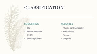
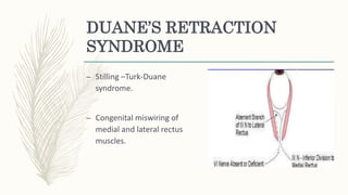
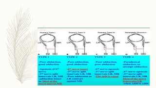
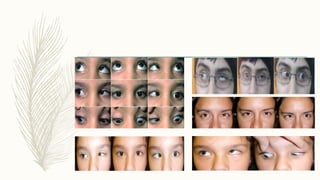
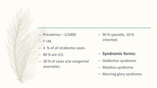
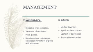
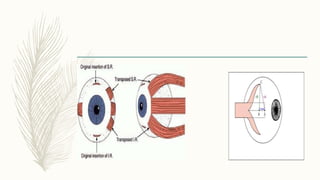
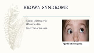
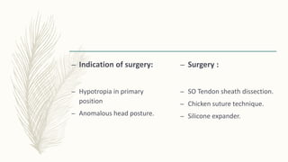
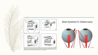
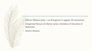
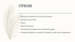
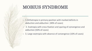
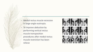
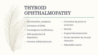
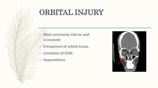
RESTRICTIVE
STRABISMUS
Dr. Debarati Dasgupta
– Characterised by limitation of movement which is out of
proportion to the ocular deviation in primary position.
– Can be mechanical or innervational anomaly.
– Medial Rectus – Adduction.
– Lateral Rectus – Abduction.
– Superior Rectus – Elevation , adduction , intortion.
– Inferior Rectus – Depression, adduction, extortion.
– Superior Oblique – Intortion , abduction, depression.
– Inferior oblique – Extortion , abduction, Elevation.
CLASSIFICATION
CONGENITAL
– DRS
– Brown’s syndrome
– CFEOM
– Mobius syndrome
ACQUIRED
– Thyroid ophthalmopathy
– Orbital injury
– Tumours
– Surgeries
DUANE’S RETRACTION
SYNDROME
– Stilling –Turk-Duane
syndrome.
– Congenital miswiring of
medial and lateral rectus
muscles.
– Prevalence – 1/1000.
– F >M.
– 4 % of all strabismic cases.
– 80 % are U/L.
– 30 % of cases a/w congenital
anomalies.
– 90 % sporadic, 10 %
inherited.
– Syndromic forms:
– Goldenhar syndrome.
– Moebius syndrome.
– Morning glory syndrome.
– Complete or partial abduction or adduction deficit.
– Retraction on adduction.
– Pseudotosis on adduction.
– Upshoot or downshoot on adduction.
– Abnormal head posture.
MANAGEMENT
NON SURGICAL
– Retractive error correction.
– Treatment of amblyopia.
– Prism glasses.
– Botulinum toxin – decreases
upshoot or downshoot of globe
with adduction.
 SURGERY
– Marked deviation.
– Significant head posture.
– Upshoot or downshoot.
– Severe globe retraction.
Type 1 & 3 with head turn –
– Recession of medial rectus or
horizontal transposition of vertical
rectus muscle.
Type 1 & 3 with upshoot or
downshoot or severe globe
retraction –
– Recession of both MR & LR with Y
splitting of LR.
Type 2 with head turn &
fixation with uninvolved
eye :
– Recession of I/L LR.
Type 2 with head turn &
fixation with involved eye:
– Recession of C/L LR.
 Type 2 with upshoot or
downshoot:
– Recession of LR with with Y
splitting.
– Congenital 6th Nerve Palsy –
– Infantile esotropia
– Mobius Syndrome
BROWN SYNDROME
– Tight or short superior
oblique tendon.
– Congenital or acquired.
– Limited elevation in adduction.
– Unaffected elevation in primary position & abduction.
– Divergnce in upgaze.
– FDT +ve.
– Widening of palpebral fissure on adduction.
– Ortho or hypo in primary position.
– Head posture.
– Indication of surgery:
– Hypotropia in primary
position
– Anomalous head posture.
– Surgery :
– SO Tendon sheath dissection.
– Chicken suture technique.
– Silicone expander.
– Inferior Oblique palsy – no divergence in upgaze, SO overaction.
– Congenital fibrosis of inferior rectus- limitation of elevation in
abduction.
– Grave’s disease.
CFEOM
– Fibrosis of extraocular muscles and tenon.
– Inelastic conjunctiva.
– Ptosis.
– Absent elevation.
– Perverted convergence on attempted upgaze.
– Choroidal coloboma, pendular nystagmus, optic nerve hypoplasia.
– supramaximal recession of I.R.
– Correction of S.O overaction.
– Correction of Exodeviations ,
– Perverted convergence – Faden procedure.
– CPEO – later onset with progression.
– Congenital ptosis
– Congenital myasthenia – fatigable ptosis.
MOBIUS SYNDROME
– 1.Orthotropia in primary position with marked deficits in
abduction and adduction (40% of cases)
– 2. Esotropia with cross-fixation and sparing of convergence and
adduction (50% of cases)
– 3. Large exotropia with absence of convergence (10% of cases)
– Medial rectus muscle recession
in large-angle esotropia.
– To improve abduction by
performing vertical rectus
muscle transposition
procedures after medial rectus
muscle restriction has been
relieve
THYROID
OPHTHALMOPATHY
– Lid retraction, proptosis.
– Limitation of EOM.
– Convergence insufficiency.
– GAG production &
deposition.
– Increase orbital pressure.
– Correction by prism or
occlusion.
– Steroid.
– Surgical decompression
– Ocular deviation by muscle
recession.
– Adjustable suture.
ORBITAL INJURY
– Most commonly inferior wall
is involved.
– Entrapment of orbital tissue.
– Limitation of EOM.
– Hypoesthesia
– Observe .
– Follow up with vision and
diplopia chart.
 Indication for surgery :
– Diplopia in primary position
or downgaze.
– Severe enophthalmos.
– Ocular deviation can be dealt
later.
– MRI
– Surgical exploration.
THANK YOU

Restrictive strabismus

  • 1.
  • 2.
    – Characterised bylimitation of movement which is out of proportion to the ocular deviation in primary position. – Can be mechanical or innervational anomaly.
  • 4.
    – Medial Rectus– Adduction. – Lateral Rectus – Abduction. – Superior Rectus – Elevation , adduction , intortion. – Inferior Rectus – Depression, adduction, extortion. – Superior Oblique – Intortion , abduction, depression. – Inferior oblique – Extortion , abduction, Elevation.
  • 5.
    CLASSIFICATION CONGENITAL – DRS – Brown’ssyndrome – CFEOM – Mobius syndrome ACQUIRED – Thyroid ophthalmopathy – Orbital injury – Tumours – Surgeries
  • 6.
    DUANE’S RETRACTION SYNDROME – Stilling–Turk-Duane syndrome. – Congenital miswiring of medial and lateral rectus muscles.
  • 9.
    – Prevalence –1/1000. – F >M. – 4 % of all strabismic cases. – 80 % are U/L. – 30 % of cases a/w congenital anomalies. – 90 % sporadic, 10 % inherited. – Syndromic forms: – Goldenhar syndrome. – Moebius syndrome. – Morning glory syndrome.
  • 10.
    – Complete orpartial abduction or adduction deficit. – Retraction on adduction. – Pseudotosis on adduction. – Upshoot or downshoot on adduction. – Abnormal head posture.
  • 11.
    MANAGEMENT NON SURGICAL – Retractiveerror correction. – Treatment of amblyopia. – Prism glasses. – Botulinum toxin – decreases upshoot or downshoot of globe with adduction.  SURGERY – Marked deviation. – Significant head posture. – Upshoot or downshoot. – Severe globe retraction.
  • 12.
    Type 1 &3 with head turn – – Recession of medial rectus or horizontal transposition of vertical rectus muscle. Type 1 & 3 with upshoot or downshoot or severe globe retraction – – Recession of both MR & LR with Y splitting of LR.
  • 13.
    Type 2 withhead turn & fixation with uninvolved eye : – Recession of I/L LR. Type 2 with head turn & fixation with involved eye: – Recession of C/L LR.  Type 2 with upshoot or downshoot: – Recession of LR with with Y splitting.
  • 15.
    – Congenital 6thNerve Palsy – – Infantile esotropia – Mobius Syndrome
  • 16.
    BROWN SYNDROME – Tightor short superior oblique tendon. – Congenital or acquired.
  • 17.
    – Limited elevationin adduction. – Unaffected elevation in primary position & abduction. – Divergnce in upgaze. – FDT +ve. – Widening of palpebral fissure on adduction. – Ortho or hypo in primary position. – Head posture.
  • 18.
    – Indication ofsurgery: – Hypotropia in primary position – Anomalous head posture. – Surgery : – SO Tendon sheath dissection. – Chicken suture technique. – Silicone expander.
  • 20.
    – Inferior Obliquepalsy – no divergence in upgaze, SO overaction. – Congenital fibrosis of inferior rectus- limitation of elevation in abduction. – Grave’s disease.
  • 21.
    CFEOM – Fibrosis ofextraocular muscles and tenon. – Inelastic conjunctiva. – Ptosis. – Absent elevation. – Perverted convergence on attempted upgaze. – Choroidal coloboma, pendular nystagmus, optic nerve hypoplasia.
  • 23.
    – supramaximal recessionof I.R. – Correction of S.O overaction. – Correction of Exodeviations , – Perverted convergence – Faden procedure.
  • 25.
    – CPEO –later onset with progression. – Congenital ptosis – Congenital myasthenia – fatigable ptosis.
  • 26.
    MOBIUS SYNDROME – 1.Orthotropiain primary position with marked deficits in abduction and adduction (40% of cases) – 2. Esotropia with cross-fixation and sparing of convergence and adduction (50% of cases) – 3. Large exotropia with absence of convergence (10% of cases)
  • 27.
    – Medial rectusmuscle recession in large-angle esotropia. – To improve abduction by performing vertical rectus muscle transposition procedures after medial rectus muscle restriction has been relieve
  • 28.
    THYROID OPHTHALMOPATHY – Lid retraction,proptosis. – Limitation of EOM. – Convergence insufficiency. – GAG production & deposition. – Increase orbital pressure. – Correction by prism or occlusion. – Steroid. – Surgical decompression – Ocular deviation by muscle recession. – Adjustable suture.
  • 29.
    ORBITAL INJURY – Mostcommonly inferior wall is involved. – Entrapment of orbital tissue. – Limitation of EOM. – Hypoesthesia
  • 30.
    – Observe . –Follow up with vision and diplopia chart.  Indication for surgery : – Diplopia in primary position or downgaze. – Severe enophthalmos. – Ocular deviation can be dealt later. – MRI – Surgical exploration.
  • 31.