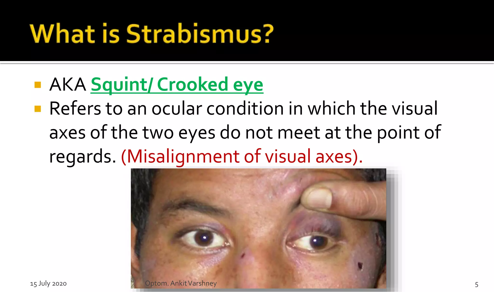
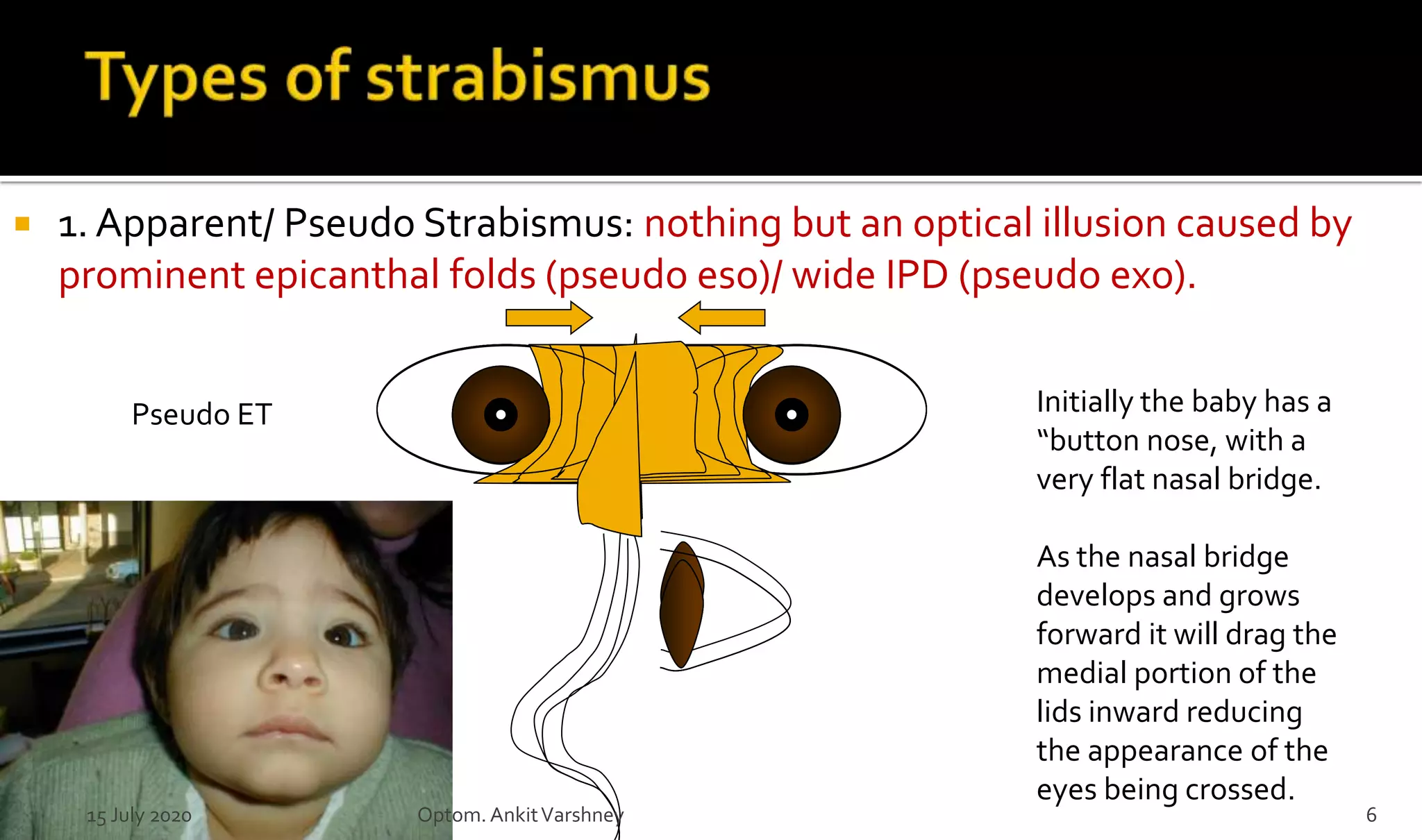
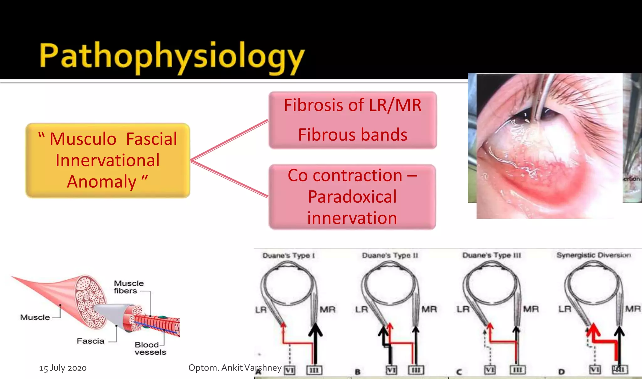
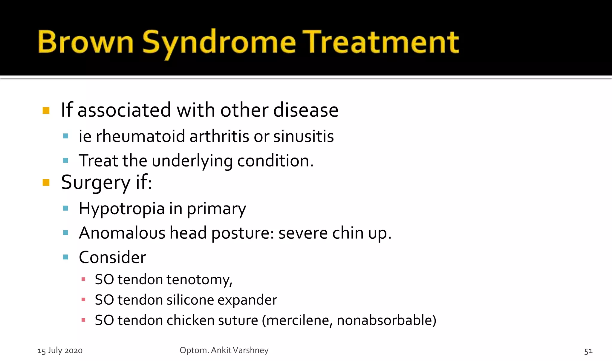
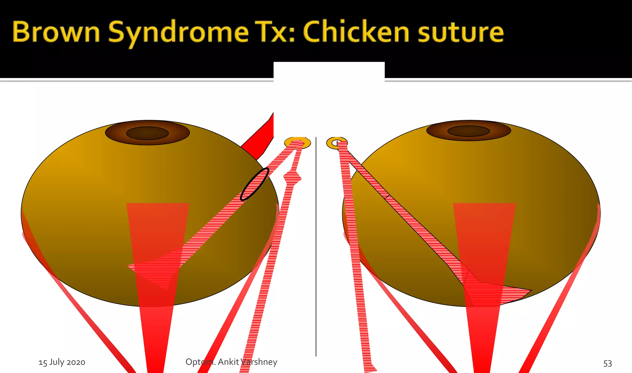
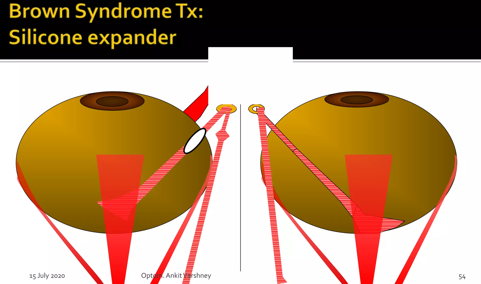
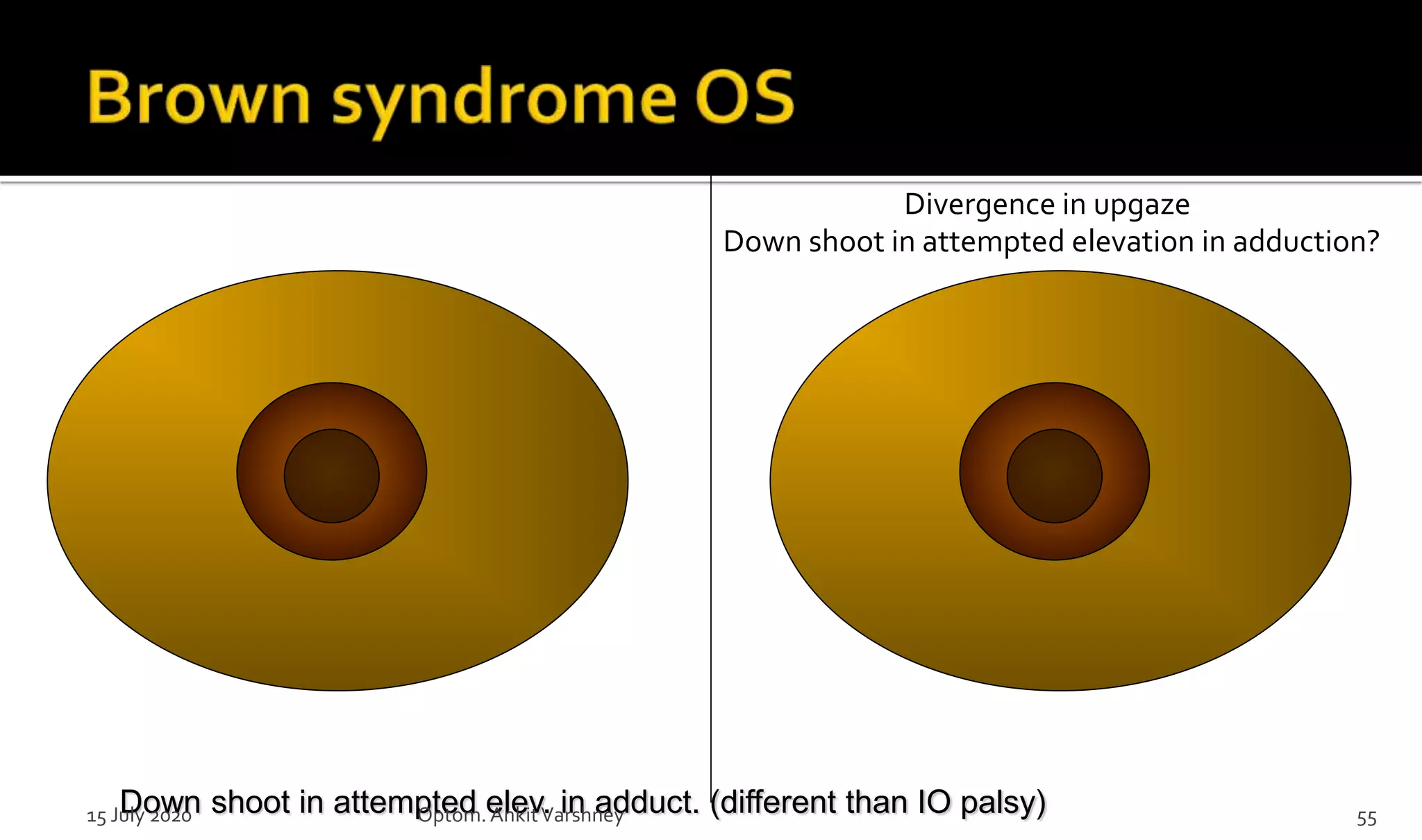
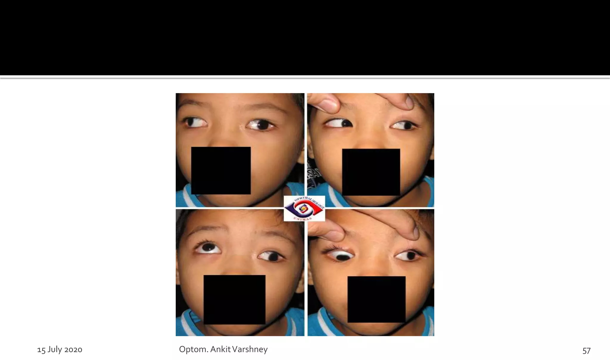
Restrictive strabismus is form of squint which is rare in origin. Here various form of restrictive squint have been explained, its diagnosis and its management is briefly explained.
VIP Call Girls Pune Vrinda 9907093804 Short 1500 Night 6000 Best call girls S...
Restrictive Strabismus by Ankit Varshney
1. Optom. Ankit S. Varshney
B.Optom, M.Optom, Ph.D. in Optometry (pursuing) Fellow of IACLE (Aus.), Fellow of ASCO(Mum.)
Prof. at (Shree Bharatimaiya College of Optometry & Physiotherapy, Surat)
Life Member of Indian Optometric Association (IOA)
Associate Member of Association of Schools and Colleges of Optometry(ASCO)
Member of Optometry Council of India(OCI)
Educator Member of International Association of Contact lense Educators (Australia)(IACLE)
Mail id: ankitsvarshney@yahoo.com
Whatsapp no. +918155955820
2. Binocular vision & strabismus –gk von
noorden
Clinical mangement of strabismus elezabeth
e.Calarossa & michael w. Rouse
Aao- section: pediatric ophthalmology &
strabismus
Strabismus simplified- pradeep sharma
Practical orthoptics in the treatment of squint-
lyle and jackson’s15 July 2020 Optom.AnkitVarshney 2
4. Eye Muscles
Left eye
Superior Oblique/Trochlear Muscle
Superior Rectus Muscle
Lateral Rectus Muscle
Inferior Rectus Muscle
Inferior Oblique Muscle
Medial Rectus Muscle
15 July 2020 Optom.AnkitVarshney 4
5. AKA Squint/ Crooked eye
Refers to an ocular condition in which the visual
axes of the two eyes do not meet at the point of
regards. (Misalignment of visual axes).
15 July 2020 Optom.AnkitVarshney 5
6. 1. Apparent/ Pseudo Strabismus: nothing but an optical illusion caused by
prominent epicanthal folds (pseudo eso)/ wide IPD (pseudo exo).
Pseudo ET Initially the baby has a
“button nose, with a
very flat nasal bridge.
As the nasal bridge
develops and grows
forward it will drag the
medial portion of the
lids inward reducing
the appearance of the
eyes being crossed.
15 July 2020 Optom.AnkitVarshney 6
7. 2. Latent Strabismus (Phoria): deviations are
present only some of the time: (interruption of
BV, fatigue, illness, stress)
Orthophoria
15 July 2020 Optom.AnkitVarshney 7
8. 3. Manifest Strabismus (tropia):
eyes deviated all the time
Concomitant ( Non-paralytic)
Incomitant strabismus (Paralytic/ Restrictive/
A &V pattern)
Esotropia
15 July 2020 Optom.AnkitVarshney 8
9. Manifest squint in which the amount of deviation
in squinting eye remains constant in all directions
of the gaze and there is no associated limitation in
ocular movement.
15 July 2020 Optom.AnkitVarshney 9
10. Manifest squint in which the amount of deviation
varies in different directions of gaze.
1. Paralytic squint: It refers to ocular deviation resulting
from complete/incomplete paralysis of one or more
EOM.
2. Restrictive squint: in this, the EOM involved is not
paralyzed but its movement is mechanically restricted.
It is differentiate by smaller amount of ocular
deviation and a Positive FDT.
15 July 2020 Optom.AnkitVarshney 10
11. Classified in two types:
1. Congenital causes (Musculo-fascial innervational anomalies):
DRS, Brown’s SO sheath Syndrome, Strabismus fixus & muscle
fibrosis syndrome.
2. Acquired causes: Dysthyroid orbito myopathy, pseudotumor,
orbital tumors, fibrosis of EOM secondary to trauma or surgery.
15 July 2020 Optom.AnkitVarshney 11
12.
13. Duane syndrome is a rare, congenital disorder of
eye movement
Stilling andTurk : described it first
Duane popularised it
Affecting the Horizontally acting muscle: so it
Horizontal strabismus.
15 July 2020 Optom.AnkitVarshney 13
14. Incidence 1-4 percent of all strabismus
Mostly Sporadic
Female > Males (3:2)
Left eye > Right eye (60-75%) where unilateral
Bilateral 18-22%
Many associated congenital anomalies
Occasionally familial
15 July 2020 Optom.AnkitVarshney 14
17. Type I (d): Failure or limitation of abduction.
Type II (dd): Failure or limitation of adduction.
Type III (ddd): Failure of abduction and adduction
Classic features
Retraction of globe on adduction
Adduction narrowing of palpebral fissure
Upshoot / downshoot of eye in adduction
15 July 2020 Optom.AnkitVarshney 17
24. Squinting
Head tilt
Loss of binocular vision
Reduced ocular movement
Facial asymmetry
Picture of paralytic squint
15 July 2020 Optom.AnkitVarshney 24
25. AHP
Strabismus ( 77% of cases)
Globe retraction
Palpebral fissure narrowing
Anisometropia /amblyopia
Sensory adaptation with excellent binocular functions
-suppression without diplopia in gaze of abnormal
muscle
15 July 2020 Optom.AnkitVarshney 25
27. 30 PD LET actually,
But can fuse in right
gaze, left head turn
Check his ductions…
Primary gaze
15 July 2020 Optom.AnkitVarshney 27
28. 30 PD LET actually,
But can fuse in right
gaze, left head turn
Notice the limited
abduction and narrow
fissure in adduction
Left gaze,
Right face turn…
15 July 2020 Optom.AnkitVarshney 28
30. DRS
ET small angle
Narrowing
Saccadic velocities
SIXTH N PALSY (LR)
ET large angle
-ve
Slow in abduction only
MOBIUS SYNDROME:
Rare condition affecting bilateral sixth and seventh (facial) cranial nerve palsy.
15 July 2020 Optom.AnkitVarshney 30
31. Treat refractive error
/amblyopia
Conservative : Age 5-6 yrs
Prisms: up to 25 error
Special seating arrangement for
children in schools
15 July 2020 Optom.AnkitVarshney 31
32. Conservative : Age 5-6 yrs
Vision therapy for secondary convergence insufficiency
Normal Convergence
Convergence Insufficiency
15 July 2020 Optom.AnkitVarshney 32
33. Anomalous head posture
Strabismus in primary gaze
Significant upshoot or downshoot in adduction
Cosmetically significant palpebral fissure
narrowing in adduction.
“ Limited Goals ”
15 July 2020 Optom.AnkitVarshney 33
34. Eso DRS
Small angle <15
PD
Single MR
recession
Larger angle
Bilateral MR
recession
Asymmetric
15 July 2020 Optom.AnkitVarshney 34
35. Exo DRS
Small angle <15
PD
Single LR
recession
Larger angle
Bilateral LR
recession
Symmetric
15 July 2020 Optom.AnkitVarshney 35
38. A very rare condition, in which both eyes are fixed
(in extreme duction), by fibrous tightening of MR
(Convergent strabismus fixus) or LR (Divergent
strabismus fixus).
FDT confirms their immobility.
Affecting the Horizontally acting muscle.
15 July 2020 Optom.AnkitVarshney 38
40. Convergent strabismus fixus: Complete
disinsertion of MR and resection of LR.
Divergent strabismus fixus: just reverse of above
15 July 2020 Optom.AnkitVarshney 40
42. 1. Absence of elevation in adduction
2. Downshoot or hypotropia in attempting elevation
in adduction
3. Unaffected elevation in primary and abducted
position
4. Y pattern with divergence in upgaze
5. Anomalous head posture (Often seen with chin up
position)
6. Positive FDT
7. 2/3 are mild and do not require treatment if ortho
in primary15 July 2020 Optom.AnkitVarshney 42
43. Trauma or
Infection ofTrochlear area
SO tucking Sx
Canine tooth syndrome: Dog bites- scarring SO
15 July 2020 Optom.AnkitVarshney 43
44. Divergence in upgaze
Down shoot in attempted elevation in adduction?
15 July 2020 Optom.AnkitVarshney 44
45. Mild: Restricted elevation in adduction but no downshoot
or hypotropia in primary position.
Moderate: Restricted elevation in adduction with
downshoot but no hypotropia in primary position.
Severe: Restricted elevation & downshoot in adduction
with hypotropia in primary position.
To understand Brown’s syndrome
We need to understand relationship between the
superior and inferior oblique.
15 July 2020 Optom.AnkitVarshney 45
50. 1-I O PALSY
2-DOUBLE ELEVATOR PALSY
3- CONGENITAL FIBROSIS SYNDROM
4-BLOW OUT FX
5-THYROID OPHTHALMOPATY
5-ADHERENCE SYNDROM
15 July 2020 Optom.AnkitVarshney 50
51. If associated with other disease
ie rheumatoid arthritis or sinusitis
Treat the underlying condition.
Surgery if:
Hypotropia in primary
Anomalous head posture: severe chin up.
Consider
▪ SO tendon tenotomy,
▪ SO tendon silicone expander
▪ SO tendon chicken suture (mercilene, nonabsorbable)
15 July 2020 Optom.AnkitVarshney 51
55. Divergence in upgaze
Down shoot in attempted elevation in adduction?
Down shoot in attempted elev. in adduct. (different than IO palsy)15 July 2020 Optom.AnkitVarshney 55
56. Rare, Nonprogressive, Autosomal dominant.
Characterized by hypoplasia and fibrosis of EOM & LPS.
Bilateral congenital Ptosis.
In primary position each eye is fixed below the horizontal
by about 10 o due to Congenital fibrosis of the inferior
rectus.
Thus hypotropic eye may be severe exotropic or esotropic
due to involvement of the horizontal recti.
15 July 2020 Optom.AnkitVarshney 56
58. Surgery is difficult and requires release of the
restricted muscles;
A good surgical result aligns the eyes in primary
position, but full ocular rotations cannot be
restored and the outcome is unpredictable.
15 July 2020 Optom.AnkitVarshney 58
59. Developmental anomaly occurring between the
EOMs and their fascial sheaths.
Abnormal connections most commonly occur
between the LR and IO, and between the SR and
SO muscles.
15 July 2020 Optom.AnkitVarshney 59
60. Diagnosis often mysterious SO LOOK for it !
Treat Ref Error , amblyopia
Risk benefit ratio of surgery to be well understood
by patients and surgeon
Tailor made surgery required for every case
15 July 2020 Optom.AnkitVarshney 60