PIGMENT DISPERSION SYNDROME 
Dr. Aditi Singh
 Described by : Sugar and Brown 1949
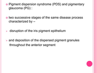
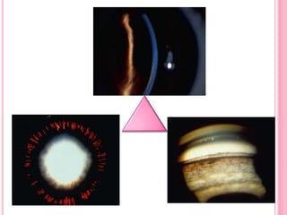
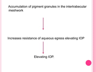
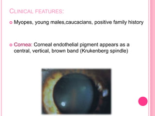
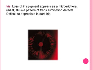
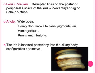
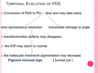
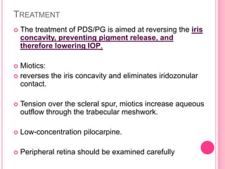
 Pigment dispersion syndrome (PDS) and pigmentary 
glaucoma (PG) : 
 two successive stages of the same disease process 
characterized by – 
 disruption of the iris pigment epithelium 
 and deposition of the dispersed pigment granules 
throughout the anterior segment
EPIDEMIOLOGY 
 Young 
 Myopic 
 Male 
 Experiences blurry vision or eye pain after exercise
PATHOPHYSIOLOGY 
 Concave iris contour that allows apposition of its 
posterior surface to the zonular bundles. 
 Friction between zonules and the peripheral iris - cause 
of the pigment liberation. 
 Reverse pupillary block mechanism may exist : “flap 
valve,”
Accumulation of pigment granules in the intertrabecular 
meshwork 
Increases resistance of aqueous egress elevating IOP 
Elevating IOP.
 Exercise (jogging, playing basketball, and bouncing 
during dancing) can cause the release of pigment 
as a result of pupillary movement. 
 Pharmacologic pupillary dilation - may result in 
significant pigment liberation into the anterior 
chamber. 
 This pigment liberation may be accompanied by 
IOP increase.
CLINICAL FEATURES: 
 Myopes, young males,caucacians, positive family history 
 Cornea: Corneal endothelial pigment appears as a 
central, vertical, brown band (Krukenberg spindle)
Iris: Loss of iris pigment appears as a midperipheral, 
radial, slit-like pattern of transillumination defects. 
Difficult to appreciate in dark iris.
 Lens / Zonules : Interrupted lines on the posterior 
peripheral surface of the lens – Zentamayer ring or 
Scheie’s stripe. 
 Angle: Wide open. 
Heavy dark brown to black pigmentation. 
Homogenous . 
Prominent inferiorly. 
 The iris is inserted posteriorly into the ciliary body, 
configuration : concave
 Posterior segment: 
lattice degeneration - 20% of patients 
retinal breaks- 11.7% 
rhegmatogenous retinal detachments 
requiring surgery may occur in 3.3% 
oOptic nerve examination: 
Size , PPA, RNFL defect, disc h’ges, NRR thinning
TEMPORAL EVOLUTION OF PDS 
 Conversion of PDS to PG – slow and may take years 
slow spontaneous resolution irreversible damage to angle 
 transillumination defects may disappear, 
 the IOP may return to normal, 
 the trabecular meshwork pigmentation may decrease. 
Pigment reversal sign ( burned out )
DIFFERENTIAL DIAGNOSIS 
 Disorders causing anterior segment pigment dispersion : 
 exfoliation syndrome (XFS), 
 diabetes, 
 herpetic eye disease, 
 iris pigment epithelitis, 
 radiation, 
 trauma, 
 iris pigment epithelial cysts, 
 ciliary 
 body cysts, 
 iris nevus, and 
 melanoma or melanocytoma of 
 the anterior and posterior segment
. 
Features Pseudoexfoliation 
syndrome 
Pigment dispersion 
syndrome 
Transillumination 
defects 
Peripupillary Radial mid peripheral 
TM pigmentation Patchy homogenous 
Age group > 60 yrs ( older) 20- 30 yrs (younger) 
whitish granular 
deposits 
Present Absent
TREATMENT 
 The treatment of PDS/PG is aimed at reversing the iris 
concavity, preventing pigment release, and 
therefore lowering IOP. 
 Miotics: 
 reverses the iris concavity and eliminates iridozonular 
contact. 
 Tension over the scleral spur, miotics increase aqueous 
outflow through the trabecular meshwork. 
 Low-concentration pilocarpine. 
 Peripheral retina should be examined carefully
 Prostaglandin analogues: Increasing uveoscleral 
outflow. 
 Agents that lower IOP by reducing aqueous production 
hypothetically – 
 may diminish the rate of clearance of the pigment from 
the trabecular meshwork, possibly exacerbating the 
disease process. 
 these agents may inhibit relative pupillary block, which 
is therapeutic in PDS.
LASER IRIDOTOMY 
 Equalizes pressures between the anterior and posterior 
chambers, 
 Flattens the iris, 
 Eliminates iridozonular contact, and 
 Occasionally decreases further liberation of pigment 
 Proper patient selection. 
 Ideally, patients should still be in the pigment liberation stage. 
 In young patients with iris concavity, active release of pigment 
and ocular hypertension, LI may be of benefit for years.
 Argon laser trabeculoplasty and selective laser 
trabeculoplasty 
 Alternative treatments to lower IOP, mostly in young 
pigmentary glaucoma patients. 
 The success rate of argon laser trabeculoplasty 
(ALT) in PG is greater in younger patients than in 
older ones and decreases with age
 Trabeculectomy : 
 Not responding to medical / laser therapy. 
 MMC to be used in lower concentration for lesser 
time. 
 Outcomes comparable /better than of POAG

Pigment dispersion syndrome

  • 1.
  • 2.
     Described by: Sugar and Brown 1949
  • 3.
     Pigment dispersionsyndrome (PDS) and pigmentary glaucoma (PG) :  two successive stages of the same disease process characterized by –  disruption of the iris pigment epithelium  and deposition of the dispersed pigment granules throughout the anterior segment
  • 5.
    EPIDEMIOLOGY  Young  Myopic  Male  Experiences blurry vision or eye pain after exercise
  • 6.
    PATHOPHYSIOLOGY  Concaveiris contour that allows apposition of its posterior surface to the zonular bundles.  Friction between zonules and the peripheral iris - cause of the pigment liberation.  Reverse pupillary block mechanism may exist : “flap valve,”
  • 7.
    Accumulation of pigmentgranules in the intertrabecular meshwork Increases resistance of aqueous egress elevating IOP Elevating IOP.
  • 8.
     Exercise (jogging,playing basketball, and bouncing during dancing) can cause the release of pigment as a result of pupillary movement.  Pharmacologic pupillary dilation - may result in significant pigment liberation into the anterior chamber.  This pigment liberation may be accompanied by IOP increase.
  • 9.
    CLINICAL FEATURES: Myopes, young males,caucacians, positive family history  Cornea: Corneal endothelial pigment appears as a central, vertical, brown band (Krukenberg spindle)
  • 10.
    Iris: Loss ofiris pigment appears as a midperipheral, radial, slit-like pattern of transillumination defects. Difficult to appreciate in dark iris.
  • 11.
     Lens /Zonules : Interrupted lines on the posterior peripheral surface of the lens – Zentamayer ring or Scheie’s stripe.  Angle: Wide open. Heavy dark brown to black pigmentation. Homogenous . Prominent inferiorly.  The iris is inserted posteriorly into the ciliary body, configuration : concave
  • 12.
     Posterior segment: lattice degeneration - 20% of patients retinal breaks- 11.7% rhegmatogenous retinal detachments requiring surgery may occur in 3.3% oOptic nerve examination: Size , PPA, RNFL defect, disc h’ges, NRR thinning
  • 13.
    TEMPORAL EVOLUTION OFPDS  Conversion of PDS to PG – slow and may take years slow spontaneous resolution irreversible damage to angle  transillumination defects may disappear,  the IOP may return to normal,  the trabecular meshwork pigmentation may decrease. Pigment reversal sign ( burned out )
  • 14.
    DIFFERENTIAL DIAGNOSIS Disorders causing anterior segment pigment dispersion :  exfoliation syndrome (XFS),  diabetes,  herpetic eye disease,  iris pigment epithelitis,  radiation,  trauma,  iris pigment epithelial cysts,  ciliary  body cysts,  iris nevus, and  melanoma or melanocytoma of  the anterior and posterior segment
  • 15.
    . Features Pseudoexfoliation syndrome Pigment dispersion syndrome Transillumination defects Peripupillary Radial mid peripheral TM pigmentation Patchy homogenous Age group > 60 yrs ( older) 20- 30 yrs (younger) whitish granular deposits Present Absent
  • 16.
    TREATMENT  Thetreatment of PDS/PG is aimed at reversing the iris concavity, preventing pigment release, and therefore lowering IOP.  Miotics:  reverses the iris concavity and eliminates iridozonular contact.  Tension over the scleral spur, miotics increase aqueous outflow through the trabecular meshwork.  Low-concentration pilocarpine.  Peripheral retina should be examined carefully
  • 17.
     Prostaglandin analogues:Increasing uveoscleral outflow.  Agents that lower IOP by reducing aqueous production hypothetically –  may diminish the rate of clearance of the pigment from the trabecular meshwork, possibly exacerbating the disease process.  these agents may inhibit relative pupillary block, which is therapeutic in PDS.
  • 18.
    LASER IRIDOTOMY Equalizes pressures between the anterior and posterior chambers,  Flattens the iris,  Eliminates iridozonular contact, and  Occasionally decreases further liberation of pigment  Proper patient selection.  Ideally, patients should still be in the pigment liberation stage.  In young patients with iris concavity, active release of pigment and ocular hypertension, LI may be of benefit for years.
  • 20.
     Argon lasertrabeculoplasty and selective laser trabeculoplasty  Alternative treatments to lower IOP, mostly in young pigmentary glaucoma patients.  The success rate of argon laser trabeculoplasty (ALT) in PG is greater in younger patients than in older ones and decreases with age
  • 21.
     Trabeculectomy :  Not responding to medical / laser therapy.  MMC to be used in lower concentration for lesser time.  Outcomes comparable /better than of POAG