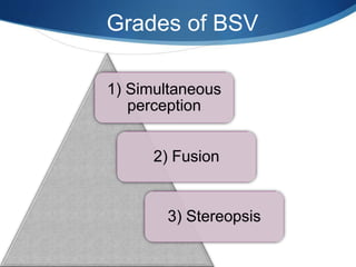
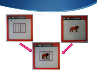
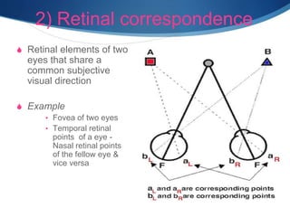
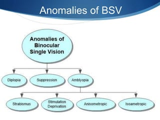
This document provides an introduction to binocular single vision (BSV) including its definition, grades, advantages, development, mechanisms, anomalies, and investigations. BSV is the coordinated use of both eyes to see a single image through the process of fusion. It develops in early childhood as the visual axes align and fusional movements are established. Maintaining BSV provides advantages like stereopsis and binocular summation. Investigations of BSV assess fusion, retinal correspondence, suppression, and stereopsis.
To know Humphrey visual field analyser
To know about various types of perimetry
To identify field defect
To recognize that field defect is due to glaucoma or neurological lesion
To know that field defect is progressive or not
Interpretation of HVFA
Detailed instumentaion and use of manual Lensometer and just a outline of automated lensometer.
I have used the picture of manual lensometer with out the parts describtion because i have explained orally by showing the picture..
Hope u all like it and may help you in learning better. :)
Accommodation/ Accommodation of Eye, Measurement of Accommodation of Eye (hea...Bikash Sapkota
CLICK HERE TO DOWNLOAD FULL PPT ❤❤ https://healthkura.com/measurement-of-accommodation-of-eye/ ❤❤
Dear viewers Check Out my other piece of works at ❤❤❤ https://healthkura.com ❤❤❤
Measurement of Accommodation of eye:
Amplitude, Facility,
Relative Accommodation, Fatigue, Lag,
Dynamic Retinoscopy
Presentation Layout:
-Introduction to accommodation of eye
-Mechanism
-Components
-Measurement of accommodation of eye
- Amplitude
- Facility
- Relative accommodation
- Lag
-Dynamic Retinoscopy
Accommodation
-dioptric adjustment of the crystalline lens of the eye
- to obtain clear vision for a given target of regard
-process by which the refractive power of eye is altered
- to ensure a clear retinal image
For further reading
-Clinical Procedures in Optometry by J.D. Bartlett, J.B. Eskridge, J.F. Amos
-Primary Care Optometry by Theodere Grosvenor
-Borish’s Clinical Refraction by W.J. Benjamin
-Clinical Procedures for Ocular examination by Carlson et al
-American Academy of Ophthalmology
-Optometric Clinical Practice Guideline by American Optometric Association
-Internet
Follow me to get in touch with optometric and ophthalmic updates
It describes about the procedure of Hess charting. it serves as a great tool to understand the concepts involved. Suitable for optometry course. This is not a routine procedure but an important procedure which is used in diagnosis.
Binocular single vision (BSV) is the ability to use both eyes together to achieve a single fused percept, even in the presence of disparity of the image seen by each eye. It is divided into five grades: simultaneous perception, superimposition, sensory fusion, motor fusion, and stereopsis.
The current study aimed to examine the contribution that motor fusion and stereoacuity make to visuomotor task performance, while addressing some of these confounding factors. Individual differences in task performance and adaptation to BSV deficits were minimized using a repeated measures design, in which participants with normal BSV and no amblyopia or strabismus had their fine visuomotor task performance assessed as their BSV was progressively degraded.
Corneal topographer is a useful tool for our clinical investigations on patient's with corneal problem. Knowing about its principle, function and interpretation is important.
Cataract management in children from optometrist perspectiveAnis Suzanna Mohamad
Congenital and childhood cataracts are uncommon but regularly seen in the clinics of most paediatric ophthalmology teams in the UK. They are often associated with profound visual loss and a large proportion have a genetic aetiology, some with significant extra-ocular comorbidities. Optimal diagnosis and treatment typically require close collaboration within multidisciplinary teams. Surgery remains the mainstay of treatment. A variety of surgical techniques, timings of intervention and options for optical correction have been advocated making management seem complex for those seeing affected children infrequently.
National Ocular Biometry Course (NOBC) 2015 An echoslide presentation Anis Suzanna Mohamad
This powerpoint presentation is basically about ocular biometry. Echo presentation is one of the method to deliver infomation that obtain from the course we attend to other staff in our Ophthalmology Department.
Defecation
Normal defecation begins with movement in the left colon, moving stool toward the anus. When stool reaches the rectum, the distention causes relaxation of the internal sphincter and an awareness of the need to defecate. At the time of defecation, the external sphincter relaxes, and abdominal muscles contract, increasing intrarectal pressure and forcing the stool out
The Valsalva maneuver exerts pressure to expel faeces through a voluntary contraction of the abdominal muscles while maintaining forced expiration against a closed airway. Patients with cardiovascular disease, glaucoma, increased intracranial pressure, or a new surgical wound are at greater risk for cardiac dysrhythmias and elevated blood pressure with the Valsalva maneuver and need to avoid straining to pass the stool.
Normal defecation is painless, resulting in passage of soft, formed stool
CONSTIPATION
Constipation is a symptom, not a disease. Improper diet, reduced fluid intake, lack of exercise, and certain medications can cause constipation. For example, patients receiving opiates for pain after surgery often require a stool softener or laxative to prevent constipation. The signs of constipation include infrequent bowel movements (less than every 3 days), difficulty passing stools, excessive straining, inability to defecate at will, and hard feaces
IMPACTION
Fecal impaction results from unrelieved constipation. It is a collection of hardened feces wedged in the rectum that a person cannot expel. In cases of severe impaction the mass extends up into the sigmoid colon.
DIARRHEA
Diarrhea is an increase in the number of stools and the passage of liquid, unformed feces. It is associated with disorders affecting digestion, absorption, and secretion in the GI tract. Intestinal contents pass through the small and large intestine too quickly to allow for the usual absorption of fluid and nutrients. Irritation within the colon results in increased mucus secretion. As a result, feces become watery, and the patient is unable to control the urge to defecate. Normally an anal bag is safe and effective in long-term treatment of patients with fecal incontinence at home, in hospice, or in the hospital. Fecal incontinence is expensive and a potentially dangerous condition in terms of contamination and risk of skin ulceration
HEMORRHOIDS
Hemorrhoids are dilated, engorged veins in the lining of the rectum. They are either external or internal.
FLATULENCE
As gas accumulates in the lumen of the intestines, the bowel wall stretches and distends (flatulence). It is a common cause of abdominal fullness, pain, and cramping. Normally intestinal gas escapes through the mouth (belching) or the anus (passing of flatus)
FECAL INCONTINENCE
Fecal incontinence is the inability to control passage of feces and gas from the anus. Incontinence harms a patient’s body image
PREPARATION AND GIVING OF LAXATIVESACCORDING TO POTTER AND PERRY,
An enema is the instillation of a solution into the rectum and sig
One of the most developed cities of India, the city of Chennai is the capital of Tamilnadu and many people from different parts of India come here to earn their bread and butter. Being a metropolitan, the city is filled with towering building and beaches but the sad part as with almost every Indian city
Navigating the Health Insurance Market_ Understanding Trends and Options.pdfEnterprise Wired
From navigating policy options to staying informed about industry trends, this comprehensive guide explores everything you need to know about the health insurance market.
Telehealth Psychology Building Trust with Clients.pptxThe Harvest Clinic
Telehealth psychology is a digital approach that offers psychological services and mental health care to clients remotely, using technologies like video conferencing, phone calls, text messaging, and mobile apps for communication.
CHAPTER 1 SEMESTER V PREVENTIVE-PEDIATRICS.pdfSachin Sharma
This content provides an overview of preventive pediatrics. It defines preventive pediatrics as preventing disease and promoting children's physical, mental, and social well-being to achieve positive health. It discusses antenatal, postnatal, and social preventive pediatrics. It also covers various child health programs like immunization, breastfeeding, ICDS, and the roles of organizations like WHO, UNICEF, and nurses in preventive pediatrics.
QA Paediatric dentistry department, Hospital Melaka 2020Azreen Aj
QA study - To improve the 6th monthly recall rate post-comprehensive dental treatment under general anaesthesia in paediatric dentistry department, Hospital Melaka
QA Paediatric dentistry department, Hospital Melaka 2020
Introduction to binocular single vision (BSV)
1. S
Introduction to BSV
An approach to understand the complexity
Prepared by
Anis Suzanna Binti Mohamad
Optometrist
Ophthalmology Department, Hospital Sultanah Bahiyah
2. Overview
S Definition
S Grades of BSV
S Advantages of BSV
S Development of BSV
S Mechanism and terminologies in BSV
S Anomalies of BSV
S Investigations of BSV
S Conclusions
S References
3. Definition
S State of simultaneous vision which is achieved by
coordinated use of both eyes so that separate and
slight dissimilar images arising in each eye
appreciated as single image by process of fusion.
(Acc. to
Romano & Romano)
5. 1) Simultaneous perception
Simultaneous perception exists when signals
transmitted from the two eyes to the visual cortex are
perceived at the same time.
It consists of the ability to see two dissimilar objects
simultaneously.
6.
7. 2) Fusion
S Defined as the cortical unification of visual objects
into a single percept that is made possible by the
simultaneous stimulation of corresponding retinal
areas.
S In simple words, it is the ability to superimpose two
incomplete but similar images to form one complete
image.
8.
9. Component of fusion
Sensory fusion
• the unification of visual excitations from corresponding retinal
images into a single visual percept, a single visual image
• Ability to unify images falling on corresponding retinal areas.
Motor fusion
• It is a vergence movement that causes similar retinal images to
fall and be maintained on corresponding retinal areas.
• Ability to align the eyes in such a manner that sensory fusion
can be maintained
• Diplopia preventing mechanism
10. Normal fusional range
Prism Distance Near
Base out
I><I
16 35/40
Base in
<II>
8 16
Source: Rowe Fiona. Clinical Orthoptics 2nd Ed, Blackwell Publishing,2004 2:
23
11. 3) Stereopsis
S It is the ability to fuse images that stimulate horizontally
disparate retinal elements within Panum’s fusional area
resulting in binocular appreciation of visual object in
depth i.e. in 3D
S Ability for depth perception
S In other words, when you look at an object or scene you
are able to perceive width, height and depth.
12. How it occurs?
S Retinal disparity (Fixation
disparity) is the basis of 3 D
perception
S Stereopsis occurs when
• Retinal disparity is large
enough to simple fusion
but small enough to cause
diplopia
13. Point to ponder
S Stereopsis & depth perception are not synonymous.
S Not a form of simple fusion.
S Normal stereoacuity is considered to be 40 sec of arc
14. Fusion Vs Stereopsis
Fusion Stereopsis
Corresponding retinal elements
are stimulated
Non corresponding retinal element
are stimulated
Motor system is required Motor system is not required
Fusion can occur without
stereopsis
Without fusion it can not occur
Fusion occurs horizontal or
vertical corresponding retinal
points
Stereopsis occurs only with
horizontal disparity
15. Monocular clues for depth perception
Relative size
Aerial
perspective
Linear
perception
16. Monocular clues for depth perception
Interposition Distribution of
lights &
shadows
Motion parallax
17. Advantages of having BSV
• Stereopsis.
• Binocular
summation.
– vision shaper,
clearer & more
sensitive
• Larger field of view.
• Spare eye.
• Compensation of
blind spot.
18. Development of BSV
S Basic visual functions are innate and therefore
present at birth.
S Their coordination, maturation and refinement take
place during early postnatal period.
19. BSV milestones
• no bifoveal fixation. Monocular fixation is present at birth, but
poor.At birth
• infant begins to make movements, turning his eyes to fixate an
object2-3 weeks
• can sustain monocular fixation of large near objects
4-5 weeks
• fixation alternates rapidly between two eyes & child begins to
fixate binocularly with conjugate pursuit movements which
are saccadic initially but become smooth and gliding by 3-
5mts of age.
6 weeks
• conjugate movements and disjugate vergence movements.
3-6 months
• fusional movements are firmly established.
1 year
• adult level of visual acuity is reached.
2-3 years
20. Maturation of binocular function
At birth- eyes act as 2
independent sense organs.
Foveas are not formed until
the 3rd month.
By trial and error the child
learns that, when the
image of an object is
brought on to the 2 foveae
simultaneously, the image
is most detailed.
Hence visual axes are
oriented in such a way that
each fovea is directed at
the object of regard.
21. Mechanism and terminologies
in BSV
1) Visual axis
2) Retinal correspondence
3) Egocentric localisation
4) Horopter
5) Pannums
fusional
area
22. 1) Visual axis
S Types
S Principal
S Fovea – area of
highest VA -carries
principal visual axis.
S Secondary
S Other retinal
elements-secondary
visual axis
A visual axis is defined as a line that connects an object
point with its image on the retina.
23. 2) Retinal correspondence
S Retinal elements of two
eyes that share a
common subjective
visual direction
S Example
• Fovea of two eyes
• Temporal retinal
points of a eye -
Nasal retinal points
of the fellow eye &
vice versa
26. 4) Horopter
S Horopter is defined as the
locus of all object points that
are imaged on
corresponding retinal
elements at a given fixing
distance.
S Horizon of vision
S Object points off the
horopter
S See double
S Object points lying on the
horopter
S seen single
27. 5) Pannums fusional area
S Panum, the Danish physiologist, first reported this phenomenon.
S Region in front and back of the horopter in which single vision is present is
known as Panum’s area of single binocular vision or Panum’s fusional area
28. Continue…
S Horizontal extent of these areas is small at the center (6 to 10 minutes
near the fovea)
S Increases toward the periphery (around 30 to 40 minutes at 12° from
the fovea)
31. Investigations of BSV
S All the tests are aimed at assessing the presence or
absence of:
S Normal or abnormal retinal correspondence
S Suppression
S Simultaneous perception
S Fusion with some amplitude
S Stereopsis
S Before any test is undertaken it is essential to assess the:
S visual acuity
S fixation in the squinting eye
S direction and size of deviation
33. Test for retinal correspondence
Principal of assessment
Based on relationship between the fovea of
fixing eye and the retinal area stimulated in
squinting eye
Synoptophore
Red
filter
test
Bagolini’s
striated
glasses test
Worth’s
4 Dots
test
Based on visual directions
of the two foveas
After images
Cuppers
binocular
visuoscopy
test
39. Test for stereopsis
S Stereo acuity is a quantitative
measure of stereopsis, it
represents the smallest
horizontal retinal image
disparity that give rise to a
sensation of depth.
S Stereopsis is measured in
seconds of arc.
S 1degree=60minutes of arc,
1minute=60seconds of arc.
S Normal stereoacuity=
<60seconds of arc.
Qualitative
Lang’s 2
pencil test
Synaptophore
Quantitativ
e
Random Dot
Test
TNO test
Lang’s stereo
test
Frisby test
45. Conclusions
S BSV is not inborn,but develops in the first decade of life
S Abnormal visual experience results in poor or no BSV
S Reversible only if intervened in the plastic period of
development
S A good understanding of mechanism of BSV is
fundamental in successive treatment of its anamolies
46. References
Books
S Kaufman FL, Alm A, Adler FH: Adler’s physiology of eye: clinical application,
10th Ed. St Louis: Mosby, 2003.
S Noorden GK von. Binocular vision and ocular motility: theory and managment
of strabismus, 5th Ed. St Louis: Mosby, 1996.
S Duane’s Clinical Ophthalmology. New York: Lippincott Williams & Wilkins,
2005.
S Kenneth Wright.W: Pediatric Ophthalmology and Strabismus, ed 95: 11:163.
S Rowe Fiona. Clinical Orthoptics 2nd Ed, Blackwell Publishing,2004 2: 23
S Khurana A. K.: Theory and Practise of Squint and Orthoptics; first ed .4:61-89.
Websites
S 1.webeye.ophth.uiowa.edu/eyeforum/tutorials/BINOCULAR-VISION.pdf
Editor's Notes
Normal correspondnce is called when both fovea have a common visual direction and the retinal elements nasal to the fovea in one eye corresponds to the retinal elements temporal to the fovea in the other eye.
Retinal correspondence is abnormal when the fove of one eye has a common visual direction with an extrafoveal area in the other eye. This is generally seen if the angle of squint is small and the extrafoveal point is close to the fovea.
Measurement of Angle of Anomaly: The angle of anomaly denotes the degree of shift in visual direction. It is determined by calculating the difference between the objective and subjective angles of deviation.
Procedure of estimating the angle of anomaly -
For this the use of SMP slides is made. The arms of the synaptophore are set at zero. Both the arms of the instrument are moved by the examiner while alternately flashing the light behind each slide until there is no further fixation movement of the patient’s eye (alternate cover test). The reading of both the arms is noted at this moment and the sum total of the reading of both the arms gives the objective angle of anomaly. The subjective angle of anomaly is the angle at which the visual targets are superimposed.
The interpretation of this test is as follows-
Angle of Anomaly = Objective Angle – Subjective Angle
If Subjective Angle = Objective Angle → NRC
If Subjective Angle < Objective Angle → ARC
If Angle of Anomaly = Objective Angle →Harmonious ARC (full sensory adaptation)
If Angle of Anomaly < Objective Angle →Unharmonious ARC
Red Filter Test: If one examines the visual field of a patient with heterophoria by placing a red filter in front of the habitually fixating eye while the patient is looking at a small light source, number of different responses can be elicited.
The patient may report that two lights are seen, a red one and a white one. In esotropia the images appear in homonymous (uncrossed) diplopia, with the red light to the right of the white one when the red filter is in front of the right eye. In exotropia the images appear in heteronymous (crossed) diplopia, with the red light to the left of the white light when the red filter is in front of the right eye. This represents NRC.
The patient may report that only one pinkish light in the position of the white fixation light is seen i.e. the red and white images appear to be superimposed. This is clearly an abnormal response in presence of heterophoria. This is termed Harmonious ARC.
The patient may report that two lights in uncrossed or crossed diplopia are seen, depending upon the direction of deviation but the measured distance between the double images proves to be smaller than expected from the magnitude of deviation. This represents unharmonious ARC.
Suppression is said to occur when the patient reports only a single light (usually the white light) but occasionally red depending upon the density of the red filter and the degree of the dominance of the fellow eye.
Bagolini's Striated Glasses Test: For this the patient fixates a small light, after being provided with plano lenses with narrow fine striations across one meridian (micro Maddox cylinders). These glasses do not affect the vision or the accommodation of the patient. The fixation light is seen as an elongated streak. The lenses are usually placed at 45 degree OS and 135 degree OD (cover the patients glasses, if he wears any) and the patient fixates for distance or near. The interpretation of this test is as follows-
Crossing of the lines at right angles to each other
If cover test reveals no shift and fixation is central, the patient has NRC
If cover test reveals a shift, harmonious ARC is present
Foveal suppression scotoma (fixation point scotoma) with peripheral fusion, if no shift occurs with cover test, NRC exists, if shift occurs, ARC exists
Single line represents suppression
Worth Four Dot Test: This is a simple test utilizing red-green color dissociation. It is more dissociating than the bagolini glasses and so less physiological. The apparatus for this test consists of a box containing four panes of glass, arranged in diamond formation, which are illuminated internally. The two internal panes are green, the upper one is red and lower one is white. The patient wears red and green goggles (as a convention red in front of right and green in front of left). The test can be performed separately for distance and near vision. The interpretation of this test is as follows-
A) The patient sees all the four dots.
normal binocular response with no manifest deviation (NRC with no heterotropia)
Harmonious ARC with manifest squint.
B) The patient sees five dots.
uncrossed diplopia with esotropia, red dots appear to the right
crossed diplopia with exotropia, red dots appear to the left of the green dots.
C) The patient sees three green dots, suppression of right eye.
D) The patient sees two red dots, suppression of left eye.
Hering Bielschowsky After-Image Test: This is a highly dissociating orthoptic test in which battery- powered camera flash is used to produce a vertical after image in one eye and a horizontal after image in the other eye. The center of flash is covered with a black mark (serves as a point of fixation and protects the fovea). Once an afterimage is created in each eye, the position of the images in relation to each other no longer depends on whether the eyes are open, closed, straight or crossed. The interpretation of this test depends on the fixation behaviour.
Procedure -
Each eye fixates on the center black mark of a glowing filament, first presented horizontally to the eye with a better visual acuity and then vertically to the poorer eye for 20 sec in a darkened room while the fellow eye is occluded. The patient indicates the relative position of the two gaps in the center of each afterimage. The gaps correspond to the visual direction of each fovea if central fixation is present.
Interpretation of results -
Cross response: A symmetrical cross with the central gaps superimposed indicates a normal bifoveal correspondence (if eccentric fixation is excluded). This is irrespective of any deviation between the two eyes, i.e. any eso or exo-deviation with NRC still gives a symmetrical cross response.
Asymmetrical crossing: In case of ARC the horizontal and vertical lines have their center separated, the amount of separation dependent on the angle of anomaly. A patient with right esotropia sees the vertical afterimage displaced to the left and a case of right exotropia sees the vertical afterimage displaced to the right.
Single line with a gap: A single line with a gap indicates suppression in the fellow eye.
Foveo-Foveal Test of Cuppers: Cuppers test for retinal correspondence determines whether the two foveas have common or different visual directions. It permits quantitative analysis of the angle of anomaly when eccentric fixation is present.
Procedure -
The patient fixates with the normal eye on the central light of a Maddox scale via a plano mirror, which for the convenience of the examiner is turned in such a manner that the amblyopic eye looks straight ahead. The visuoscope asterisk is projected by the examiner onto the fovea of the amblyopic eye. The figure of the Maddox scale on which the patient sees the asterisk indicated the angle of anomaly.
Modification -
To determine which parts of the peripheral retina in the deviating eye have acquired a common visual direction with the fovea of the fixating eye, the patient is asked to guide the Visuoscope until he sees the asterisk superimposed on the central light of the Maddox cross.The examiner views the fundus when this task is completed and notes the position of the asterisk, which indicates the location of retinal elements having a common visual direction with the fovea of the sound eye.
Stereograms: Stereogram with three concentric circles and a check dot for each eye is to be seen with both eyes together. Stereograms with three eccentric circles are to be seen with each eye separately. If the patient reports seeing concentric circles, it means stereopsis is present. If they are seen eccentrically one may ask whether the inner circles are closer to the right or left of the outer circle. It determines whether the disparate elements are suppressed in the right or the left eye.
Vectographs: Consists of Polaroid material on which the two targets are imprinted so that each target is polarized at 90 degrees with respect to the other. Patient is provided with Polaroid spectacles so that each target is seen separately with the two eyes.
Titmus stereo test – A gross stereoscopic pattern representing a housefly is provided to orient the patient and check for gross stereopsis (threshold 3000 sec of arc). Can be used in young children. Disadvantage of this test is that it can test only near stereopsis.
Polaroid test – Two common types:
A) Contain three rows of animals, one animal in each row imaged disparately (threshold 100, 200 and 400 sec. of arc respectively). The child is asked which one of the animals stands out. The animal figures contain a misleading clue. In each row one of the animals correspondingly imaged in two eyes is printed heavily black. A child without stereopsis will name this animal as the one that standsout.
B) Contains nine sets of four circles arranged in the form of a diamond. In this sequence the upper, lower, left or right are disparately imaged at random with thresholds ranging from 800 to 40 sec of arc. The child is now asked to push down the circle that stands out, beginning with the first set. A child with limited stereopsis will make mistakes or find no circle to push down.
E- Random Dot Test - This test consists of two cards, one with a ‘E’ stereo figure and the other stereoblank. A model of the figure is shown to the child before the test. The child is provided with Polaroid glasses and seated at 50 cms from the cards is required to point out the card which contains the "E".
Random Dot Stereogram of Julesz - Random dot stereogram, when viewed monocularly,convey no visual information and is seen as scattered random dots. When viewed binocularly, a square pattern appears in vivid depth above or below the level of the page. This test exposes the child to visual demands that are more difficult than those that occur under more casual conditions of seeing.