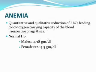
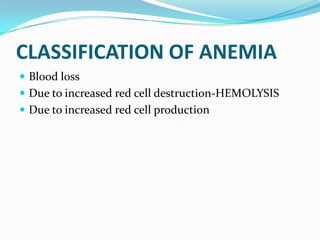
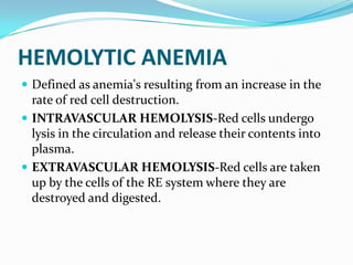
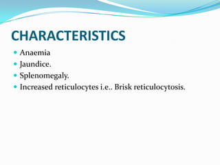
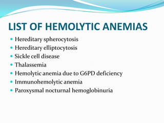
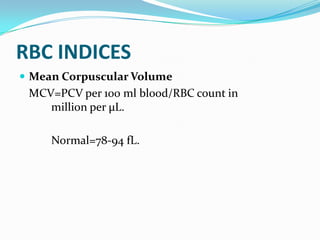
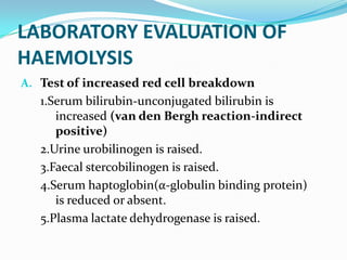
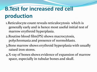
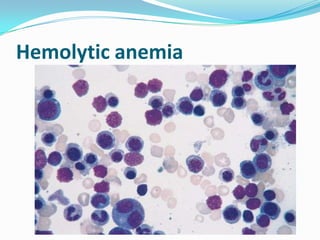
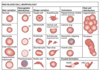
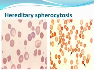
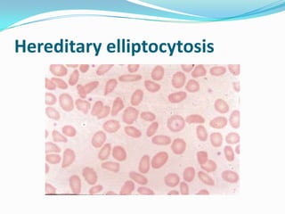
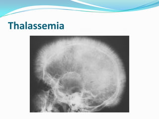
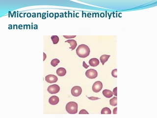
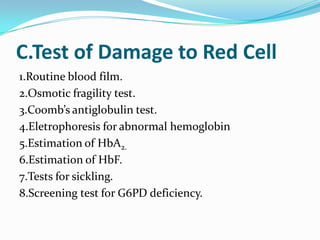
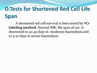
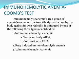
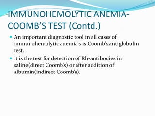
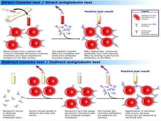
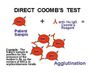
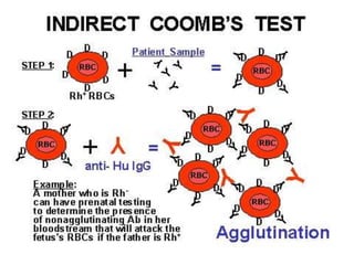
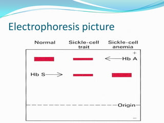
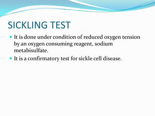
INVESTIGATION OF HAEMOLYTIC
           ANAEMIA
                  BY,


   MOHANKUMAR.M
                        43RD VZ
ANEMIA
 Quantitative and qualitative reduction of RBCs leading
  to low oxygen carrying capacity of the blood
  irrespective of age & sex.
 Normal Hb:
         Males: 14-18 gm/dl
         Females:12-15.5 gm/dl
CLASSIFICATION OF ANEMIA
 Blood loss
 Due to increased red cell destruction-HEMOLYSIS
 Due to increased red cell production
HEMOLYTIC ANEMIA
 Defined as anemia's resulting from an increase in the
  rate of red cell destruction.
 INTRAVASCULAR HEMOLYSIS-Red cells undergo
  lysis in the circulation and release their contents into
  plasma.
 EXTRAVASCULAR HEMOLYSIS-Red cells are taken
  up by the cells of the RE system where they are
  destroyed and digested.
CHARACTERISTICS
 Anaemia
 Jaundice.
 Splenomegaly.
 Increased reticulocytes i.e.. Brisk reticulocytosis.
LIST OF HEMOLYTIC ANEMIAS
 Hereditary spherocytosis
 Hereditary elliptocytosis
 Sickle cell disease
 Thalassemia
 Hemolytic anemia due to G6PD deficiency
 Immunohemolytic anemia
 Paroxysmal nocturnal hemoglobinuria
RBC INDICES
 Mean Corpuscular Volume
 MCV=PCV per 100 ml blood/RBC count in
    million per µL.

     Normal=78-94 fL.
 Mean Corpuscular Hemoglobin


 MCH=Hemoglobin in gm per dL/RBC count in
   million per µL.

     Normal=28-32 pg.
 Mean Corpuscular Hemoglobin Concentration


 MCHC=Hb in gm per dL/PCV per 100 ml blood

     Normal=30-37%
LABORATORY EVALUATION OF
HAEMOLYSIS
A. Test of increased red cell breakdown
   1.Serum bilirubin-unconjugated bilirubin is
      increased (van den Bergh reaction-indirect
      positive)
   2.Urine urobilinogen is raised.
   3.Faecal stercobilinogen is raised.
   4.Serum haptoglobin(α-globulin binding protein)
      is reduced or absent.
   5.Plasma lactate dehydrogenase is raised.
B.Test for increased red cell
production
1.Reticulocyte count reveals reticulocytosis which is
  generally early and is hence most useful initial test of
  marrow erythroid hyperplasia.
2.Routine blood film(PS) shows macrocytosis,
  polychromasia and presence of normoblasts.
3.Bone marrow shows erythroid hyperplasia with usually
  raised iron stores.
4.X-ray of bones shows evidence of expansion of marrow
  space, especially in tubular bones and skull.
Hemolytic anemia
Hereditary spherocytosis
Hereditary elliptocytosis
G6PD deficiency
Sickle cell disease
Sickle cell disease
Thalassemia
Thalassemia
Microangiopathic hemolytic
anemia
C.Test of Damage to Red Cell
1.Routine blood film.
2.Osmotic fragility test.
3.Coomb’s antiglobulin test.
4.Eletrophoresis for abnormal hemoglobin
5.Estimation of HbA2.
6.Estimation of HbF.
7.Tests for sickling.
8.Screening test for G6PD deficiency.
D.Tests for Shortened Red Cell Life
Span
      A shortened red cell survival is best tested by 51Cr
 labeling method. Normal RBC life span of 120 is
 shortened to 20-40 days in moderate haemolysis and
 to 5-20 days in severe haemolysis.
E.OTHER TESTS-
FLOWCYTOMETRTY
IMMUNOHEMOLYTIC ANEMIA-
COOMB’S TEST
      Immunohemolytic anemia's are a group of
 anemia's occurring due to antibody production by the
 body against its own red cells. It is induced by one of
 the following three types of antibodies:
      1.Autoimmune hemolytic anemia
             a. Warm antibody AIHA
             b. Cold antibody AIHA
      2.Drug induced immunohemolytic anemia
      3.Isoimmune hemolytic anemia
IMMUNOHEMOLYTIC ANEMIA-
COOMB’S TEST (Contd.)
 An important diagnostic tool in all cases of
  immunohemolytic anemia's is Coomb’s antiglobulin
  test.
 It is the test for detection of Rh-antibodies in
  saline(direct Coomb’s) or after addition of
  albumin(indirect Coomb’s).
Electrophoresis picture
SICKLING TEST
 It is done under condition of reduced oxygen tension
  by an oxygen consuming reagent, sodium
  metabisulfate.
 It is a confirmatory test for sickle cell disease.
Hemolytic Anemia Investigation - By Mohan kumar

Hemolytic Anemia Investigation - By Mohan kumar

  • 1.
    INVESTIGATION OF HAEMOLYTIC ANAEMIA BY, MOHANKUMAR.M 43RD VZ
  • 2.
    ANEMIA  Quantitative andqualitative reduction of RBCs leading to low oxygen carrying capacity of the blood irrespective of age & sex.  Normal Hb:  Males: 14-18 gm/dl  Females:12-15.5 gm/dl
  • 3.
    CLASSIFICATION OF ANEMIA Blood loss  Due to increased red cell destruction-HEMOLYSIS  Due to increased red cell production
  • 4.
    HEMOLYTIC ANEMIA  Definedas anemia's resulting from an increase in the rate of red cell destruction.  INTRAVASCULAR HEMOLYSIS-Red cells undergo lysis in the circulation and release their contents into plasma.  EXTRAVASCULAR HEMOLYSIS-Red cells are taken up by the cells of the RE system where they are destroyed and digested.
  • 5.
    CHARACTERISTICS  Anaemia  Jaundice. Splenomegaly.  Increased reticulocytes i.e.. Brisk reticulocytosis.
  • 6.
    LIST OF HEMOLYTICANEMIAS  Hereditary spherocytosis  Hereditary elliptocytosis  Sickle cell disease  Thalassemia  Hemolytic anemia due to G6PD deficiency  Immunohemolytic anemia  Paroxysmal nocturnal hemoglobinuria
  • 7.
    RBC INDICES  MeanCorpuscular Volume MCV=PCV per 100 ml blood/RBC count in million per µL. Normal=78-94 fL.
  • 8.
     Mean CorpuscularHemoglobin MCH=Hemoglobin in gm per dL/RBC count in million per µL. Normal=28-32 pg.
  • 9.
     Mean CorpuscularHemoglobin Concentration MCHC=Hb in gm per dL/PCV per 100 ml blood Normal=30-37%
  • 10.
    LABORATORY EVALUATION OF HAEMOLYSIS A.Test of increased red cell breakdown 1.Serum bilirubin-unconjugated bilirubin is increased (van den Bergh reaction-indirect positive) 2.Urine urobilinogen is raised. 3.Faecal stercobilinogen is raised. 4.Serum haptoglobin(α-globulin binding protein) is reduced or absent. 5.Plasma lactate dehydrogenase is raised.
  • 11.
    B.Test for increasedred cell production 1.Reticulocyte count reveals reticulocytosis which is generally early and is hence most useful initial test of marrow erythroid hyperplasia. 2.Routine blood film(PS) shows macrocytosis, polychromasia and presence of normoblasts. 3.Bone marrow shows erythroid hyperplasia with usually raised iron stores. 4.X-ray of bones shows evidence of expansion of marrow space, especially in tubular bones and skull.
  • 12.
  • 14.
  • 15.
  • 16.
  • 17.
  • 18.
  • 19.
  • 20.
  • 21.
  • 22.
    C.Test of Damageto Red Cell 1.Routine blood film. 2.Osmotic fragility test. 3.Coomb’s antiglobulin test. 4.Eletrophoresis for abnormal hemoglobin 5.Estimation of HbA2. 6.Estimation of HbF. 7.Tests for sickling. 8.Screening test for G6PD deficiency.
  • 23.
    D.Tests for ShortenedRed Cell Life Span A shortened red cell survival is best tested by 51Cr labeling method. Normal RBC life span of 120 is shortened to 20-40 days in moderate haemolysis and to 5-20 days in severe haemolysis.
  • 24.
  • 26.
    IMMUNOHEMOLYTIC ANEMIA- COOMB’S TEST Immunohemolytic anemia's are a group of anemia's occurring due to antibody production by the body against its own red cells. It is induced by one of the following three types of antibodies: 1.Autoimmune hemolytic anemia a. Warm antibody AIHA b. Cold antibody AIHA 2.Drug induced immunohemolytic anemia 3.Isoimmune hemolytic anemia
  • 27.
    IMMUNOHEMOLYTIC ANEMIA- COOMB’S TEST(Contd.)  An important diagnostic tool in all cases of immunohemolytic anemia's is Coomb’s antiglobulin test.  It is the test for detection of Rh-antibodies in saline(direct Coomb’s) or after addition of albumin(indirect Coomb’s).
  • 31.
  • 32.
    SICKLING TEST  Itis done under condition of reduced oxygen tension by an oxygen consuming reagent, sodium metabisulfate.  It is a confirmatory test for sickle cell disease.

Editor's Notes

  • #13 Marrow smear from a patient with hemolytic anemia. The marrow reveals greatly increased numbers of maturing erythroid progenitors (normoblasts).
  • #15 Hereditary spherocytosis (peripheral smear). Note the anisocytosis and several dark-appearing spherocytes with no central pallor. Howell-Jolly bodies (small dark nuclear remnants) are also present in red cells of this asplenic patient
  • #17 G6PD deficiency: effects of oxidant drug exposure (peripheral blood smear). Inset, Red cells with precipitates of denatured globin (Heinz bodies) revealed by supravital staining. As the splenic macrophages pluck out these inclusions, “bite cells” like the one in this smear are produced
  • #18 Sickle cell disease (peripheral blood smear). A, Low magnification shows sickle cells, anisocytosis, and poikilocytosis. B, Higher magnification shows an irreversibly sickled cell in the center.
  • #19 A, Spleen in sickle cell disease (low power). Red pulp cords and sinusoids are markedly congested; between the congested areas, pale areas of fibrosis resulting from ischemic damage are evident. B, Under high power, splenic sinusoids are dilated and filled with sickled red cells
  • #21 Thalassemia: x-ray film of the skull showing new bone formation on the outer table, producing perpendicular radiations resembling a crewcut
  • #22 Microangiopathic hemolytic anemia. A peripheral blood smear from a person with hemolytic-uremic syndrome shows several fragmented red cells