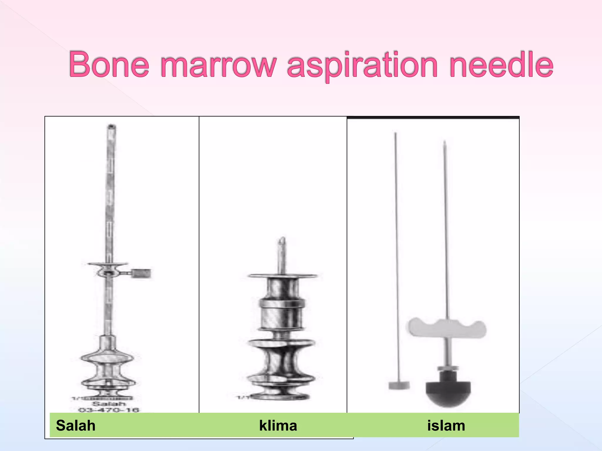
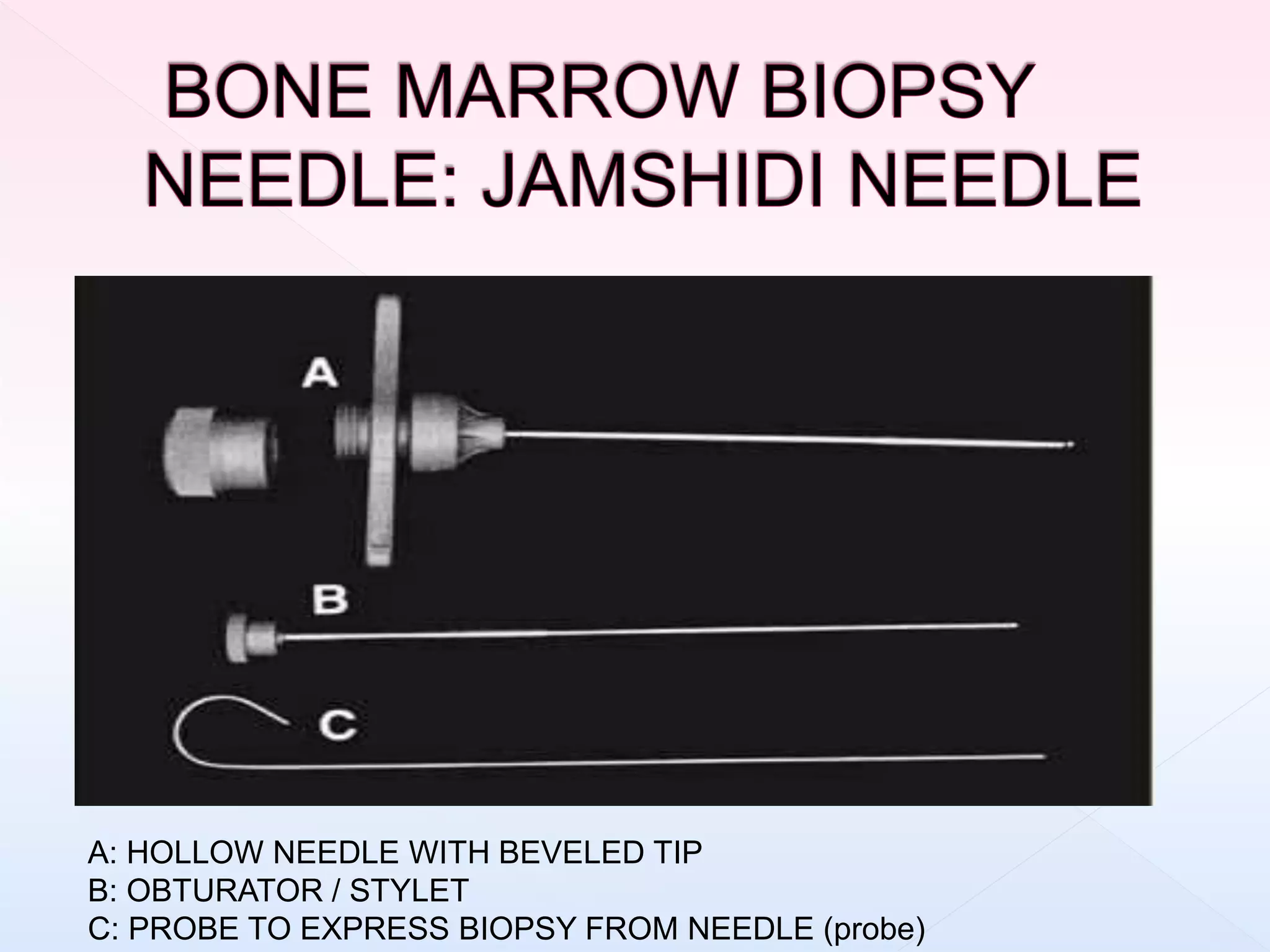
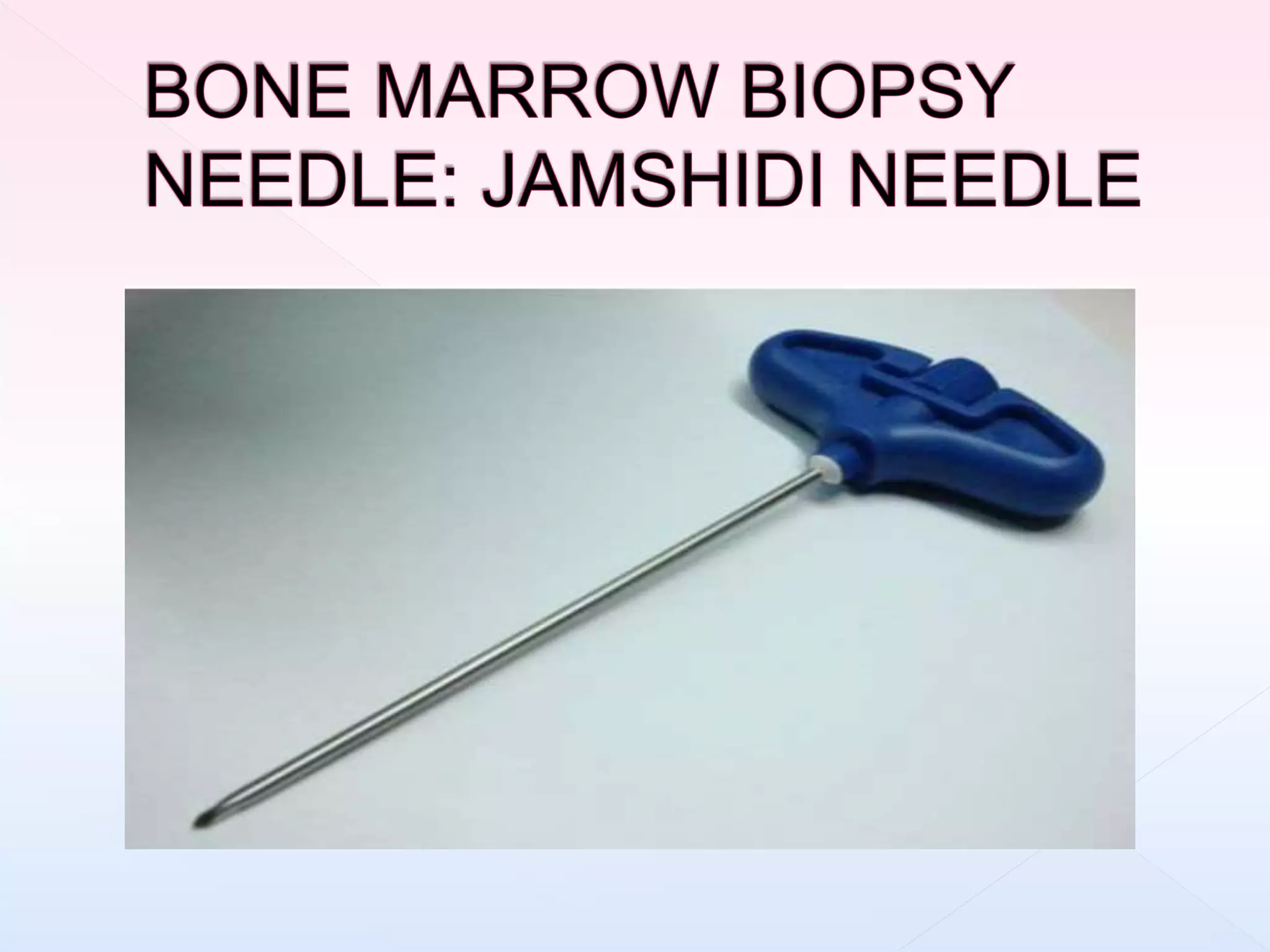
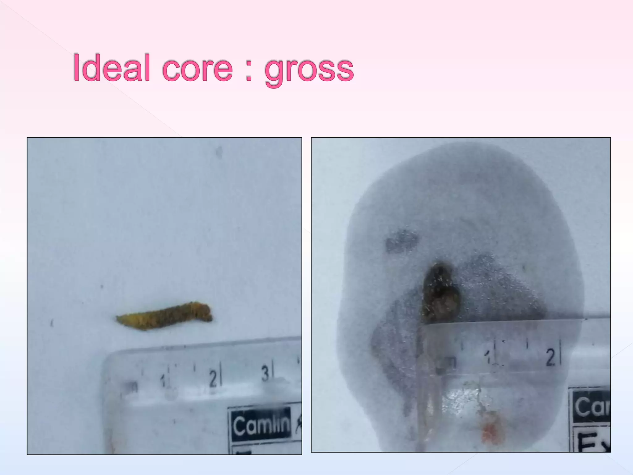
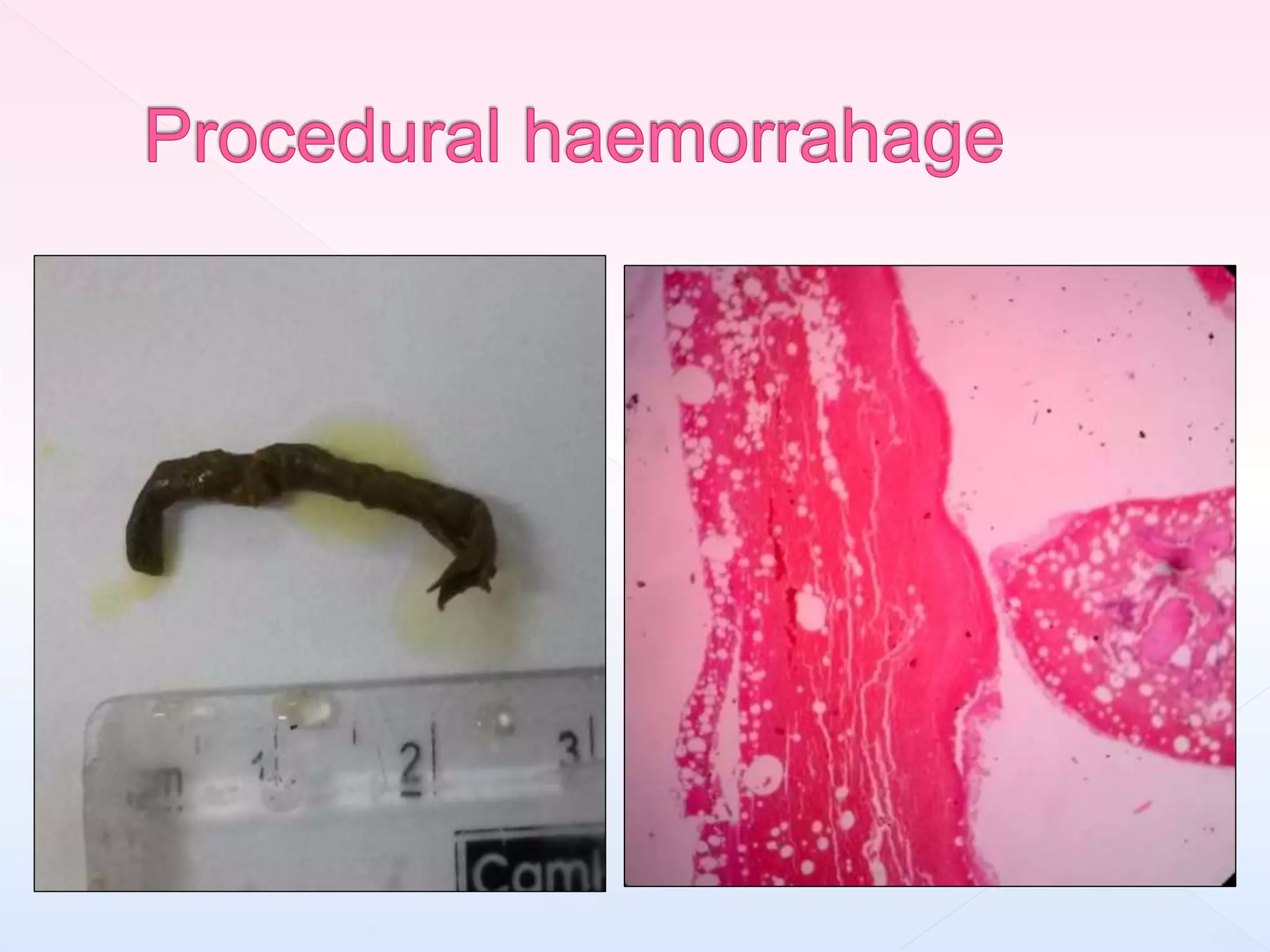
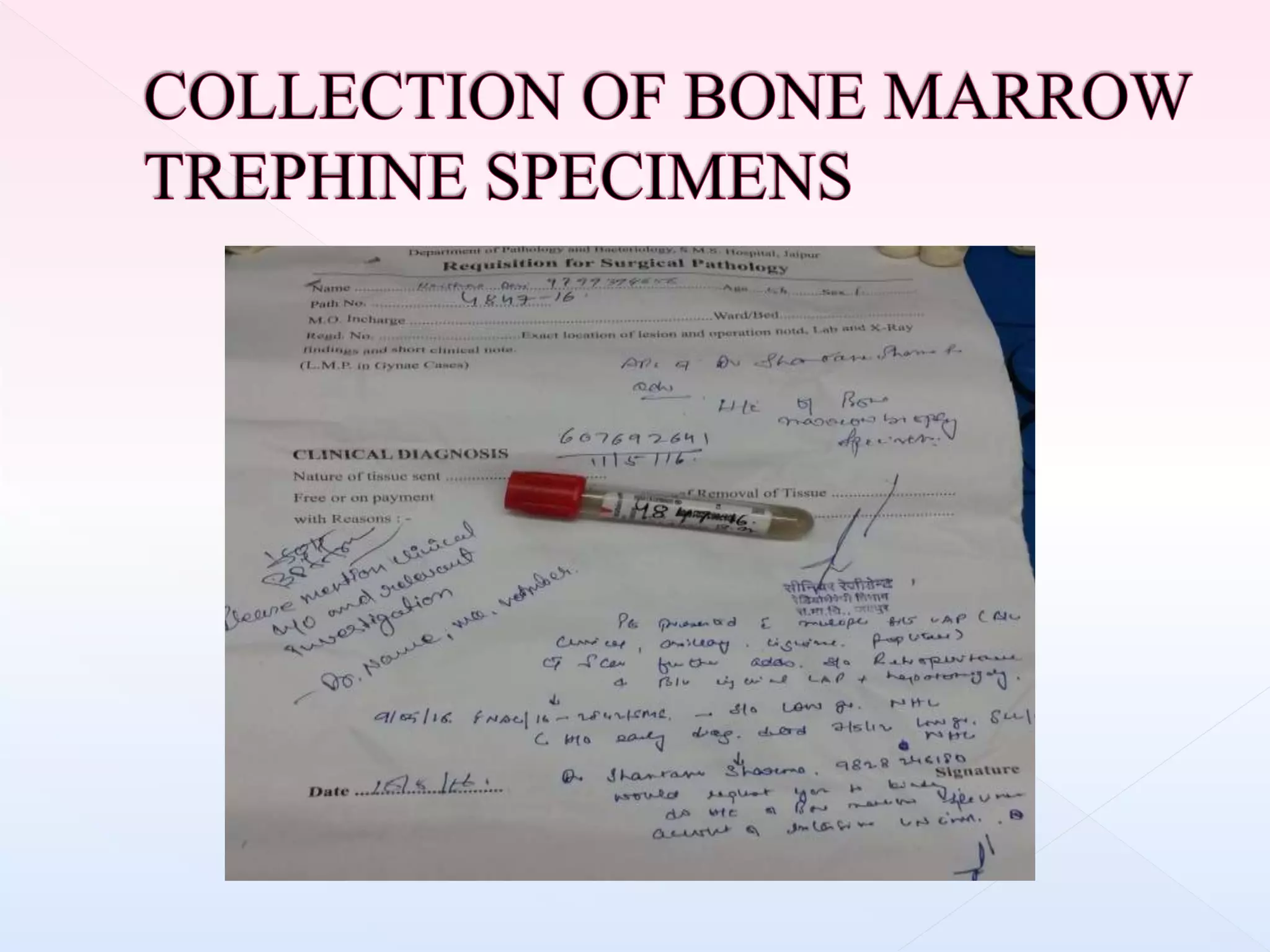
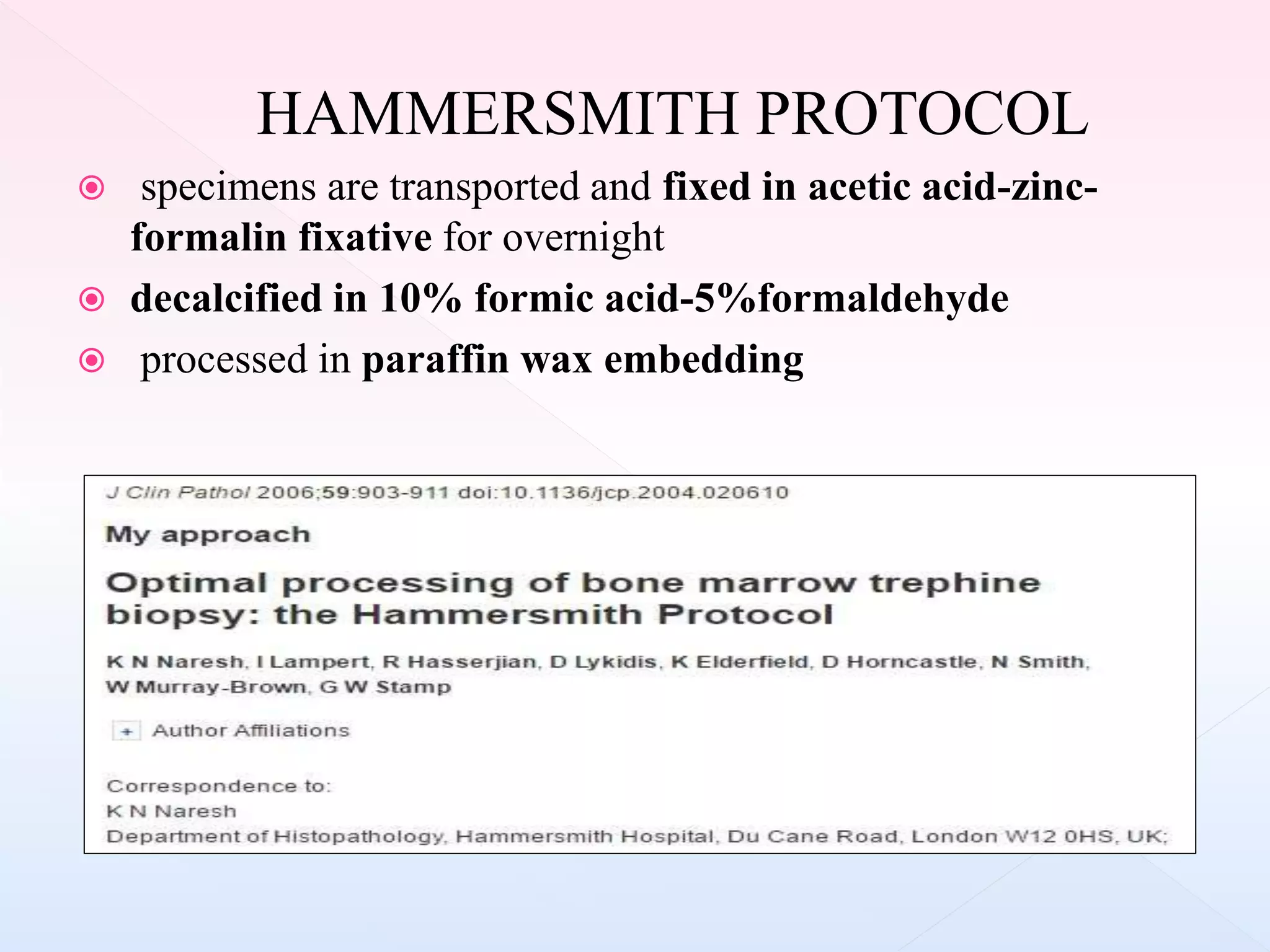
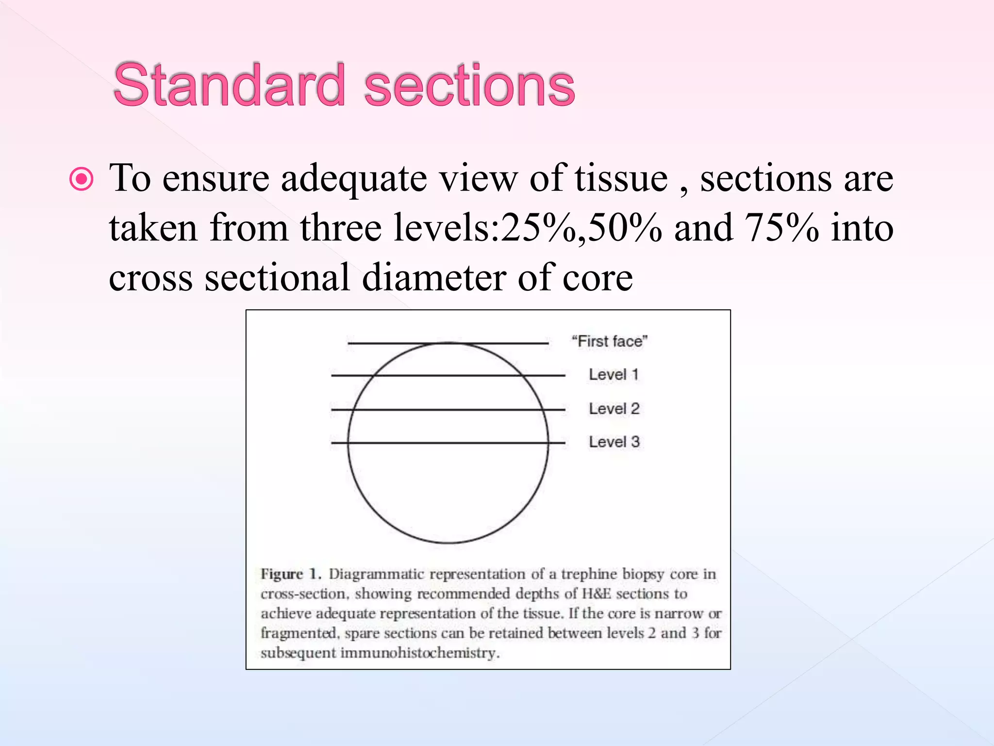
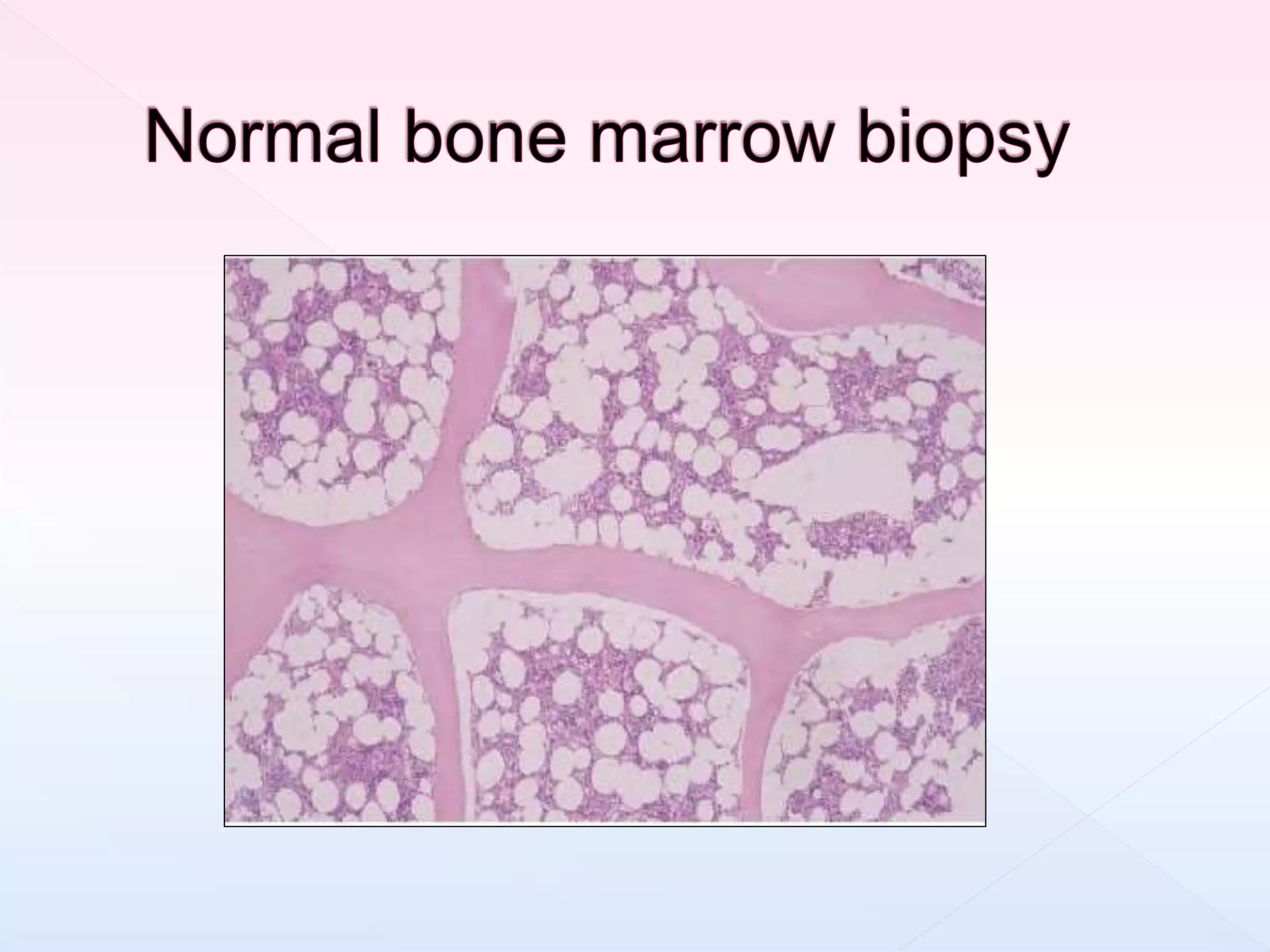
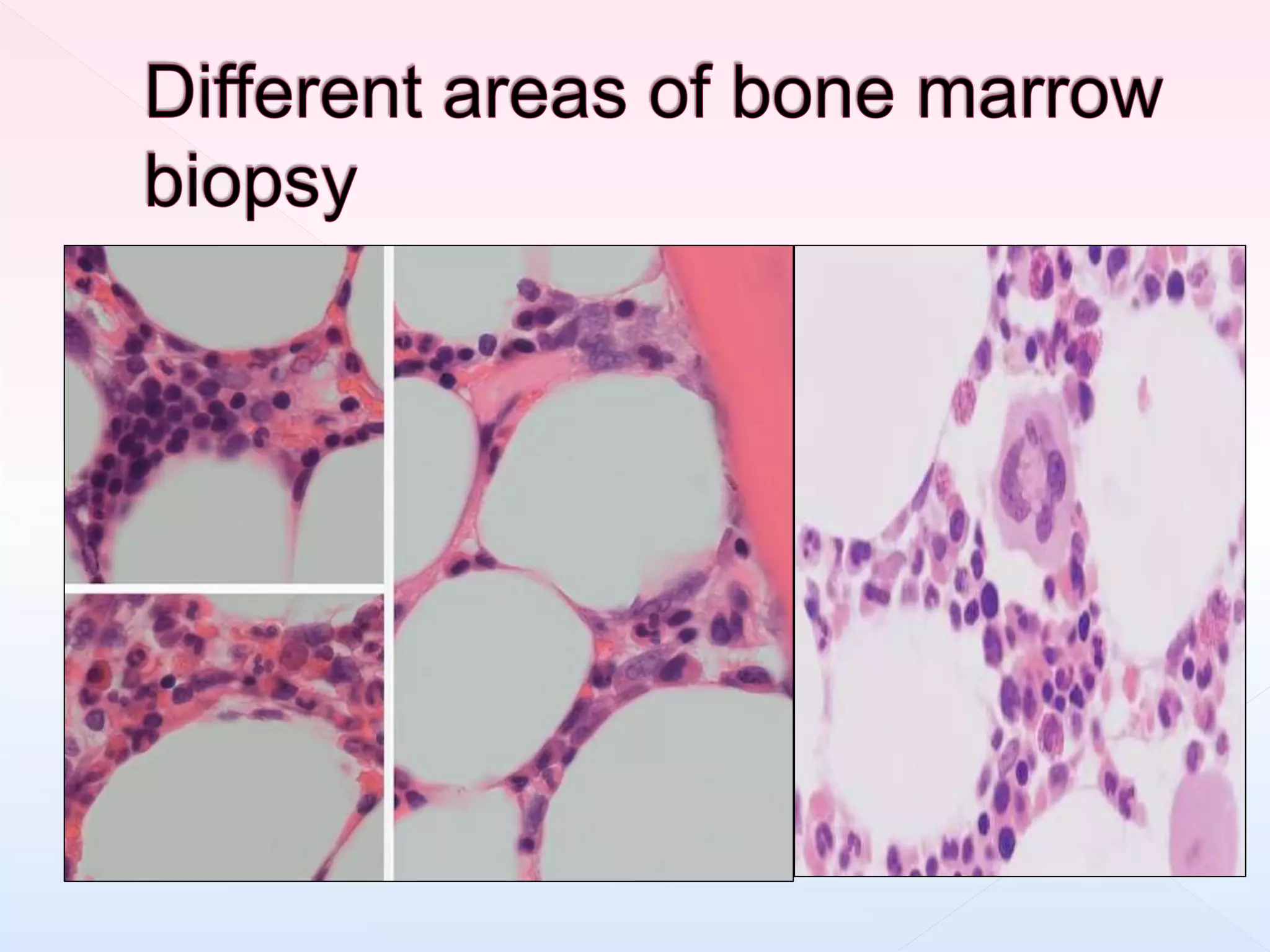
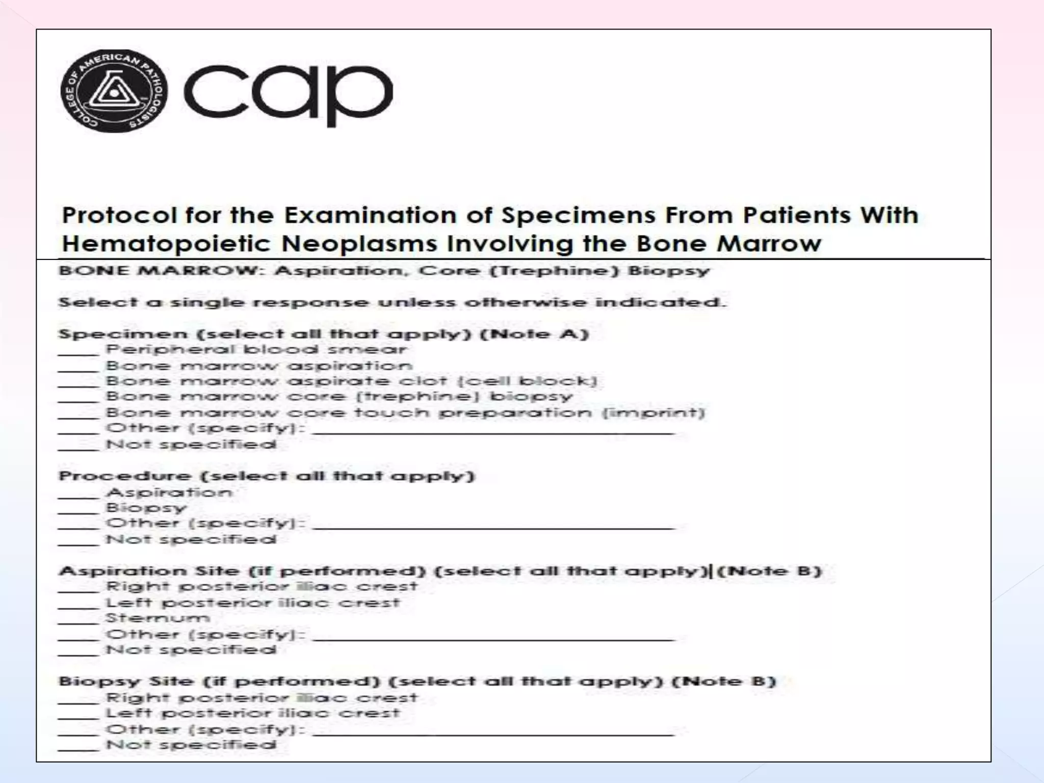
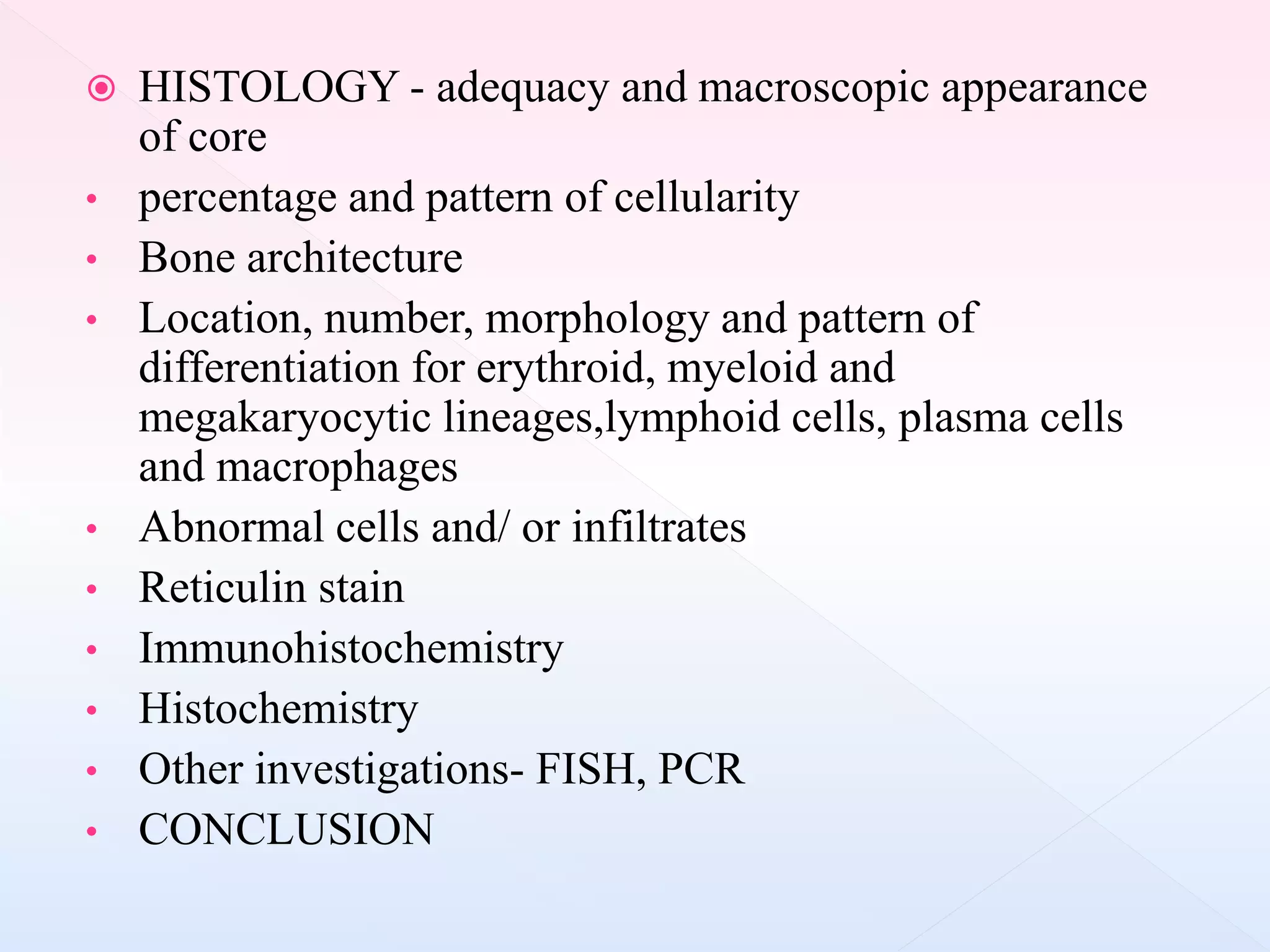
Consists of
1) BONE MARROW ASPIRATION: cell
morphology and enumeration of marrow cellular
elements
2) BONE MARROW BIOPSY: cellularity, fibrosis,
marrow architecture or vasculature, infections or
infiltrative disorders
1) Unexplained anaemia, abnormal red cell indices, cytopenia
or cytoses
2) Investigation of abnormal peripheral blood smear morphology
e.g. leukoerythroblastic picture suggestive of bone marrow
pathology
3) Diagnosis, staging and follow up of malignant haematological
disorders ( acute and chronic leukemias, myelodysplastic
syndromes, chronic myeloproliferative disorders, lymphomas,
plasma cell myeloma, amyloidosis, mastocytosis)
4) Investigation of suspected bone marrow metastasis
5) Unexplained focal bony lesions on radiography
6) Diagnosis, staging and follow up of small round cell tumours
of childhood
7) Pyrexia of unknown origin or specific infections e.g. miliary
tuberculosis, leishmaniasis, malaria
8) Evaluation of iron stores
9) Investigation of lipid/glycogen storage disorders
10) Exclusion of haematological disease in potential allogenic
stem cell transplant donors
 Sternal aspirate is absolutely contraindicated in
patients with diseases associated with marked
osteoporosis including multiple myeloma
 Coagulopathies : BMB is contraindicated
 Isolated thrombocytopenia is not a
contraindication . Adequate post procedure
supervision is must.
 POSTERIOR ILIAC CREST- most common
site
 ANTERIOR ILIAC CREST- in cases where
previous radiation, surgery or discomfort
 STERNUM AT SECOND INTERCOSTAL
SPACE –don’t allow biopsies
 Either the aspirate or biopsy can be performed first.
 If aspirate is performed first, trephine biopsy should be
performed through same incision, approximately 0.5-1 cm
away from the site of aspiration to avoid damaged or
haemmorhagic trephine biopsy.
Salah klima islam
A: HOLLOW NEEDLE WITH BEVELED TIP
B: OBTURATOR / STYLET
C: PROBE TO EXPRESS BIOPSY FROM NEEDLE (probe)
 https://www.youtube.com/watch?v=NkdsLHB
CreI
 Core of atleast 1.5cm – atleast three intact
intertrabecular marrow spaces- free of artefacts
 Biopsy specimen shrinks by 20% after
processing
 for focal pathologies (lymphoma infiltration /
metastatic deposits)- bilateral trephine biopsies
may be obtained ; totalling length of 20-30 mm
 Inadequate clinical, hematological, genetic and radiological
information
 Inadequate specimen- too small, too crushed, both, poorly
decalcified
 Inadequate sections- thickness, number of levels
 Inadequate stains- poor technical quality, limited stains
 COLLECTION OF BONE MARROW
TREPHINE SPECIMENS
 FIXATION
 DECALCIFICATION
 PROCESSING
 STAINING
 MICROSCOPY
 SUPPLEMENTARY INVESTIGATIONS
 Various fixatives –
1) neutral buffered formalin -6hrs
2) zinc formaldehyde
3) B5 (mercuric chloride, sodium acetate and formalin)
4) AZF ( acetic acid-zinc-formalin)- 6hrs
5) IBF (isotonic buffered formalin)
6) Bouin’s fixative ( picric acid, acetic acid and formaldehyde)-
4-12 hrs
7) Zenkers fixative(mercuric chloride, potassium
dichromate,sodium sulfate and glacial acetic acid) -4h
 Fixation times varies from 1h to maximum of >24h
depending upon the fixative used
 Trephine biopsy cores should be fixed in formol-saline for a
minimum of 18 h but fixation for longer periods does not
affect subsequent processing or morphology and is desirable
for large samples
 Mercurial fixatives such as B5 and Zenker’s fixative result
in improved morphology over formalin fixation
 The reactivity of antibodies used for immunohistochemical
staining may also be affected by the choice of fixative and
Zenker’s fixative can destroy chloroacetate esterase activity.
 EDTA(5%)
 Formic acid(5%)
 Acetic acid
 Picric acid
 Nitric acid (5%)
 Commercial decalcifying agents
Decalcification chelates storage iron,affects morphology
and cytological details, ability to perform histochemistry
and IHC and to retrieve material suitable for molecular
analysis
 Decalcification time varies from 15 min to 72 hours
 depending on decalcifying agent and size of
biopsy
 Decalcification with EDTA is slower(24-48 hrs) than
other agents but result in better preservation of nucleic
acids
 Warm incubation with agitation or microwave heating
can be employed to increase efficiency and reduce time
required for decalcification
 Weak acids like aqueous 10% formic acid are also slow
in action (2-3 days) and also cause tissue distortion
 Strong acids like fresh aqueous nitric acid 5%
are rapid decalcifiers
 Using inorganic acids, such as hydrochloric or
nitric acid, should be avoided as this affects
morphological preservation adversely and
impairs metachromatic staining of sections,
e.g. with Giemsa or toluidine blue.
 There is NO SINGLE BEST METHOD
 Fixation and decalcification schedules varies
widely, depending on local priorities for speed,
IHC and preservation of nucleic acids for
molecular studies
 Priniciple- to ensure proper fixation prior to
exposure of tissue to acidic or chelating
decalcifying agents
International Council for Standardization in
Hematology (ICSH) recommends neutral buffered
formalin with EDTA decalcification - adequate
pathology , preserves antigens for IHC and nucleic
acids for molecular studies
HAMMERSMITH PROTOCOL
 specimens are transported and fixed in acetic acid-zinc-
formalin fixative for overnight
 decalcified in 10% formic acid-5%formaldehyde
 processed in paraffin wax embedding
Wilkins et al. also recommends use of AZF for 6hrs.
 Addition of zinc in fixative stabilize nucleic acids, can protect
against hydrolysis in presence of weak acid
 AZF fixative results in superior antigen preservation, well
preserved RNA and DNA for FISH and molecular analysis
 Hence, AZF preparation can effectively speed decalcification
without detriment to morphology or antigen preservation
 AZF : preparation
zinc chloride – 12.5g
Glacial acetic acid- 7.5ml
Concentrated formaldehyde 150ml
Distilled water 1000ml
Hematologist is instructed to place the freshly
obtained BMT specimens directly into AZF fixative
and transport it
 FIXATION - in AZF overnight; next day biopsy is
washed with distilled water for 30 min
 DECALCIFICATION- 10% formic acid and 5%
formaldehyde – decalcify in 6hrs
 Processing and embedding in paraffin wax
 Sectioning and staining
PROCEDURE TIME(HRS) PROCEDURE
COMPLETION ON DAY
FIXATION 20-24 hrs 1
DECALCIFICATION 6 1
PROCESSING AND
EMBEDDING
12-16 2
SECTIONING FOR H&E 1 2
 After decalcification biopsy specimen is embedded
in paraffin wax and sections cut on microtome
 Recommended thickness 2-3 microns
 To allow high quality morphological assessment
including cytological evaluation using oil immersion
lens
 For DNA extraction two 15 micron thick sections are
cut
 To ensure adequate view of tissue , sections are
taken from three levels:25%,50% and 75% into
cross sectional diameter of core
 Spare sections on poly-l-lysine coated slides are
retained between levels 2 and 3 - suitable for use
IHC
PLASTIC EMBEDDING
 Some laboratories embed trephine cores in plastic resins
such as glycol methacrylate or methyl methacrylate
without decalcification .
 here section cutting requires glass knives or tungsten
carbide knives
 It gives good cytological details but is technically more
difficult and limits the range of immunohistochemical
studies and FISH
 It may be useful for the evaluation of metabolic bone
diseases and histochemical stains ablated by
decalcification process
 Hematoxylin and eosin
 Additional-
1) GEIMSA may be done; helpful in identifying
plasma cells, mast cells, lymphoid cells and eosinophils
and for distinguishing between myeloblasts and
proerythroblasts
2) RETICULIN STAIN – silver impregnation
method (gomori method and gorden and sweets
method)
3) Prussion blue (perls’) stain – iron
 Most enzyme histochemistry is unsuccessful because
of irreversible denaturation of the enzymes during
decalcification and processing.
 One exception is acid phosphatase activity which is
sometimes retained. When hairy cell leukaemia is
suspected, demonstration of tartrate-resistant acid
phosphatase (TRAP) activity may be useful
 HISTOLOGY - adequacy and macroscopic appearance
of core
• percentage and pattern of cellularity
• Bone architecture
• Location, number, morphology and pattern of
differentiation for erythroid, myeloid and
megakaryocytic lineages,lymphoid cells, plasma cells
and macrophages
• Abnormal cells and/ or infiltrates
• Reticulin stain
• Immunohistochemistry
• Histochemistry
• Other investigations- FISH, PCR
• CONCLUSION
processing of bone marrow trephine biopsy

processing of bone marrow trephine biopsy

  • 2.
    Consists of 1) BONEMARROW ASPIRATION: cell morphology and enumeration of marrow cellular elements 2) BONE MARROW BIOPSY: cellularity, fibrosis, marrow architecture or vasculature, infections or infiltrative disorders
  • 3.
    1) Unexplained anaemia,abnormal red cell indices, cytopenia or cytoses 2) Investigation of abnormal peripheral blood smear morphology e.g. leukoerythroblastic picture suggestive of bone marrow pathology 3) Diagnosis, staging and follow up of malignant haematological disorders ( acute and chronic leukemias, myelodysplastic syndromes, chronic myeloproliferative disorders, lymphomas, plasma cell myeloma, amyloidosis, mastocytosis) 4) Investigation of suspected bone marrow metastasis
  • 4.
    5) Unexplained focalbony lesions on radiography 6) Diagnosis, staging and follow up of small round cell tumours of childhood 7) Pyrexia of unknown origin or specific infections e.g. miliary tuberculosis, leishmaniasis, malaria 8) Evaluation of iron stores 9) Investigation of lipid/glycogen storage disorders 10) Exclusion of haematological disease in potential allogenic stem cell transplant donors
  • 5.
     Sternal aspirateis absolutely contraindicated in patients with diseases associated with marked osteoporosis including multiple myeloma  Coagulopathies : BMB is contraindicated  Isolated thrombocytopenia is not a contraindication . Adequate post procedure supervision is must.
  • 6.
     POSTERIOR ILIACCREST- most common site  ANTERIOR ILIAC CREST- in cases where previous radiation, surgery or discomfort  STERNUM AT SECOND INTERCOSTAL SPACE –don’t allow biopsies
  • 7.
     Either theaspirate or biopsy can be performed first.  If aspirate is performed first, trephine biopsy should be performed through same incision, approximately 0.5-1 cm away from the site of aspiration to avoid damaged or haemmorhagic trephine biopsy.
  • 9.
  • 10.
    A: HOLLOW NEEDLEWITH BEVELED TIP B: OBTURATOR / STYLET C: PROBE TO EXPRESS BIOPSY FROM NEEDLE (probe)
  • 13.
  • 14.
     Core ofatleast 1.5cm – atleast three intact intertrabecular marrow spaces- free of artefacts  Biopsy specimen shrinks by 20% after processing  for focal pathologies (lymphoma infiltration / metastatic deposits)- bilateral trephine biopsies may be obtained ; totalling length of 20-30 mm
  • 17.
     Inadequate clinical,hematological, genetic and radiological information  Inadequate specimen- too small, too crushed, both, poorly decalcified  Inadequate sections- thickness, number of levels  Inadequate stains- poor technical quality, limited stains
  • 18.
     COLLECTION OFBONE MARROW TREPHINE SPECIMENS  FIXATION  DECALCIFICATION  PROCESSING  STAINING  MICROSCOPY  SUPPLEMENTARY INVESTIGATIONS
  • 20.
     Various fixatives– 1) neutral buffered formalin -6hrs 2) zinc formaldehyde 3) B5 (mercuric chloride, sodium acetate and formalin) 4) AZF ( acetic acid-zinc-formalin)- 6hrs 5) IBF (isotonic buffered formalin) 6) Bouin’s fixative ( picric acid, acetic acid and formaldehyde)- 4-12 hrs 7) Zenkers fixative(mercuric chloride, potassium dichromate,sodium sulfate and glacial acetic acid) -4h
  • 21.
     Fixation timesvaries from 1h to maximum of >24h depending upon the fixative used  Trephine biopsy cores should be fixed in formol-saline for a minimum of 18 h but fixation for longer periods does not affect subsequent processing or morphology and is desirable for large samples  Mercurial fixatives such as B5 and Zenker’s fixative result in improved morphology over formalin fixation  The reactivity of antibodies used for immunohistochemical staining may also be affected by the choice of fixative and Zenker’s fixative can destroy chloroacetate esterase activity.
  • 22.
     EDTA(5%)  Formicacid(5%)  Acetic acid  Picric acid  Nitric acid (5%)  Commercial decalcifying agents Decalcification chelates storage iron,affects morphology and cytological details, ability to perform histochemistry and IHC and to retrieve material suitable for molecular analysis
  • 23.
     Decalcification timevaries from 15 min to 72 hours  depending on decalcifying agent and size of biopsy  Decalcification with EDTA is slower(24-48 hrs) than other agents but result in better preservation of nucleic acids  Warm incubation with agitation or microwave heating can be employed to increase efficiency and reduce time required for decalcification  Weak acids like aqueous 10% formic acid are also slow in action (2-3 days) and also cause tissue distortion
  • 24.
     Strong acidslike fresh aqueous nitric acid 5% are rapid decalcifiers  Using inorganic acids, such as hydrochloric or nitric acid, should be avoided as this affects morphological preservation adversely and impairs metachromatic staining of sections, e.g. with Giemsa or toluidine blue.
  • 25.
     There isNO SINGLE BEST METHOD  Fixation and decalcification schedules varies widely, depending on local priorities for speed, IHC and preservation of nucleic acids for molecular studies  Priniciple- to ensure proper fixation prior to exposure of tissue to acidic or chelating decalcifying agents
  • 26.
    International Council forStandardization in Hematology (ICSH) recommends neutral buffered formalin with EDTA decalcification - adequate pathology , preserves antigens for IHC and nucleic acids for molecular studies
  • 27.
    HAMMERSMITH PROTOCOL  specimensare transported and fixed in acetic acid-zinc- formalin fixative for overnight  decalcified in 10% formic acid-5%formaldehyde  processed in paraffin wax embedding
  • 28.
    Wilkins et al.also recommends use of AZF for 6hrs.  Addition of zinc in fixative stabilize nucleic acids, can protect against hydrolysis in presence of weak acid  AZF fixative results in superior antigen preservation, well preserved RNA and DNA for FISH and molecular analysis  Hence, AZF preparation can effectively speed decalcification without detriment to morphology or antigen preservation
  • 29.
     AZF :preparation zinc chloride – 12.5g Glacial acetic acid- 7.5ml Concentrated formaldehyde 150ml Distilled water 1000ml Hematologist is instructed to place the freshly obtained BMT specimens directly into AZF fixative and transport it
  • 30.
     FIXATION -in AZF overnight; next day biopsy is washed with distilled water for 30 min  DECALCIFICATION- 10% formic acid and 5% formaldehyde – decalcify in 6hrs  Processing and embedding in paraffin wax  Sectioning and staining
  • 31.
    PROCEDURE TIME(HRS) PROCEDURE COMPLETIONON DAY FIXATION 20-24 hrs 1 DECALCIFICATION 6 1 PROCESSING AND EMBEDDING 12-16 2 SECTIONING FOR H&E 1 2
  • 32.
     After decalcificationbiopsy specimen is embedded in paraffin wax and sections cut on microtome  Recommended thickness 2-3 microns  To allow high quality morphological assessment including cytological evaluation using oil immersion lens  For DNA extraction two 15 micron thick sections are cut
  • 33.
     To ensureadequate view of tissue , sections are taken from three levels:25%,50% and 75% into cross sectional diameter of core
  • 34.
     Spare sectionson poly-l-lysine coated slides are retained between levels 2 and 3 - suitable for use IHC
  • 35.
    PLASTIC EMBEDDING  Somelaboratories embed trephine cores in plastic resins such as glycol methacrylate or methyl methacrylate without decalcification .  here section cutting requires glass knives or tungsten carbide knives  It gives good cytological details but is technically more difficult and limits the range of immunohistochemical studies and FISH  It may be useful for the evaluation of metabolic bone diseases and histochemical stains ablated by decalcification process
  • 36.
     Hematoxylin andeosin  Additional- 1) GEIMSA may be done; helpful in identifying plasma cells, mast cells, lymphoid cells and eosinophils and for distinguishing between myeloblasts and proerythroblasts
  • 37.
    2) RETICULIN STAIN– silver impregnation method (gomori method and gorden and sweets method) 3) Prussion blue (perls’) stain – iron
  • 38.
     Most enzymehistochemistry is unsuccessful because of irreversible denaturation of the enzymes during decalcification and processing.  One exception is acid phosphatase activity which is sometimes retained. When hairy cell leukaemia is suspected, demonstration of tartrate-resistant acid phosphatase (TRAP) activity may be useful
  • 42.
     HISTOLOGY -adequacy and macroscopic appearance of core • percentage and pattern of cellularity • Bone architecture • Location, number, morphology and pattern of differentiation for erythroid, myeloid and megakaryocytic lineages,lymphoid cells, plasma cells and macrophages • Abnormal cells and/ or infiltrates • Reticulin stain • Immunohistochemistry • Histochemistry • Other investigations- FISH, PCR • CONCLUSION

Editor's Notes

  • #42 BM trephine biopsy section showing normal bone structure; there are anastomosing bony trabeculae