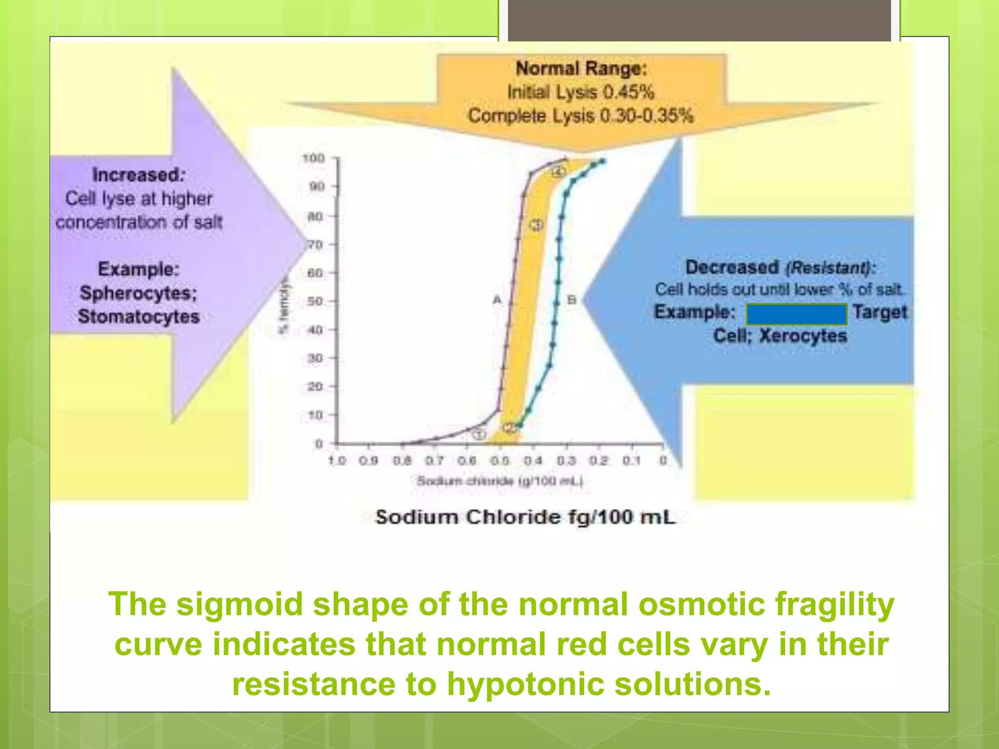
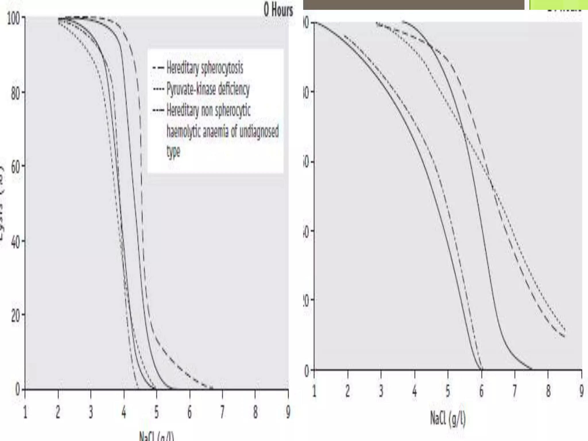
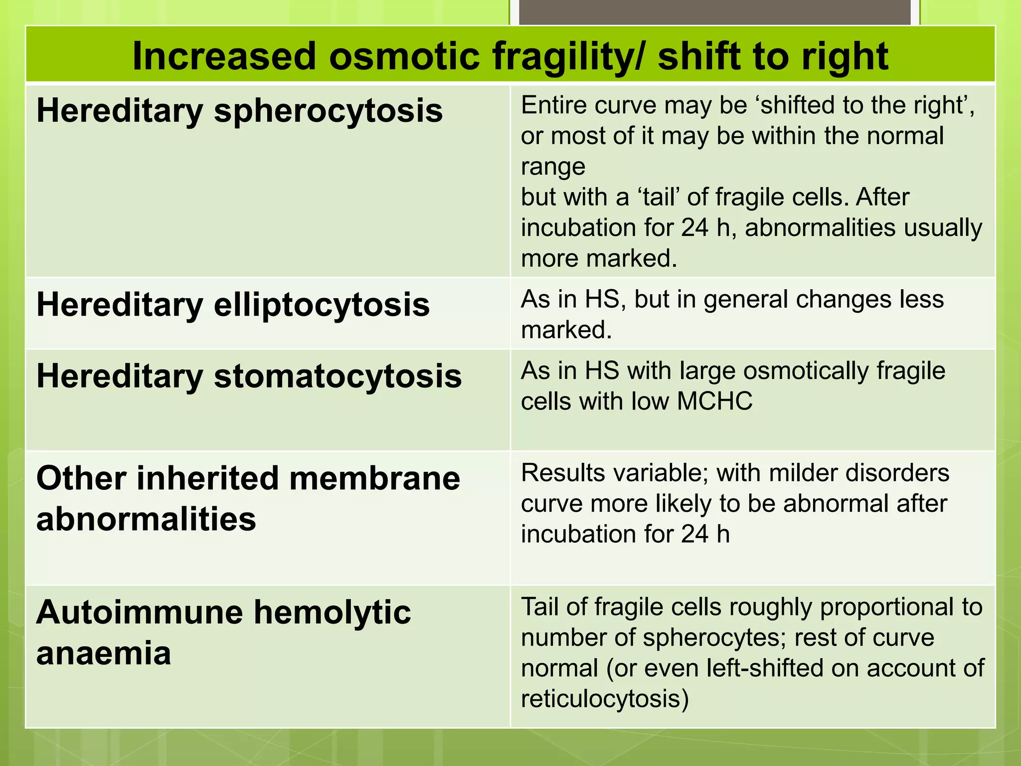
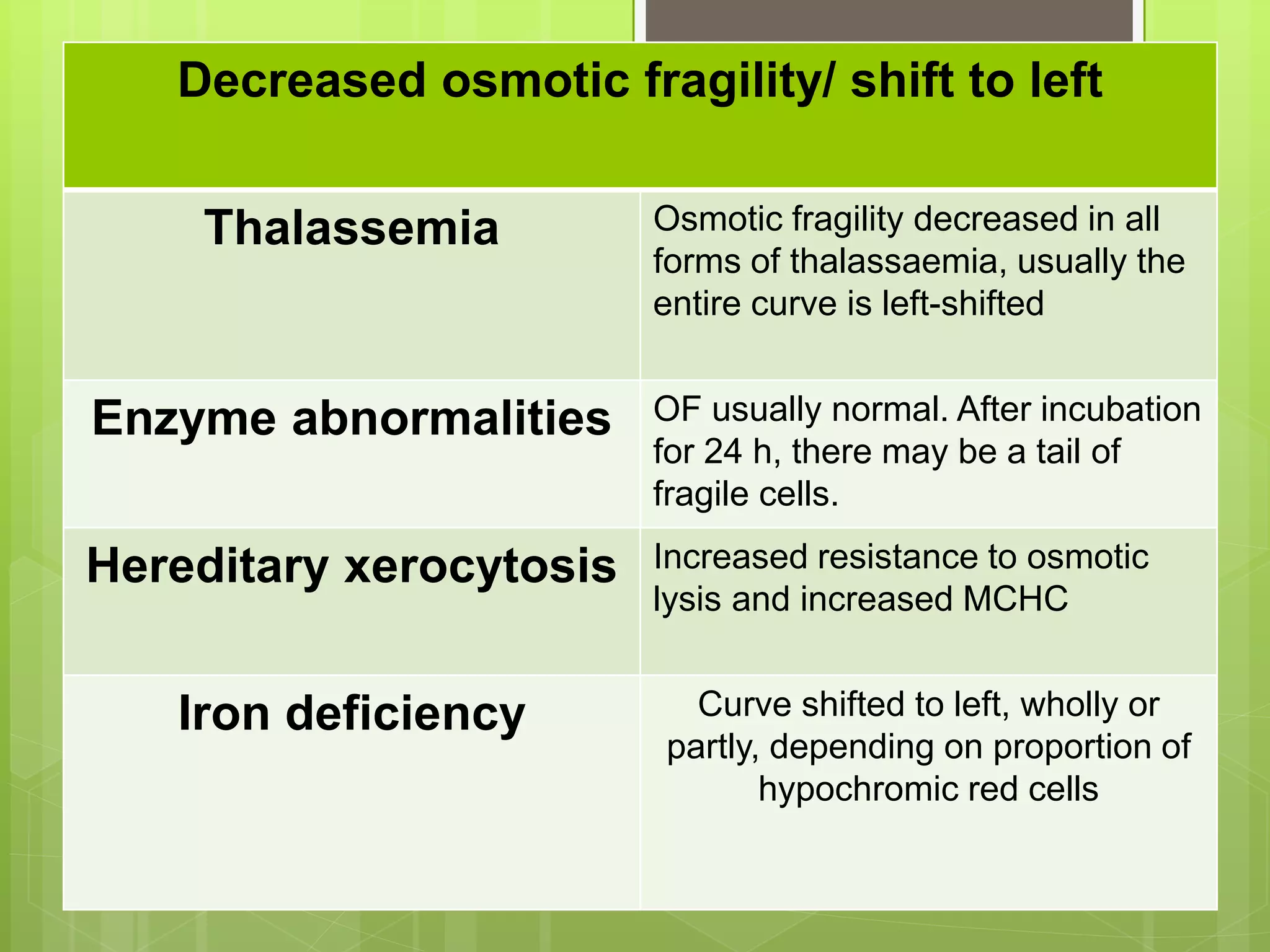
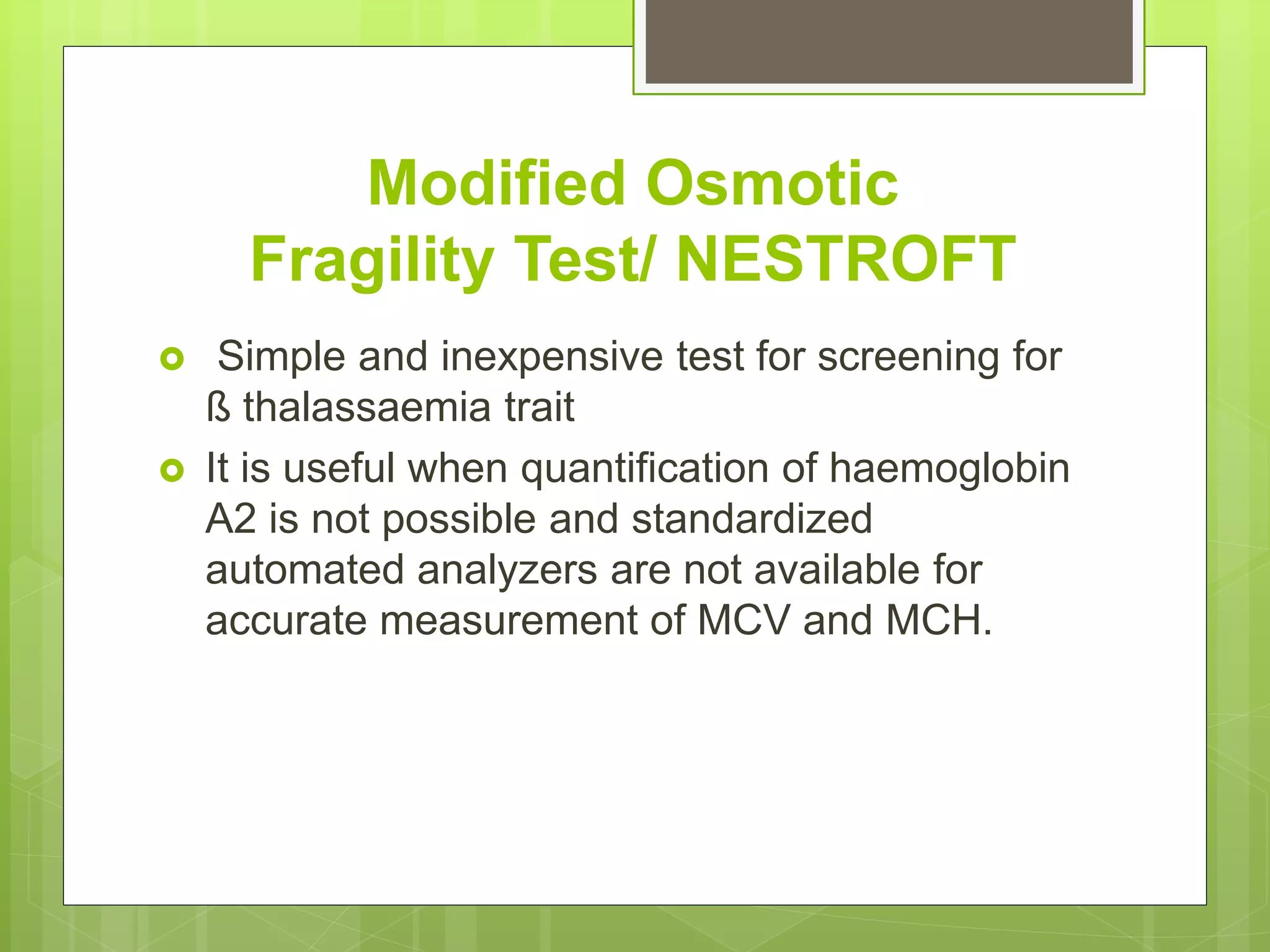
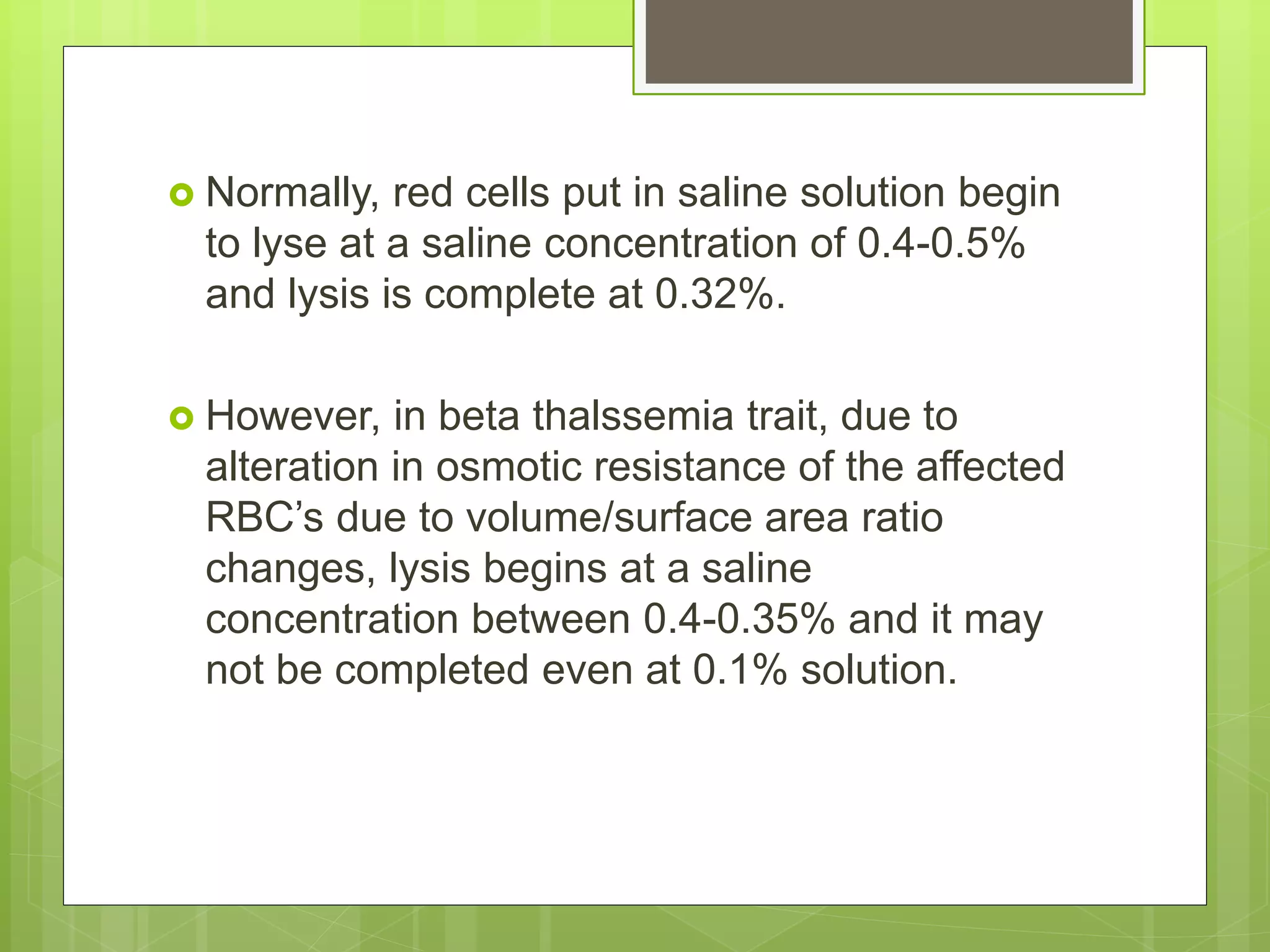
I have listed out the LE cells structure and Microscopical examinaton of LE CELLS, Difference between tart cells and le cells, clinical symptoms and diagnostic procedure.
An absolute eosinophil count is a blood test that measures the number of one type of white blood cells called eosinophils.
Eosinophils become active when you have certain allergic diseases, infections, and other medical conditions.
An absolute eosinophil count is a blood test that measures the number of one type of white blood cells called eosinophils.
Eosinophils become active when you have certain allergic diseases, infections, and other medical conditions.
Hemolytic anemias share the following features:
A shortened red cell life span below the normal 120 days
Elevated erythropoietin levels and a compensatory increase in erythropoiesis
Accumulation of hemoglobin degradation products that are created as part of the process of red cell hemolysis
Classification of Thrombocyte Disorders Using Platelet Aggregation AssaysChristine Joyce Javier
IF YOU ARE GOING TO DOWNLOAD THIS FILE, PLEASE NOTIFY me by sending a message via Facebook.
It's a pleasure to help you through my presentation. Thank you so much!
NOTE: This presentation is in PDF format. If you want to have a copy of this file in PPT format, kindly message me thru Facebook also.
Rasamanikya is a excellent preparation in the field of Rasashastra, it is used in various Kushtha Roga, Shwasa, Vicharchika, Bhagandara, Vatarakta, and Phiranga Roga. In this article Preparation& Comparative analytical profile for both Formulationon i.e Rasamanikya prepared by Kushmanda swarasa & Churnodhaka Shodita Haratala. The study aims to provide insights into the comparative efficacy and analytical aspects of these formulations for enhanced therapeutic outcomes.
Local Advanced Lung Cancer: Artificial Intelligence, Synergetics, Complex Sys...Oleg Kshivets
Overall life span (LS) was 1671.7±1721.6 days and cumulative 5YS reached 62.4%, 10 years – 50.4%, 20 years – 44.6%. 94 LCP lived more than 5 years without cancer (LS=2958.6±1723.6 days), 22 – more than 10 years (LS=5571±1841.8 days). 67 LCP died because of LC (LS=471.9±344 days). AT significantly improved 5YS (68% vs. 53.7%) (P=0.028 by log-rank test). Cox modeling displayed that 5YS of LCP significantly depended on: N0-N12, T3-4, blood cell circuit, cell ratio factors (ratio between cancer cells-CC and blood cells subpopulations), LC cell dynamics, recalcification time, heparin tolerance, prothrombin index, protein, AT, procedure type (P=0.000-0.031). Neural networks, genetic algorithm selection and bootstrap simulation revealed relationships between 5YS and N0-12 (rank=1), thrombocytes/CC (rank=2), segmented neutrophils/CC (3), eosinophils/CC (4), erythrocytes/CC (5), healthy cells/CC (6), lymphocytes/CC (7), stick neutrophils/CC (8), leucocytes/CC (9), monocytes/CC (10). Correct prediction of 5YS was 100% by neural networks computing (error=0.000; area under ROC curve=1.0).
Basavarajeeyam is an important text for ayurvedic physician belonging to andhra pradehs. It is a popular compendium in various parts of our country as well as in andhra pradesh. The content of the text was presented in sanskrit and telugu language (Bilingual). One of the most famous book in ayurvedic pharmaceutics and therapeutics. This book contains 25 chapters called as prakaranas. Many rasaoushadis were explained, pioneer of dhatu druti, nadi pareeksha, mutra pareeksha etc. Belongs to the period of 15-16 century. New diseases like upadamsha, phiranga rogas are explained.
Integrating Ayurveda into Parkinson’s Management: A Holistic ApproachAyurveda ForAll
Explore the benefits of combining Ayurveda with conventional Parkinson's treatments. Learn how a holistic approach can manage symptoms, enhance well-being, and balance body energies. Discover the steps to safely integrate Ayurvedic practices into your Parkinson’s care plan, including expert guidance on diet, herbal remedies, and lifestyle modifications.
Antimicrobial stewardship to prevent antimicrobial resistanceGovindRankawat1
India is among the nations with the highest burden of bacterial infections.
India is one of the largest consumers of antibiotics worldwide.
India carries one of the largest burdens of drug‑resistant pathogens worldwide.
Highest burden of multidrug‑resistant tuberculosis,
Alarmingly high resistance among Gram‑negative and Gram‑positive bacteria even to newer antimicrobials such as carbapenems.
NDM‑1 ( New Delhi Metallo Beta lactamase 1, an enzyme which inactivates majority of Beta lactam antibiotics including carbapenems) was reported in 2008
These simplified slides by Dr. Sidra Arshad present an overview of the non-respiratory functions of the respiratory tract.
Learning objectives:
1. Enlist the non-respiratory functions of the respiratory tract
2. Briefly explain how these functions are carried out
3. Discuss the significance of dead space
4. Differentiate between minute ventilation and alveolar ventilation
5. Describe the cough and sneeze reflexes
Study Resources:
1. Chapter 39, Guyton and Hall Textbook of Medical Physiology, 14th edition
2. Chapter 34, Ganong’s Review of Medical Physiology, 26th edition
3. Chapter 17, Human Physiology by Lauralee Sherwood, 9th edition
4. Non-respiratory functions of the lungs https://academic.oup.com/bjaed/article/13/3/98/278874
Knee anatomy and clinical tests 2024.pdfvimalpl1234
This includes all relevant anatomy and clinical tests compiled from standard textbooks, Campbell,netter etc..It is comprehensive and best suited for orthopaedicians and orthopaedic residents.
Here is the updated list of Top Best Ayurvedic medicine for Gas and Indigestion and those are Gas-O-Go Syp for Dyspepsia | Lavizyme Syrup for Acidity | Yumzyme Hepatoprotective Capsules etc
2. Reagents
Prepare a stock solution of buffered sodium
chloride, osmotically equivalent to 100 g/l
In preparing hypotonic solutions for use, it is
convenient to make first a 10 g/l solution from the
100 g/l NaCl stock solution by dilution with water.
Dilutions equivalent to 9.0, 7.5, 6.5, 6.0, 5.5, 5.0,
4.0, 3.5, 3.0, 2.0 and 1.0 g/l are then made.
3. Preparation of serial dilutions
Prepare a stock solution dissolving 10g NaCl in
1litre of distilled water;
i.e. 10g in 1000mL or 10x1000 mg in 1000 mL
1mL of such a solution will contain 10mg NaCl
From this stock solution prepare further dilutions.
5. Procedure
• Deliver 5.0 ml of each of the 11 saline
solutions into 12 test tubes.
• Add 5.0 ml of water to the 12th tube.
• Add to each tube 50 µl of well-mixed
blood and mix immediately by inverting
the tubes several times, avoiding foam.
• Leave the suspensions for 30 min at room
temperature.
• Mix again and then centrifuge for 5 min at
1200 rpm.
6. The sigmoid shape of the normal osmotic fragility
curve indicates that normal red cells vary in their
resistance to hypotonic solutions.
7. Osmotic Fragility after incubating the
Blood at 370C for 24 Hours
Defibrinated blood should be used, care being
taken to ensure that sterility is maintained.
Incubate 1 ml or 2 ml volumes of blood in
sterile 5 ml bottles.
After 24 h, if no infection is evident, after
thoroughly mixing the sedimented red cells in
the overlying serum, estimate the fragility as
previously described.
8. Because the fragility may be markedly
increased set up additional hypotonic
solutions containing 7.0 g/l and 8.0 g/l NaCl.
In addition, use a solution equivalent to 12.0
g/l NaCl because sometimes, as in HS, lysis
may take place in 9.0 g/l NaCl.
In this case, use the supernatant of the tube
containing 12.0 g/l NaCl as the blank in the
colorimetric estimation.
11. The osmotic fragility of freshly taken red cells
reflects their ability to take up a certain
amount of water before lysing.
This is determined by their volume-to-surface
area ratio.
The ability of the normal red cell to withstand
hypotonicity results from its biconcave shape,
which allows the cell to increase its volume by
about 70% before the surface membrane is
stretched; once this limit is reached lysis
occurs.
13. Spherocytes have an increased volume-to-
surface area ratio.
Their ability to take in water before stretching
the surface membrane is thus more limited
than normal and they are therefore
particularly susceptible to osmotic lysis.
The increase in osmotic fragility is a property
of the spheroidal shape of the cell and is
independent of the cause of the
spherocytosis.
14. Decreased osmotic fragility indicates the
presence of unusually flattened red cells
(leptocytes) in which the volume-to-surface
area ratio is decreased.
Such a change occurs in iron deficiency
anaemia and thalassaemia in which the red
cells with a low mean cell haemoglobin (MCH)
and mean cell volume (MCV) are unusually
resistant to osmotic lysis
15. Reticulocytes and red cells from patients who
have been splenectomized also tend to have
a greater amount of membrane compared
with normal cells and are osmotically
resistant.
In liver disease, target cells may be produced
by passive accumulation of lipid and these
cells, too, are resistant to osmotic lysis.
16. The osmotic fragility of red cells that have an
abnormal membrane or have enzyme defects,
increases abnormally after incubation.
In thalassaemia major and minor, osmotic
fragility is frequently markedly reduced after
incubation, again owing to a marked loss of
potassium.
A similar, although usually less marked,
change is seen in iron deficiency anaemia.
17. The increased osmotic fragility of normal red
cells, which occurs after incubation is mainly
caused by swelling of the cells associated
with an accumulation of sodium that exceeds
loss of potassium.
During incubation for 24 hr., the metabolism of
the red cell becomes stressed and the
pumping mechanisms tend to fail, one factor
being a relative lack of glucose in the
medium.
18. Increased osmotic fragility/ shift to right
Hereditary spherocytosis Entire curve may be ‘shifted to the right’,
or most of it may be within the normal
range
but with a ‘tail’ of fragile cells. After
incubation for 24 h, abnormalities usually
more marked.
Hereditary elliptocytosis As in HS, but in general changes less
marked.
Hereditary stomatocytosis As in HS with large osmotically fragile
cells with low MCHC
Other inherited membrane
abnormalities
Results variable; with milder disorders
curve more likely to be abnormal after
incubation for 24 h
Autoimmune hemolytic
anaemia
Tail of fragile cells roughly proportional to
number of spherocytes; rest of curve
normal (or even left-shifted on account of
reticulocytosis)
19. Decreased osmotic fragility/ shift to left
Thalassemia Osmotic fragility decreased in all
forms of thalassaemia, usually the
entire curve is left-shifted
Enzyme abnormalities OF usually normal. After incubation
for 24 h, there may be a tail of
fragile cells.
Hereditary xerocytosis Increased resistance to osmotic
lysis and increased MCHC
Iron deficiency Curve shifted to left, wholly or
partly, depending on proportion of
hypochromic red cells
20. Modified Osmotic
Fragility Test/ NESTROFT
Simple and inexpensive test for screening for
ß thalassaemia trait
It is useful when quantification of haemoglobin
A2 is not possible and standardized
automated analyzers are not available for
accurate measurement of MCV and MCH.
22. Normally, red cells put in saline solution begin
to lyse at a saline concentration of 0.4-0.5%
and lysis is complete at 0.32%.
However, in beta thalssemia trait, due to
alteration in osmotic resistance of the affected
RBC’s due to volume/surface area ratio
changes, lysis begins at a saline
concentration between 0.4-0.35% and it may
not be completed even at 0.1% solution.
23. Reagents
NESTROFT is done at a saline concentration
of 0.36%.
0.36% buffered saline (BS) prepared by
diluting 36ml of 1% buffered saline with 64ml
of distilled water (DW) to make 100ml
24. Procedure
Two test tubes labelled as BS (2ml) and DW
(2ml) are taken and a drop of blood is added
to each of the tubes, which are then left
undisturbed for half an hour at room
temperature.
25. The line is clearly visible through DW tube,
If it is the same in BS tube; it is considered negative,
otherwise test result is interpreted as positive
26. The tubes are then left undisturbed for 3
hours.
At the end of 3 hours, the DW tube is usually
seen to be homogeneously pink with no
sediments.
In the BS tube the negative test shows similar
findings as DW tube where as in a positive
case, a clear supernatant and a sediment
at bottom is observed.
27. Because the false-positive rate is around 10%,
confirmation of a positive result requires referral of a
sample to a laboratory able to quantitate haemoglobin
A2.
The test can also be used to screen for alpha
thalassaemia trait, with positive samples being referred
to a reference centre for DNA analysis.
28. About 50% of samples containing
haemoglobin E also give a positive result.
This is an advantage rather than a
disadvantage because detection of
haemoglobin E is important in predicting the
possibility of thalassaemia major or
intermedia in compound heterozygotes with ß
thalassaemia.
29. Summary
The osmotic fragility test gives an indication of
the surface area/volume ratio of erythrocytes.
Its greatest usefulness is in the diagnosis of
hereditary spherocytosis.
The test may also be used in screening for
thalassaemia.
30. References
Dacie and Lewis practical haematology.
Piplani M. NESTROFT - A Valuable, Cost
Effective Screening Test for Beta Thalassemia
Trait in North Indian Punjabi Population.
Journal of clinical and diagnostic research.
2013.