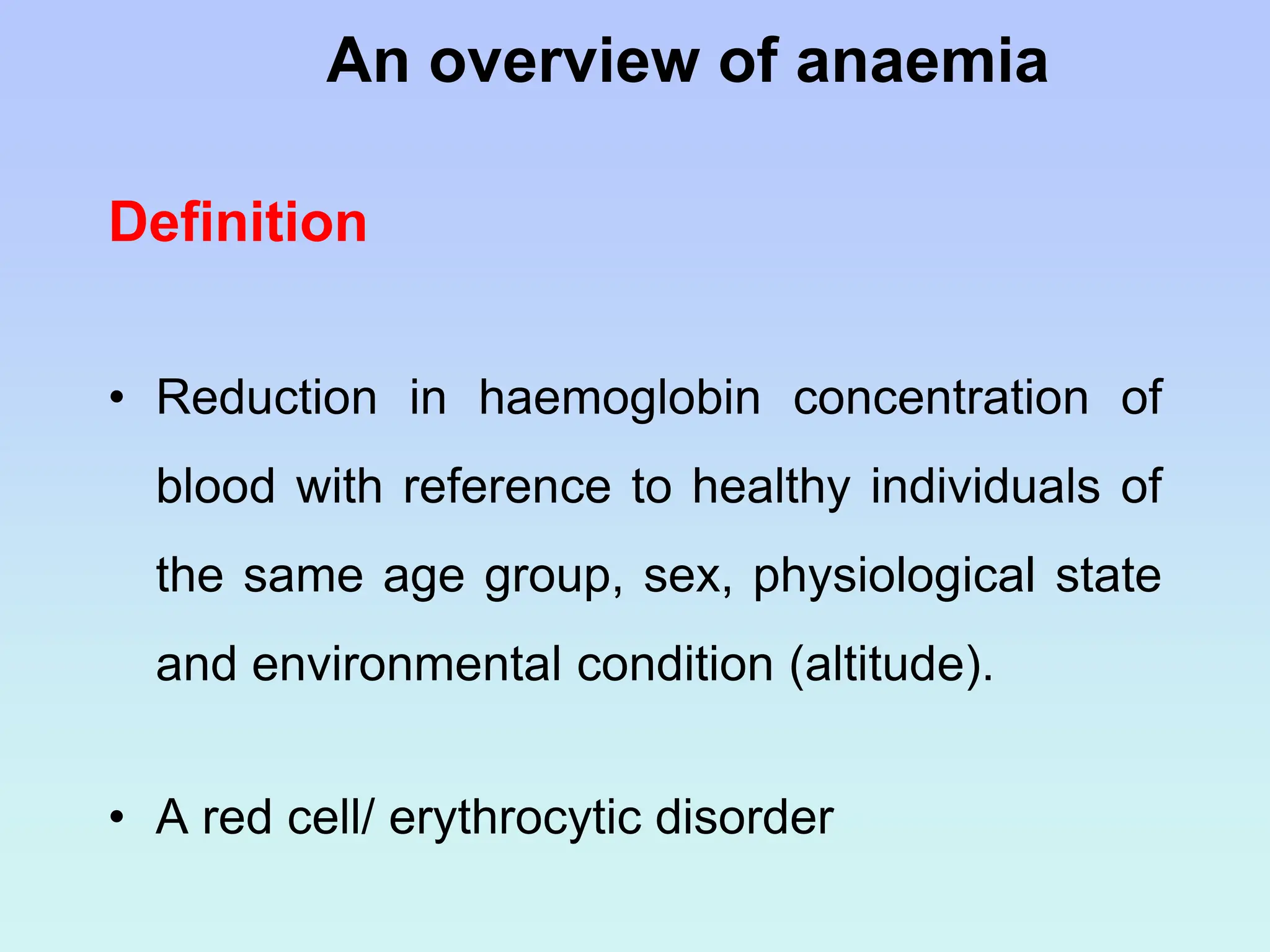
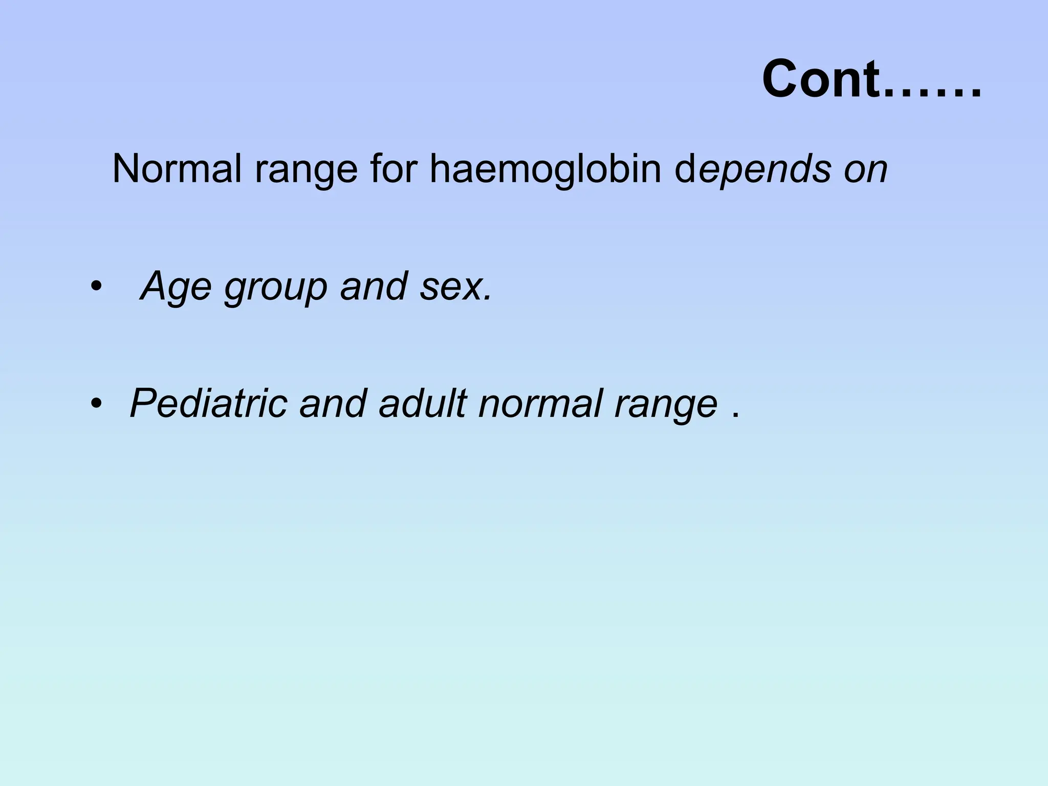
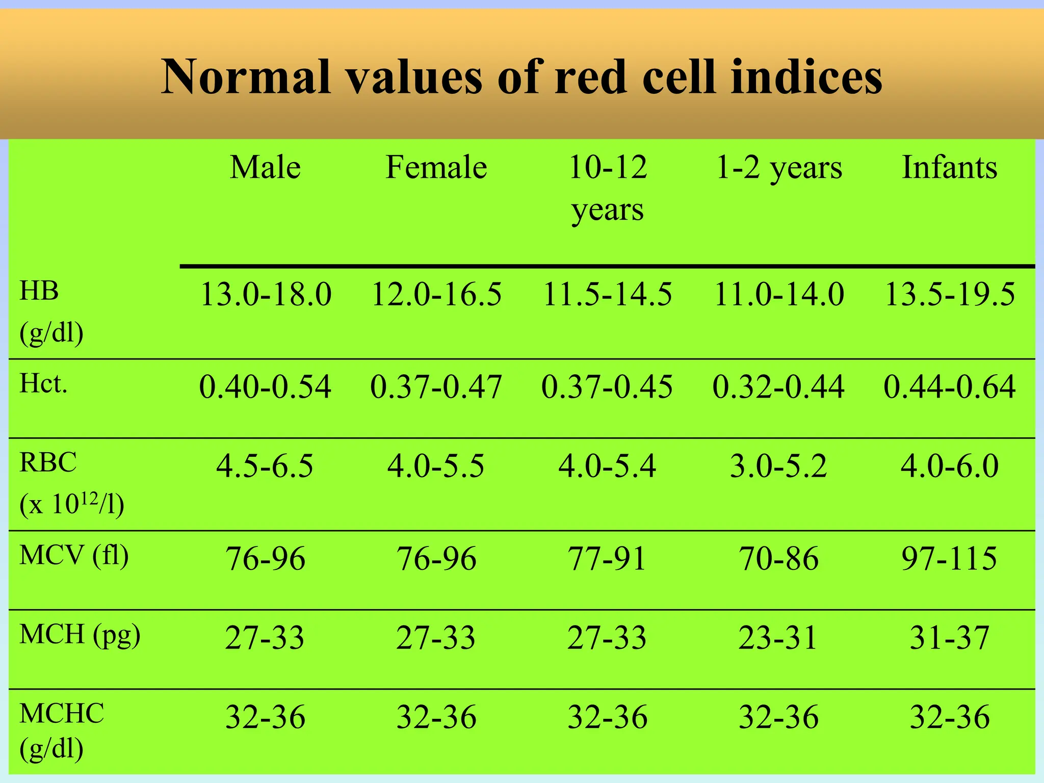
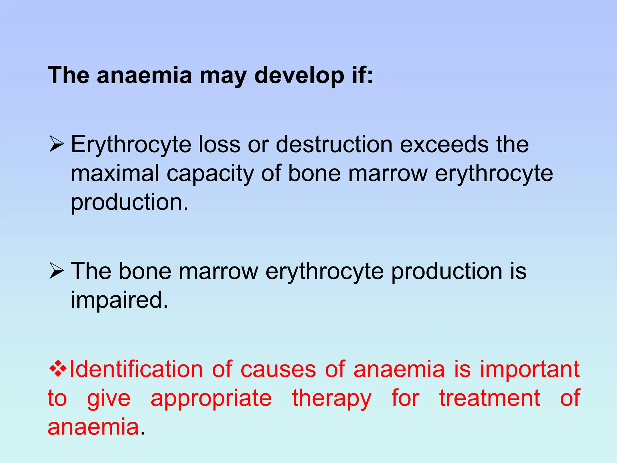
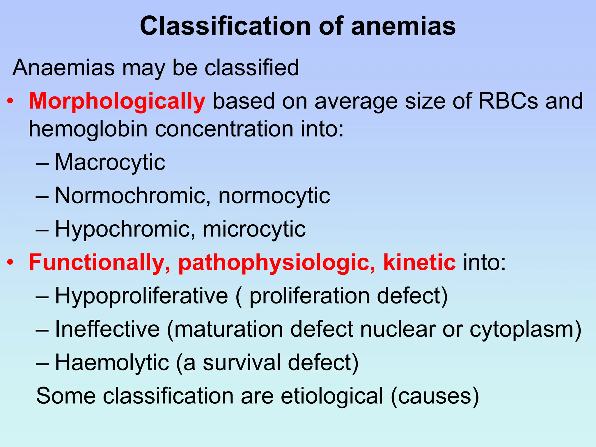
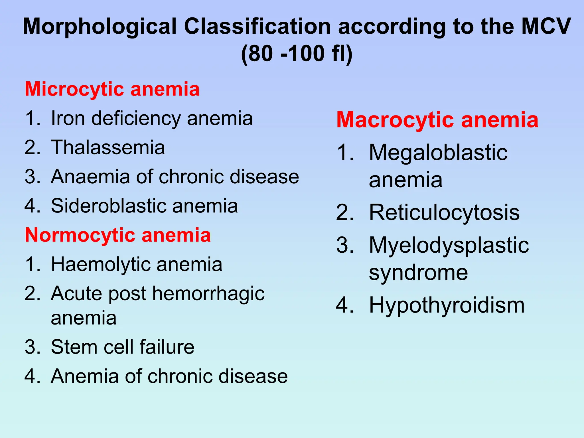
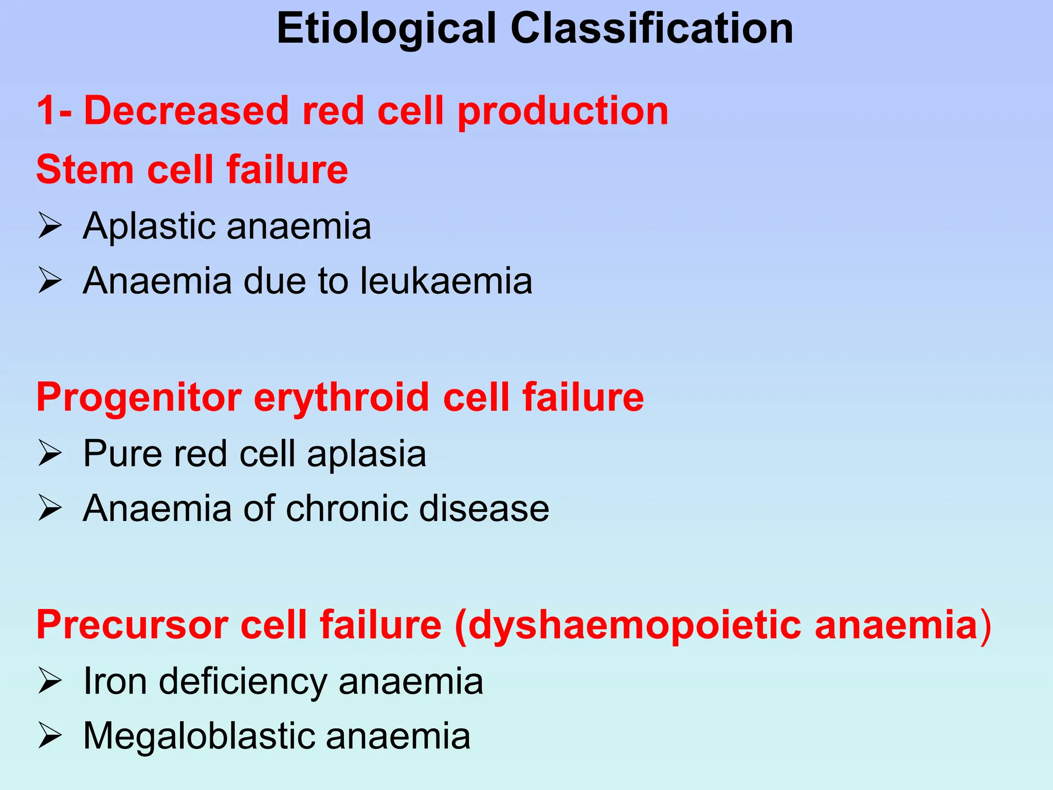
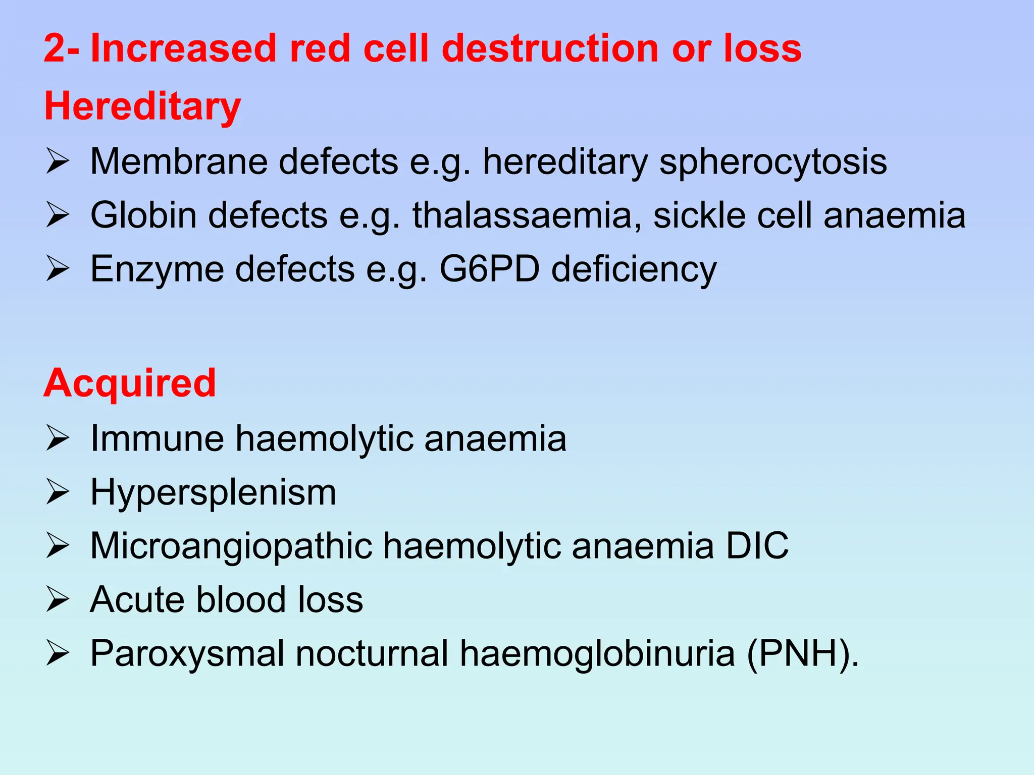
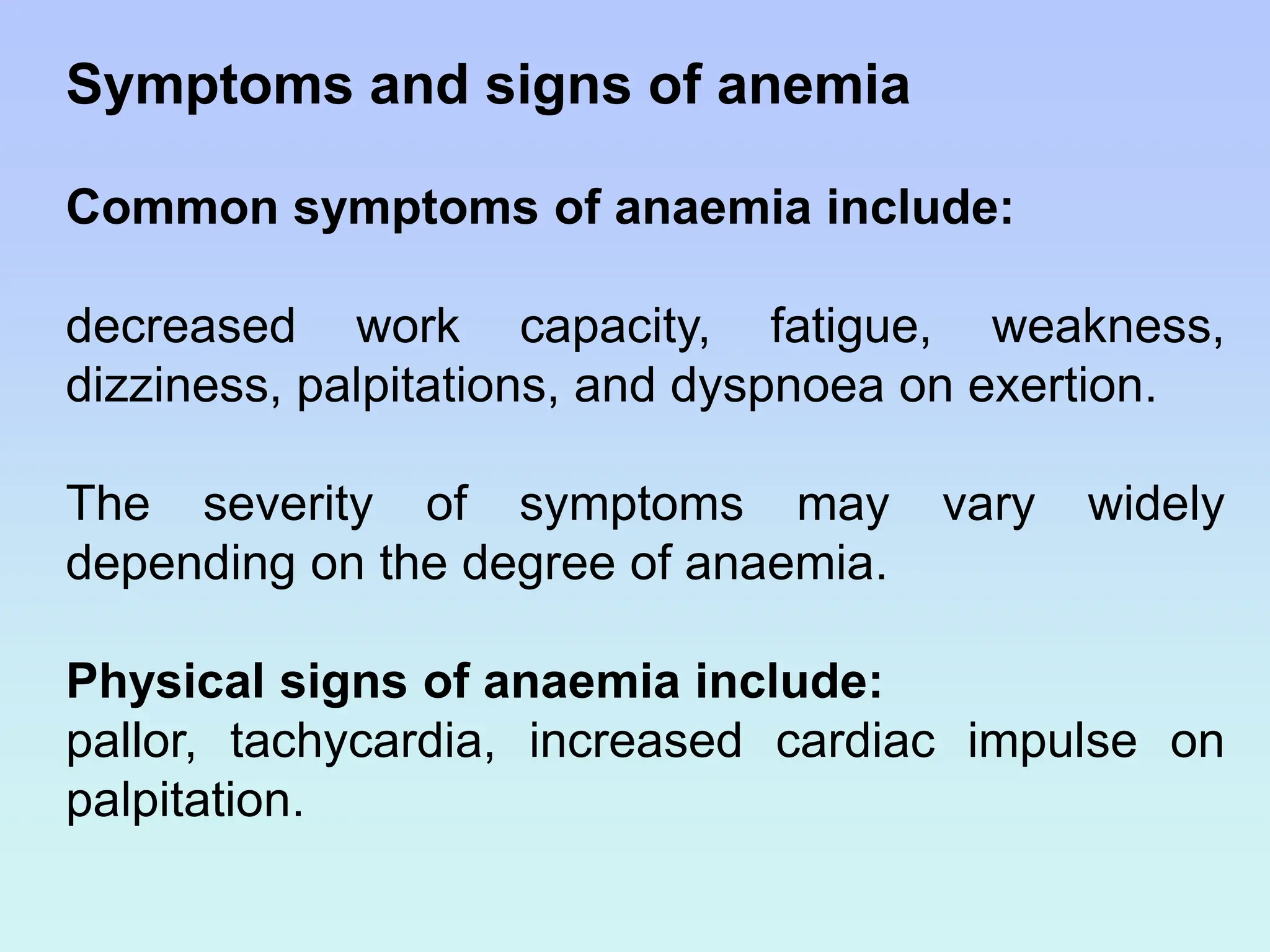
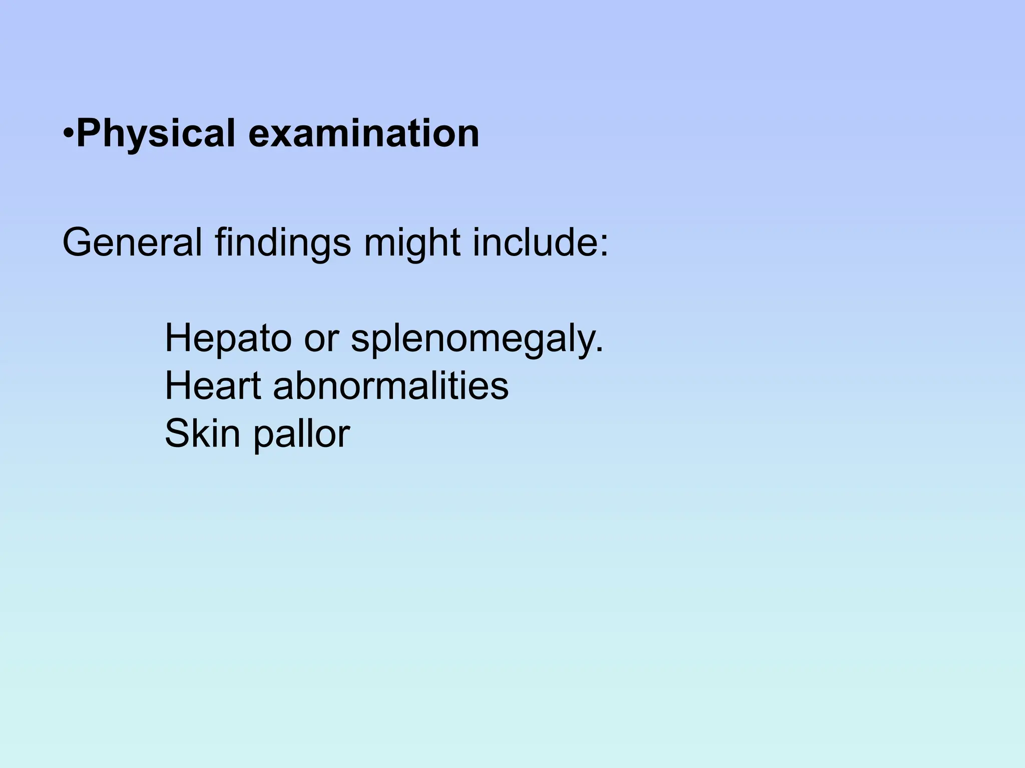
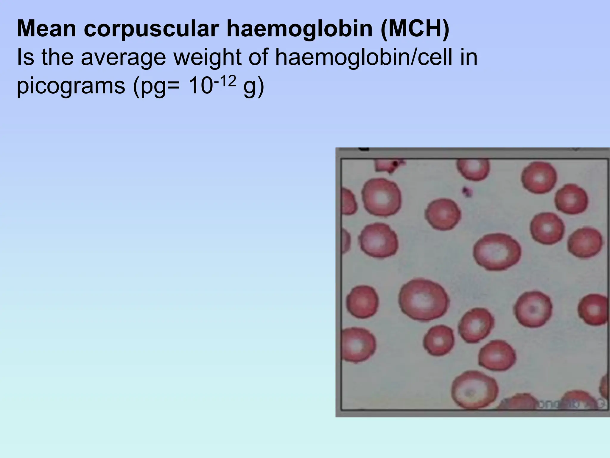
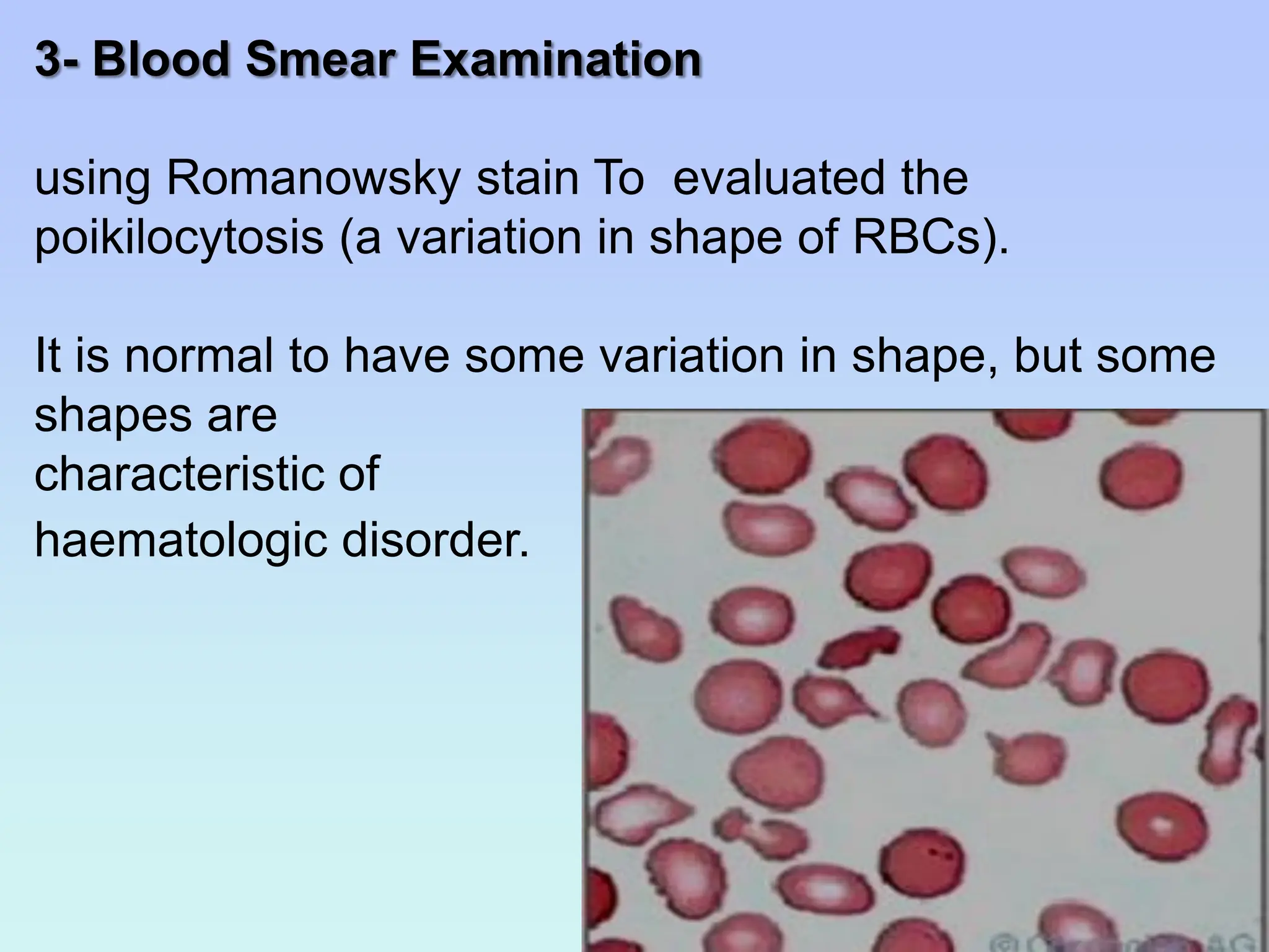
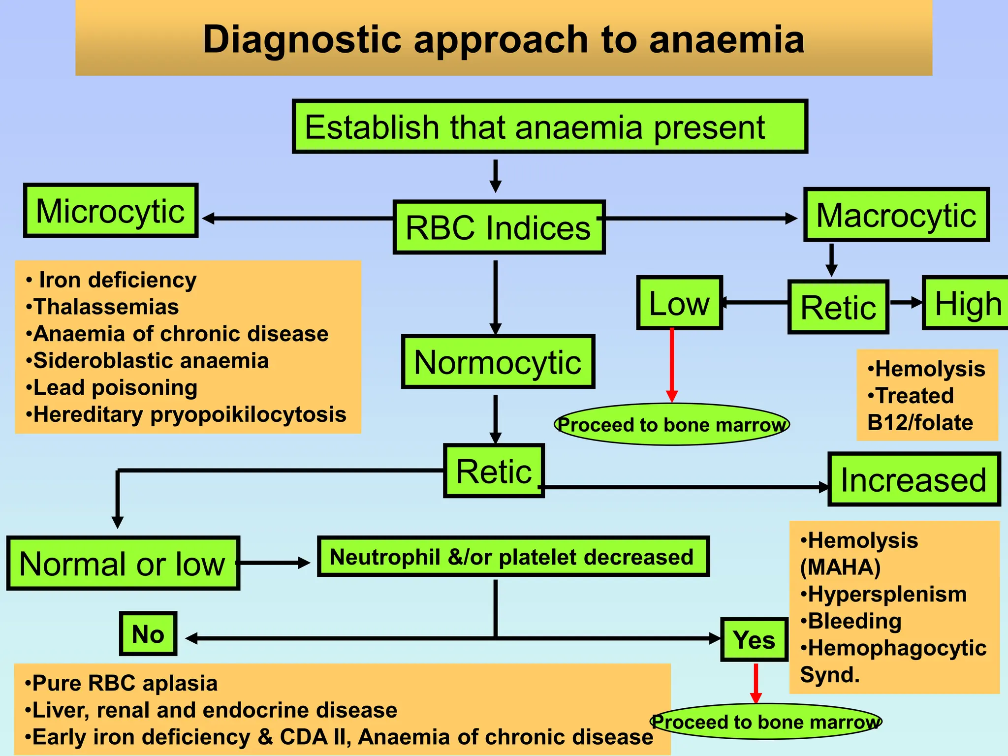
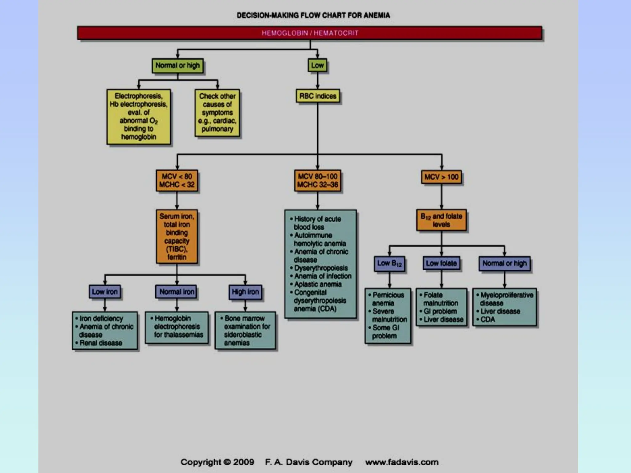
In The Name of ALLAH
The Most Gracious , The
Most Merciful.
ANAEMIAS
Introduction & classification
Mohammed SeedAhmed Abdoelateef
Lecturer at PAU
Haematology & Blood Transifusion
Outline
Outline
1. Definition
2. Classification
3. Diagnosis
4. Symptoms
5. Physical examination
6. Laboratory investigation
Definition
• Reduction in haemoglobin concentration of
blood with reference to healthy individuals of
the same age group, sex, physiological state
and environmental condition (altitude).
• A red cell/ erythrocytic disorder
An overview of anaemia
Normal range for haemoglobin depends on
• Age group and sex.
• Pediatric and adult normal range .
Cont……
Normal values of red cell indices
Male Female 10-12
years
1-2 years Infants
HB
(g/dl)
13.0-18.0 12.0-16.5 11.5-14.5 11.0-14.0 13.5-19.5
Hct. 0.40-0.54 0.37-0.47 0.37-0.45 0.32-0.44 0.44-0.64
RBC
(x 1012/l)
4.5-6.5 4.0-5.5 4.0-5.4 3.0-5.2 4.0-6.0
MCV (fl) 76-96 76-96 77-91 70-86 97-115
MCH (pg) 27-33 27-33 27-33 23-31 31-37
MCHC
(g/dl)
32-36 32-36 32-36 32-36 32-36
The anaemia may develop if:
 Erythrocyte loss or destruction exceeds the
maximal capacity of bone marrow erythrocyte
production.
 The bone marrow erythrocyte production is
impaired.
Identification of causes of anaemia is important
to give appropriate therapy for treatment of
anaemia.
Classification of anemias
Anaemias may be classified
• Morphologically based on average size of RBCs and
hemoglobin concentration into:
– Macrocytic
– Normochromic, normocytic
– Hypochromic, microcytic
• Functionally, pathophysiologic, kinetic into:
– Hypoproliferative ( proliferation defect)
– Ineffective (maturation defect nuclear or cytoplasm)
– Haemolytic (a survival defect)
Some classification are etiological (causes)
Morphological Classification according to the MCV
(80 -100 fl)
Microcytic anemia
1. Iron deficiency anemia
2. Thalassemia
3. Anaemia of chronic disease
4. Sideroblastic anemia
Normocytic anemia
1. Haemolytic anemia
2. Acute post hemorrhagic
anemia
3. Stem cell failure
4. Anemia of chronic disease
Macrocytic anemia
1. Megaloblastic
anemia
2. Reticulocytosis
3. Myelodysplastic
syndrome
4. Hypothyroidism
Etiological Classification
1- Decreased red cell production
Stem cell failure
 Aplastic anaemia
 Anaemia due to leukaemia
Progenitor erythroid cell failure
 Pure red cell aplasia
 Anaemia of chronic disease
Precursor cell failure (dyshaemopoietic anaemia)
 Iron deficiency anaemia
 Megaloblastic anaemia
2- Increased red cell destruction or loss
Hereditary
 Membrane defects e.g. hereditary spherocytosis
 Globin defects e.g. thalassaemia, sickle cell anaemia
 Enzyme defects e.g. G6PD deficiency
Acquired
 Immune haemolytic anaemia
 Hypersplenism
 Microangiopathic haemolytic anaemia DIC
 Acute blood loss
 Paroxysmal nocturnal haemoglobinuria (PNH).
Symptoms and signs of anemia
Common symptoms of anaemia include:
decreased work capacity, fatigue, weakness,
dizziness, palpitations, and dyspnoea on exertion.
The severity of symptoms may vary widely
depending on the degree of anaemia.
Physical signs of anaemia include:
pallor, tachycardia, increased cardiac impulse on
palpitation.
•Physical examination
General findings might include:
Hepato or splenomegaly.
Heart abnormalities
Skin pallor
Specific findings may help to establish the
underlying cause:
1- In vitamin B12 deficiency there may be signs of
malnutrition and neurological changes.
2- In iron deficiency there may be severe pallor,
spoon nail and a smooth tongue.
3- In haemolytic anaemias there may be jaundice
due to the increased levels of bilirubin from
increased RBC destruction.
Laboratory investigations
1- Complete Blood Count (CBC)
Mean corpuscular volume (MCV)
Is the average volume / RBC in fL ( femtoliters ) (1015 /L)
•The MCV is used to classify RBCs as:
Normocytic (80-100)
Microcytic (<80)
Macrocytic (>100)
Hypochromic
microcytic red
cells
Mean corpuscular haemoglobin concentration
(MCHC)
Is the average concentration of haemoglobin/unit of RBCs in
g/dl (or %) .
MCHC used to classify RBCs as:
Normochromic (31-35) Hypochromic (<31)
Some RBCs called hyperchromic, but they don’t really have
a higher Hb concentration, they just have decreased the
membrane size.
Mean corpuscular haemoglobin (MCH)
Is the average weight of haemoglobin/cell in
picograms (pg= 10-12 g)
2- Reticulocyte Count
Gives indication of bone marrow activity, when
increased called reticulocytosis or polychromasia .
•The number of reticulocytes per 1000 RBC ÷10
counted areas and reported as %.
•Relative absolute count (% retics x patient RBC
count).
• Reticulocyte production index (RPI) which is a true
indication of real BM activity: Corrected retic count ÷
maturation time (correction factor) (PCV factor)
3- Blood Smear Examination
using Romanowsky stain To evaluated the
poikilocytosis (a variation in shape of RBCs).
It is normal to have some variation in shape, but some
shapes are
characteristic of
haematologic disorder.
•The peripheral smear should also be examined for
abnormalities in leukocytes or platelets can be affected
depending on the underlying cause.
•The peripheral smear should also examined for RBCs
distribution like agglutination or rouleaux formation.
4- Erythrocyte variation in size and shape
5- Bone marrow (BM) smear and biopsy
BM is used when other tests are not conclusive.
In a BM sample, the following things should be noted:
• Maturation of RBC and WBC series
• Ratio of myeloid to erythroid series
• Abundance of iron stores (ringed sideroblasts)
• Presence or absence of granulomas or tumor cells.
• Red to yellow marrow ratio
• Presence of megakaryocytes.
6- Haemoglobin Electrophoresis
Used to identify the presence of abnormal
haemoglobin type during the movement of Hb by
electrophoresis to the different locations of the gel.
Diagnostic approach to anaemia
Establish that anaemia present
RBC Indices
Microcytic Macrocytic
• Iron deficiency
•Thalassemias
•Anaemia of chronic disease
•Sideroblastic anaemia
•Lead poisoning
•Hereditary pryopoikilocytosis
Normocytic
Retic
Normal or low Neutrophil &/or platelet decreased
No
•Pure RBC aplasia
•Liver, renal and endocrine disease
•Early iron deficiency & CDA II, Anaemia of chronic disease
Yes
Increased
•Hemolysis
(MAHA)
•Hypersplenism
•Bleeding
•Hemophagocytic
Synd.
Proceed to bone marrow
Retic
Low
Proceed to bone marrow
High
•Hemolysis
•Treated
B12/folate
Thank you

classification of aneamias by class1-1.pdf

  • 1.
    In The Nameof ALLAH The Most Gracious , The Most Merciful.
  • 2.
    ANAEMIAS Introduction & classification MohammedSeedAhmed Abdoelateef Lecturer at PAU Haematology & Blood Transifusion
  • 3.
    Outline Outline 1. Definition 2. Classification 3.Diagnosis 4. Symptoms 5. Physical examination 6. Laboratory investigation
  • 4.
    Definition • Reduction inhaemoglobin concentration of blood with reference to healthy individuals of the same age group, sex, physiological state and environmental condition (altitude). • A red cell/ erythrocytic disorder An overview of anaemia
  • 5.
    Normal range forhaemoglobin depends on • Age group and sex. • Pediatric and adult normal range . Cont……
  • 6.
    Normal values ofred cell indices Male Female 10-12 years 1-2 years Infants HB (g/dl) 13.0-18.0 12.0-16.5 11.5-14.5 11.0-14.0 13.5-19.5 Hct. 0.40-0.54 0.37-0.47 0.37-0.45 0.32-0.44 0.44-0.64 RBC (x 1012/l) 4.5-6.5 4.0-5.5 4.0-5.4 3.0-5.2 4.0-6.0 MCV (fl) 76-96 76-96 77-91 70-86 97-115 MCH (pg) 27-33 27-33 27-33 23-31 31-37 MCHC (g/dl) 32-36 32-36 32-36 32-36 32-36
  • 7.
    The anaemia maydevelop if:  Erythrocyte loss or destruction exceeds the maximal capacity of bone marrow erythrocyte production.  The bone marrow erythrocyte production is impaired. Identification of causes of anaemia is important to give appropriate therapy for treatment of anaemia.
  • 8.
    Classification of anemias Anaemiasmay be classified • Morphologically based on average size of RBCs and hemoglobin concentration into: – Macrocytic – Normochromic, normocytic – Hypochromic, microcytic • Functionally, pathophysiologic, kinetic into: – Hypoproliferative ( proliferation defect) – Ineffective (maturation defect nuclear or cytoplasm) – Haemolytic (a survival defect) Some classification are etiological (causes)
  • 9.
    Morphological Classification accordingto the MCV (80 -100 fl) Microcytic anemia 1. Iron deficiency anemia 2. Thalassemia 3. Anaemia of chronic disease 4. Sideroblastic anemia Normocytic anemia 1. Haemolytic anemia 2. Acute post hemorrhagic anemia 3. Stem cell failure 4. Anemia of chronic disease Macrocytic anemia 1. Megaloblastic anemia 2. Reticulocytosis 3. Myelodysplastic syndrome 4. Hypothyroidism
  • 10.
    Etiological Classification 1- Decreasedred cell production Stem cell failure  Aplastic anaemia  Anaemia due to leukaemia Progenitor erythroid cell failure  Pure red cell aplasia  Anaemia of chronic disease Precursor cell failure (dyshaemopoietic anaemia)  Iron deficiency anaemia  Megaloblastic anaemia
  • 11.
    2- Increased redcell destruction or loss Hereditary  Membrane defects e.g. hereditary spherocytosis  Globin defects e.g. thalassaemia, sickle cell anaemia  Enzyme defects e.g. G6PD deficiency Acquired  Immune haemolytic anaemia  Hypersplenism  Microangiopathic haemolytic anaemia DIC  Acute blood loss  Paroxysmal nocturnal haemoglobinuria (PNH).
  • 12.
    Symptoms and signsof anemia Common symptoms of anaemia include: decreased work capacity, fatigue, weakness, dizziness, palpitations, and dyspnoea on exertion. The severity of symptoms may vary widely depending on the degree of anaemia. Physical signs of anaemia include: pallor, tachycardia, increased cardiac impulse on palpitation.
  • 13.
    •Physical examination General findingsmight include: Hepato or splenomegaly. Heart abnormalities Skin pallor
  • 14.
    Specific findings mayhelp to establish the underlying cause: 1- In vitamin B12 deficiency there may be signs of malnutrition and neurological changes. 2- In iron deficiency there may be severe pallor, spoon nail and a smooth tongue. 3- In haemolytic anaemias there may be jaundice due to the increased levels of bilirubin from increased RBC destruction.
  • 15.
  • 16.
    Mean corpuscular volume(MCV) Is the average volume / RBC in fL ( femtoliters ) (1015 /L) •The MCV is used to classify RBCs as: Normocytic (80-100) Microcytic (<80) Macrocytic (>100) Hypochromic microcytic red cells
  • 17.
    Mean corpuscular haemoglobinconcentration (MCHC) Is the average concentration of haemoglobin/unit of RBCs in g/dl (or %) . MCHC used to classify RBCs as: Normochromic (31-35) Hypochromic (<31) Some RBCs called hyperchromic, but they don’t really have a higher Hb concentration, they just have decreased the membrane size.
  • 18.
    Mean corpuscular haemoglobin(MCH) Is the average weight of haemoglobin/cell in picograms (pg= 10-12 g)
  • 19.
    2- Reticulocyte Count Givesindication of bone marrow activity, when increased called reticulocytosis or polychromasia . •The number of reticulocytes per 1000 RBC ÷10 counted areas and reported as %. •Relative absolute count (% retics x patient RBC count). • Reticulocyte production index (RPI) which is a true indication of real BM activity: Corrected retic count ÷ maturation time (correction factor) (PCV factor)
  • 20.
    3- Blood SmearExamination using Romanowsky stain To evaluated the poikilocytosis (a variation in shape of RBCs). It is normal to have some variation in shape, but some shapes are characteristic of haematologic disorder.
  • 21.
    •The peripheral smearshould also be examined for abnormalities in leukocytes or platelets can be affected depending on the underlying cause. •The peripheral smear should also examined for RBCs distribution like agglutination or rouleaux formation.
  • 22.
    4- Erythrocyte variationin size and shape
  • 23.
    5- Bone marrow(BM) smear and biopsy BM is used when other tests are not conclusive. In a BM sample, the following things should be noted: • Maturation of RBC and WBC series • Ratio of myeloid to erythroid series • Abundance of iron stores (ringed sideroblasts) • Presence or absence of granulomas or tumor cells. • Red to yellow marrow ratio • Presence of megakaryocytes.
  • 24.
    6- Haemoglobin Electrophoresis Usedto identify the presence of abnormal haemoglobin type during the movement of Hb by electrophoresis to the different locations of the gel.
  • 25.
    Diagnostic approach toanaemia Establish that anaemia present RBC Indices Microcytic Macrocytic • Iron deficiency •Thalassemias •Anaemia of chronic disease •Sideroblastic anaemia •Lead poisoning •Hereditary pryopoikilocytosis Normocytic Retic Normal or low Neutrophil &/or platelet decreased No •Pure RBC aplasia •Liver, renal and endocrine disease •Early iron deficiency & CDA II, Anaemia of chronic disease Yes Increased •Hemolysis (MAHA) •Hypersplenism •Bleeding •Hemophagocytic Synd. Proceed to bone marrow Retic Low Proceed to bone marrow High •Hemolysis •Treated B12/folate
  • 27.