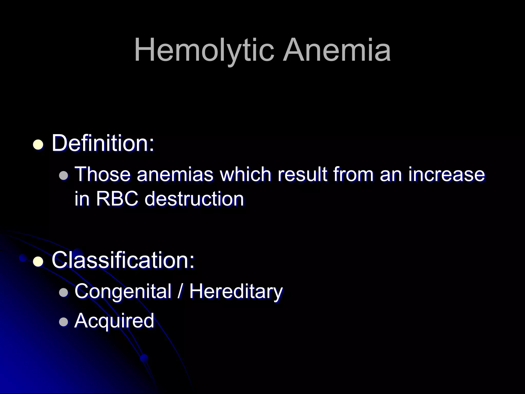
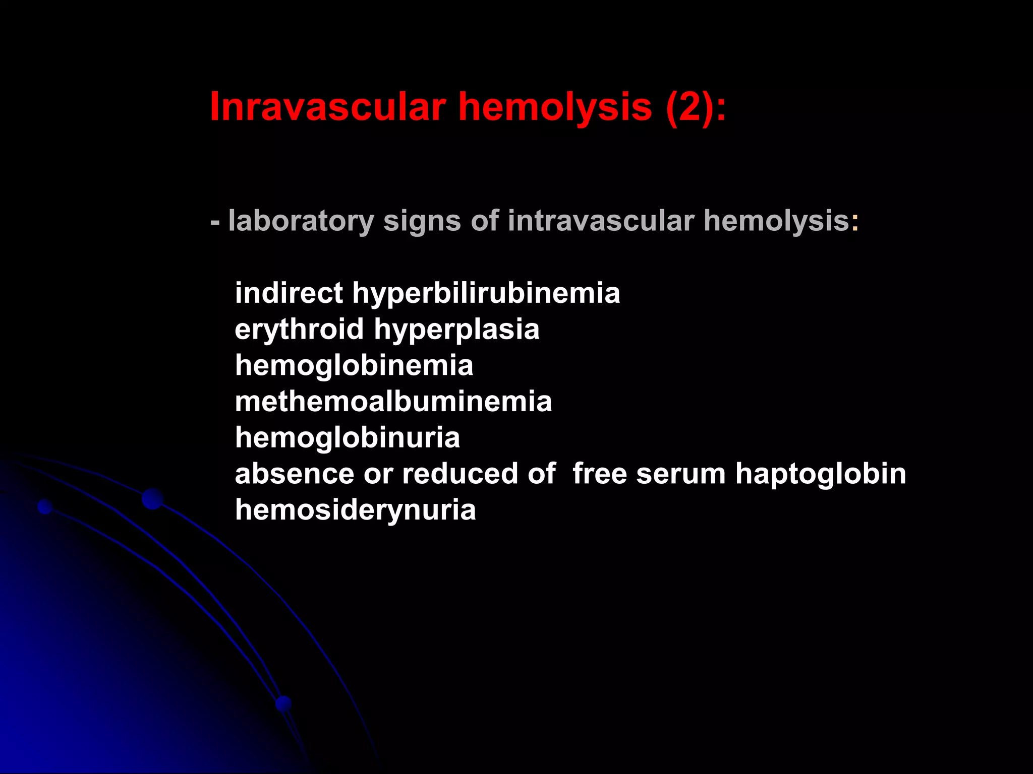
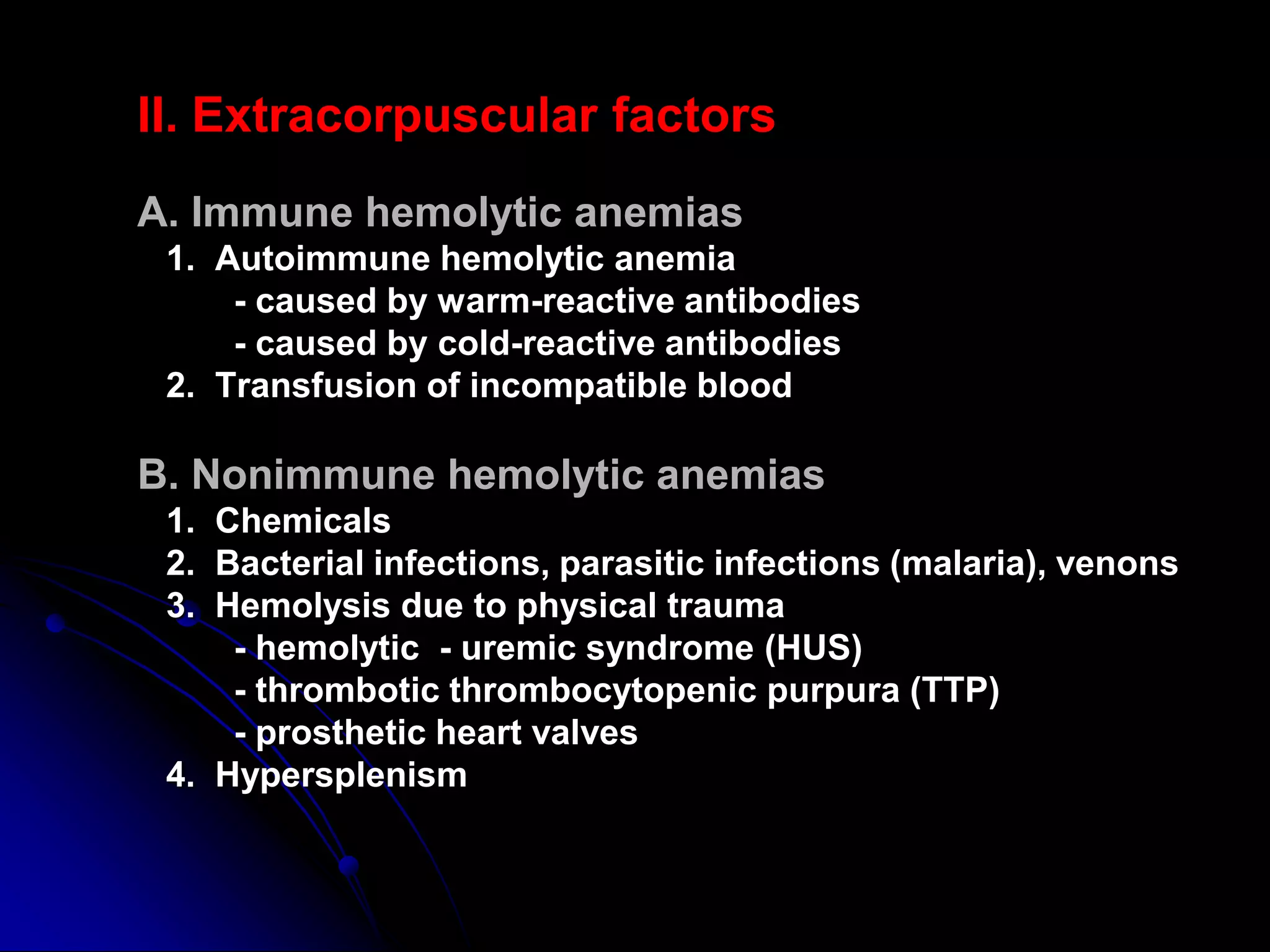
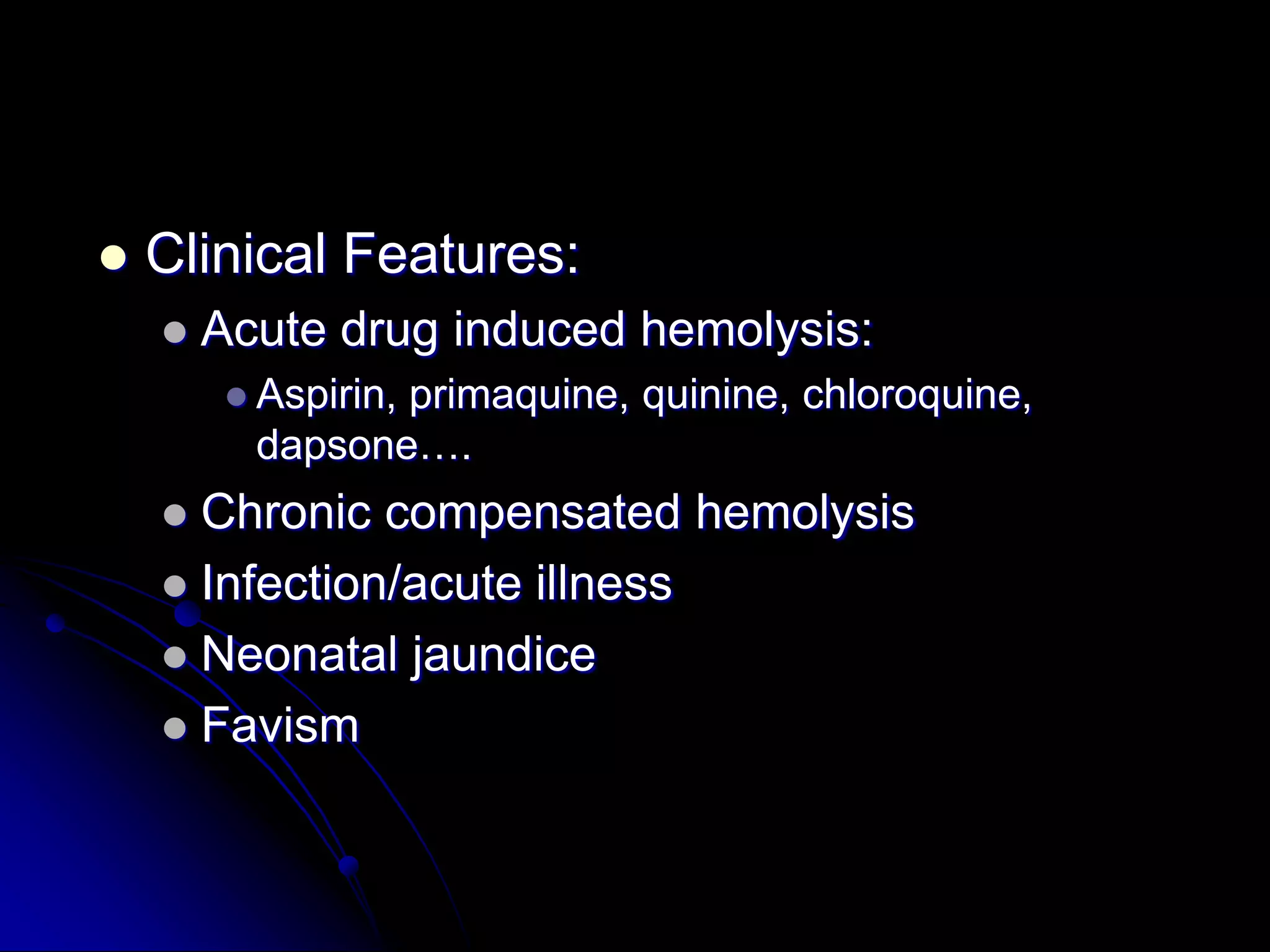
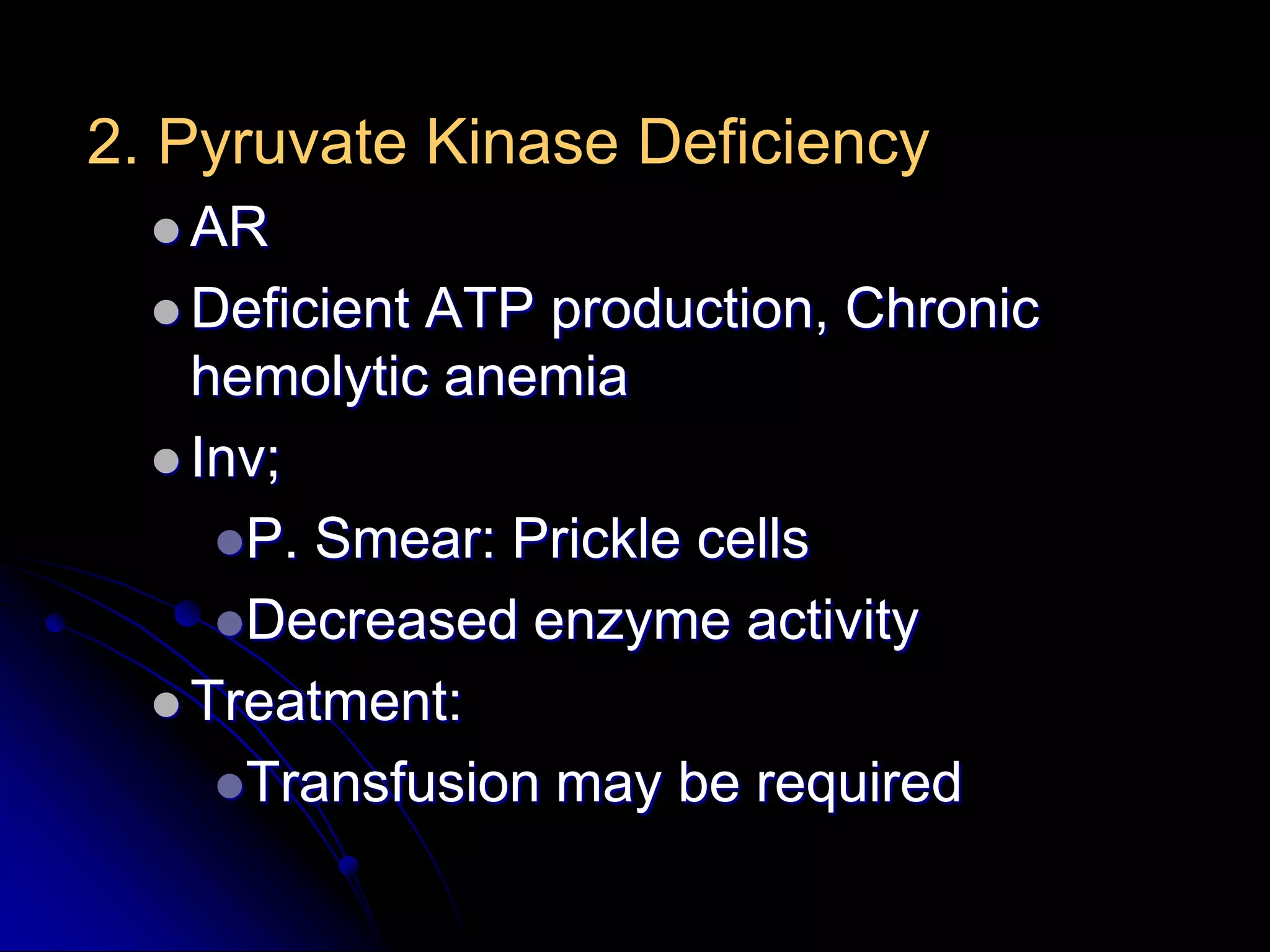
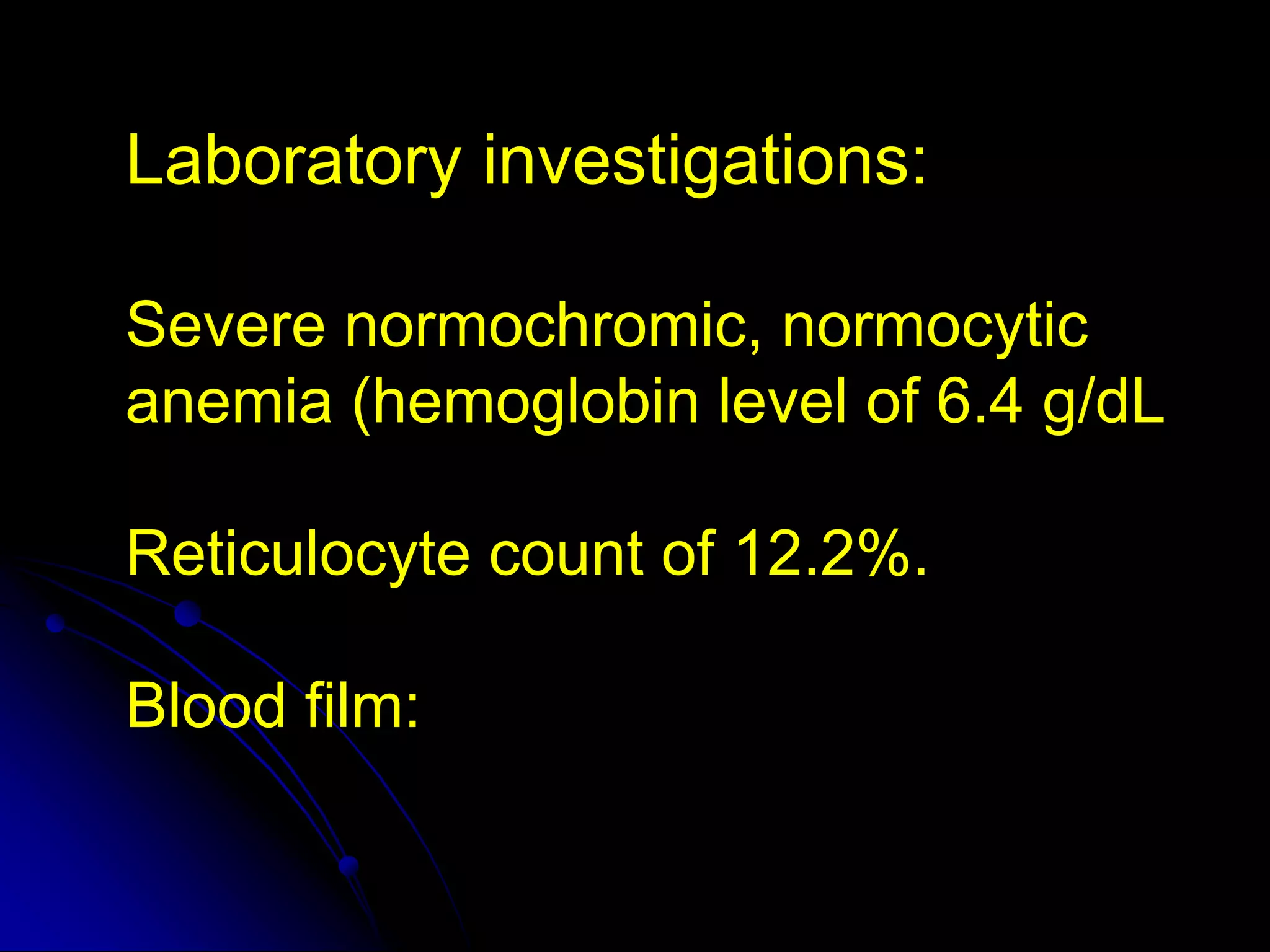
HEMOLYTIC ANAEMIA
         BY
    THEJUS.K.THILAK
HEMOLYTIC ANEMIAS
Hemolytic anemias = reduced red-cell life
                span
Hemolytic Anemia

   Definition:
       Those anemias which result from an increase
        in RBC destruction


   Classification:
     Congenital / Hereditary
     Acquired
Mechanisms of hemolysis:

  - intravascular
  - extravascular
Inravascular hemolysis (1):

- red cells destruction occurs in vascular space
- clinical states associated with Intravascular hemolysis:
   acute hemolytic transfusion reactions
   severe and extensive burns
   paroxysmal nocturnal hemoglobinuria
   severe microangiopathic hemolysis
   physical trauma
   bacterial infections and parasitic infections (sepsis)
Inravascular hemolysis (2):

- laboratory signs of intravascular hemolysis:

 indirect hyperbilirubinemia
 erythroid hyperplasia
 hemoglobinemia
 methemoalbuminemia
 hemoglobinuria
 absence or reduced of free serum haptoglobin
 hemosiderynuria
II. Extracorpuscular factors
A. Immune hemolytic anemias
 1. Autoimmune hemolytic anemia
     - caused by warm-reactive antibodies
     - caused by cold-reactive antibodies
 2. Transfusion of incompatible blood

B. Nonimmune hemolytic anemias
 1. Chemicals
 2. Bacterial infections, parasitic infections (malaria), venons
 3. Hemolysis due to physical trauma
     - hemolytic - uremic syndrome (HUS)
     - thrombotic thrombocytopenic purpura (TTP)
     - prosthetic heart valves
 4. Hypersplenism
Extravascular hemolysis :
- red cells destruction occurs in reticuloendothelial system
- clinical states associated with extravascular hemolysis :
   autoimmune hemolysis
   delayed hemolytic transfusion reactions
   hemoglobinopathies
   hereditary spherocytosis
   hypersplenism
   hemolysis with liver disease
- laboratory signs of extravascular hemolysis:
   indirect hyperbilirubinemia
   increased excretion of bilirubin by bile
   erythroid hyperplasia
   hemosiderosis
Hemolytic anemia - clinical features:

   - pallor
   - jaundice
   - splenomegaly
Laboratory features:
1. Laboratory features
   - normocytic/macrocytic, hyperchromic anemia
   - reticulocytosis
   - increased serum iron
   - antiglobulin Coombs’ test is positive

2. Blood smear
   - anisopoikilocytosis, spherocytes
   - erythroblasts
   - schistocytes

3. Bone marrow smear
  - erythroid hyperplasia
3. PNH –laboratory features:
  - pancytopenia
  - chronic urinary iron loss
  - serum iron concentration decreased
  - hemoglobinuria
  - hemosiderinuria
  - positive Ham’s test (acid hemolysis test)
  - positive sugar-water test
  - specific immunophenotype of erytrocytes (CD59,
CD55)

4. Treatment:
  - washed RBC transfusion
  - iron therapy
  - allogenic bone marrow transplantation
Introduction

 Mean life span of a RBC-120days
 Removed Extravascularly by- Macrophages
  of RE system
Classification of Hemolytic Anemias

Hereditary    1. Abnormalities of RBC interior
                 a.Enzyme defects: G-6-PD def,PK def
                 b.Hemoglobinopathies
              2. RBC membrane abnormalities
                 a. Hereditary spherocytosis etc.
                 b. PNH


Acquired      c. Spur cell anemia
              3. Extrinsic factors
                a. Hypersplenism
                b. Antibody: immune hemolysis
                c. Mechanical trauma: MAHA
                d. Infections, toxins, etc

                                                Ref : Harrison’s
Features of HEMOLYSIS
                                 Bilirubin
                                  LDH
                          Reticulocytes, n-RBC
                             Haptoglobulins
                  +ve Urinary hemosiderin, Urobilinogen

                                     Blood Film

         Spherocytes             No spherocytes           Fragmentation

DCT +ve               DCT –ve

AI Hemolysis   H. Sherocytosis   Malaria,
                                 Clostidium
                                       Hereditery enzymopathies Microangiopathic,
                                                                Traumatic
Red Cell Membrane Defects

1.Hereditary Spherocytosis
   Usually inherited as AD disorder
   Defect: Deficiency of Beta Spectrin or Ankyrin
     Loss of membrane in Spleen & RES
    becomes more spherical Destruction in
    Spleen
Spherocytes
 C/F:
  Asymptomatic
  Fluctuating
             hemolysis
  Splenomegaly
  Pigmented gall stones- 50%
RBC Membrane
Complications
   Clinical course may be complicated with
    Crisis:
     Hemolytic Crisis: associated with infection
     Aplastic crisis: associated with Parvovirus
      infection
   Inv:
     Test will confirm Hemolysis
     P Smear: Spherocytes
     Osmotic Fragility: Increased


    Screen Family members
   Management:
     Folic Acid 5mg weekly, prophylaxis life long
     Spleenectomy
     Blood transfusion in Ac, severe hemolytic crisis
2.Hereditary Elliptocytosis
   Equatorial Africa, SE Asia
   AD / AR
   Functional abnormality in one or more anchor
    proteins in RBC membrane- Alpha spectrin ,
    Protein 4.1
   Usually asymptomatic
   Mx: Similar to H. spherocytosis
   Variant:
    3.SE-Asian ovalocytosis:
        Common in Malaysia , Indonesia…
        Asymptomatic-usually
        Cells oval , rigid ,resist invasion by malarial
         parasites
Elliptocytosis
Red Cell Enzymopathies

   Physiology:
     EM pathway: ATP production
     HMP shunt pathway: NADPH & Glutathione
      production
1. Glucose-6-Phosphate Dehydrogenase
  ( G6PD ) Deficiency
  Pivotalenzyme in HMP Shunt & produces
   NADPH to protect RBC against oxidative
   stress
  Most  common enzymopathy -10%
   world’s population
  Protection against Malaria
  X-linked
   Clinical Features:
       Acute drug induced hemolysis:
          Aspirin,
                 primaquine, quinine, chloroquine,
          dapsone….
     Chronic compensated hemolysis
     Infection/acute illness
     Neonatal jaundice
     Favism
   Inv:
      e/o non-spherocytic intravascular
       hemolyis
      P. Smear: Bite cells, blister cells,
       irregular small cells, Heinz bodies,
       polychromasia
      G-6-PD level


   Treatment:
     Stop the precipitating drug or treat the
      infection
     Acute transfusions if required
2. Pyruvate Kinase Deficiency
   AR
   DeficientATP production, Chronic
    hemolytic anemia
   Inv;
     P. Smear: Prickle cells
     Decreased enzyme activity
   Treatment:
     Transfusion may be required
AQUIRED
Autoimmune Hemolytic Anemia
 Result from RBC destruction due to RBC
  autoantibodies: Ig G, M, E, A
 Most commonly-idiopathic
 Classification
     Warm AI hemolysis:Ab binds at 37degree
      Celsius
     Cold AI Hemolysis: Ab binds at 4 degree
      Celsius
1.Warm AI Hemolysis:
   Can occurs at all age groups
  F>M
   Causes:
      50% Idiopathic
      Rest - secondary causes:
        1.Lymphoid neoplasm: CLL, Lymphoma,
         Myeloma
        2.Solid Tumors: Lung, Colon, Kidney, Ovary,
         Thymoma
        3.CTD: SLE,RA
        4.Drugs: Alpha methyl DOPA, Penicillin ,
         Quinine, Chloroquine
        5.Misc: UC, HIV
   Inv:
     e/o hemolysis, MCV
     P Smear: Microspherocytosis, n-RBC
     Confirmation: Coomb’s Test / Antiglobulin test


   Treatment
     Correct the underlying cause
     Prednisolone 1mg/kg po until Hb reaches
      10mg/dl then taper slowly and stop
     Transfusion: for life threatening problems
     If no response to steroids  Spleenectomy or,
     Immunosuppressive: Azathioprine,
      Cyclophosphamide
2. Cold AI Hemolysis
    Usually Ig M
    Acute or Chronic form
    Chronic:
         C/F:
            Elderly patients
            Cold , painful & often blue fingers, toes,
             ears, or nose ( Acrocyanosis)
   Inv:
     e/o hemolysis
     P Smear: Microspherocytosis
     Ig M with specificity to I or I Ag
   Other causes of Cold Agglutination:
     Infection: Mycoplasma pneumonia, Infec
      Mononucleosis
     PCH : Rare cause seen in children in
      association with cong syphilis
   Treatment:
     Treatment of the underlying cause
     Keep extremities warm
     Steroids treatment
     Blood transfusion
Non-Immune Acquired Hemolytic
           Anemia
1. Mechanical Trauma
  A). Mechanical heart valves, Arterial grafts:
   cause shear stress damage
  B).March hemoglobinuria: Red cell damage in
   capillaries of feet
  C). Thermal injury: burns
  D). Microangiopathic hemolytic anemia (MAHA):
   by passage of RBC through fibrin strands
   deposited in small vessels  disruption of
   RBC eg: DIC,PIH, Malignant HTN,TTP,HUS
Acquired hemolysis
2.Infection
  F. malaria: intravascular hemolysis: severe
  called ‘Blackwater fever’
  Cl. perfringens septicemia
3.Chemical/Drugs: oxidant denaturation of
  hemoglobin
  Eg: Dapsone, sulphasalazine, Arsenic
  gas, Cu, Nitrates & Nitrobenzene
Paroxysmal Nocturnal Hemoglobinuria

   Hematopoietic stem cell disorder
   Mutation of phosphatidylinositol glycan class A
    (PIG-A) gene
   Glycosylphosphatidylinositol (GPI) anchors
    membrane proteins
   Without GPI, unable to regulate completment
    activities on membrane
   Hemolysis is pH dependent
   Thrombosis can occur
Lab Tests for PNH
   Acidified serum lysis test (Ham’s test): PNH cells
    lyse due to complement activation in acidified
    serm
   Sugar water (sucrose hemolysis) test: RBCs
    sensitive to complement will lyse in sucrose and
    serum
   Flow cytometry: lack of CD59 on RBCs, or lack
    of CD59 or CD55 on granulocytes
Microangiopathy
   Schistocytes
     Triangular or helmet shaped RBC fragments
   Destruction of RBC as they move through
    damaged blood vessels
     Endocarditis
     Hemangiosarcoma
     Caval Syndrome – Heartworm Disease
     Thrombosed IV catheter
     Vasculitis
     Hemolytic-uremic syndrome
     DIC
   Schistocytes are also seen with osmotic
    fragility
     Liver disease, iron deficiency, water
        intoxication, congenital, zinc toxicity
Young man of 19

  Complains of giddiness
    weakness, pallor

Examination reveals a spleen
  mild lemon yellow sclera
Laboratory Evaluation of Hemolysis
                        Extravascular Intravascular
HEMATOLOGIC
Routine blood film      Polychromatophilia    Polychromatophilia
Reticulocyte count
Bone marrow             Erythroid             Erythroid
examination             hyperplasia           hyperplasia


PLASMA OR SERUM
Bilirubin                 Unconjugated       Unconjugated
Haptoglobin              , Absent            Absent
Plasma hemoglobin          N/
Lactate dehydrogenase    (Variable)           (Variable)

URINE
Bilirubin               0                    0
Hemosiderin             0                    +
Hemoglobin              0                    + severe cases
How shall you investigate to find
 out the cause of the problem?
Laboratory investigations:

Severe normochromic, normocytic
anemia (hemoglobin level of 6.4 g/dL

Reticulocyte count of 12.2%.

Blood film:
Bilirubin level of 2.5 mg/dL,

Lactate dehydrogenase (LDH) of
2140 IU/L,

Haptoglobin below 7 mg/dL
The direct antiglobulin test was
positive for complement (C3d) (++),
and IgG (++-).

Also was positive for agglutinins of
IgM type and had a titer of 1:1024.
complement


Direct antiglobulin test
demonstrating the presence of autoantibodies (shown
here) or complement on the surface of the red blood
cell.
Serologies for human
immunodeficiency virus, hepatitis B
and C viruses, and Mycoplasma
pneumoniae were negative.

Rheumatoid factor and antinuclear
antibodies were undetectable.
Prednisone therapy was started at
 a dose of 1 mg/kg intravenously,
  daily. Hemoglobin level rose to
  11 g/dL, concomitantly with the
 improvement of hemolytic signs.
A reduction of positivity of both
direct and indirect antiglobulin tests
(polyvalent serum + ; C3d + ;
IgG+ ), as well as a reduction of
cold agglutinin titers (1:128), was
observed 8 weeks after
corticosteroid therapy.
Three months later, corticosteroids
were tapered to a maintenance
dose of 25 mg daily.

Hemolysis recurred again with the
fall of hemoglobin to 7 g/dL.
The direct antiglobulin test recurred
positive for polyvalent serum (+++),
complement (+++), and IgG (+++),
while cold agglutinin titers again
became strongly positive (1:256).
Immunophenotyping of bone
marrow cells showed that 10% of
all the cells were CD20 and CD19
positive.
CD20 is widely expressed on B-
cells.

CD20 could play a role in Ca2+
influx across plasma membranes,
maintaining intracellular Ca2+
concentration and allowing
activation of B cells.
Hemoglobin value reached
13.5 g/dL just before the third dose,
although biochemical signs of
hemolysis remained substantially
unaltered.
At the end of therapy, the hemolytic
 signs disappeared, the direct and
 indirect antiglobulin tests became
negative, and cold agglutinin titers
              fell to 1:32

  Immunophenotyping of bone
marrow cells showed the absence
   of CD20 and CD19 B cells.
Hemolytic Anemia  Classification - By Thejus K. Thilak

Hemolytic Anemia Classification - By Thejus K. Thilak

  • 1.
    HEMOLYTIC ANAEMIA BY THEJUS.K.THILAK
  • 2.
    HEMOLYTIC ANEMIAS Hemolytic anemias= reduced red-cell life span
  • 3.
    Hemolytic Anemia  Definition:  Those anemias which result from an increase in RBC destruction  Classification:  Congenital / Hereditary  Acquired
  • 4.
    Mechanisms of hemolysis: - intravascular - extravascular
  • 5.
    Inravascular hemolysis (1): -red cells destruction occurs in vascular space - clinical states associated with Intravascular hemolysis: acute hemolytic transfusion reactions severe and extensive burns paroxysmal nocturnal hemoglobinuria severe microangiopathic hemolysis physical trauma bacterial infections and parasitic infections (sepsis)
  • 7.
    Inravascular hemolysis (2): -laboratory signs of intravascular hemolysis: indirect hyperbilirubinemia erythroid hyperplasia hemoglobinemia methemoalbuminemia hemoglobinuria absence or reduced of free serum haptoglobin hemosiderynuria
  • 8.
    II. Extracorpuscular factors A.Immune hemolytic anemias 1. Autoimmune hemolytic anemia - caused by warm-reactive antibodies - caused by cold-reactive antibodies 2. Transfusion of incompatible blood B. Nonimmune hemolytic anemias 1. Chemicals 2. Bacterial infections, parasitic infections (malaria), venons 3. Hemolysis due to physical trauma - hemolytic - uremic syndrome (HUS) - thrombotic thrombocytopenic purpura (TTP) - prosthetic heart valves 4. Hypersplenism
  • 9.
    Extravascular hemolysis : -red cells destruction occurs in reticuloendothelial system - clinical states associated with extravascular hemolysis : autoimmune hemolysis delayed hemolytic transfusion reactions hemoglobinopathies hereditary spherocytosis hypersplenism hemolysis with liver disease - laboratory signs of extravascular hemolysis: indirect hyperbilirubinemia increased excretion of bilirubin by bile erythroid hyperplasia hemosiderosis
  • 11.
    Hemolytic anemia -clinical features: - pallor - jaundice - splenomegaly
  • 12.
    Laboratory features: 1. Laboratoryfeatures - normocytic/macrocytic, hyperchromic anemia - reticulocytosis - increased serum iron - antiglobulin Coombs’ test is positive 2. Blood smear - anisopoikilocytosis, spherocytes - erythroblasts - schistocytes 3. Bone marrow smear - erythroid hyperplasia
  • 13.
    3. PNH –laboratoryfeatures: - pancytopenia - chronic urinary iron loss - serum iron concentration decreased - hemoglobinuria - hemosiderinuria - positive Ham’s test (acid hemolysis test) - positive sugar-water test - specific immunophenotype of erytrocytes (CD59, CD55) 4. Treatment: - washed RBC transfusion - iron therapy - allogenic bone marrow transplantation
  • 14.
    Introduction  Mean lifespan of a RBC-120days  Removed Extravascularly by- Macrophages of RE system
  • 15.
    Classification of HemolyticAnemias Hereditary 1. Abnormalities of RBC interior a.Enzyme defects: G-6-PD def,PK def b.Hemoglobinopathies 2. RBC membrane abnormalities a. Hereditary spherocytosis etc. b. PNH Acquired c. Spur cell anemia 3. Extrinsic factors a. Hypersplenism b. Antibody: immune hemolysis c. Mechanical trauma: MAHA d. Infections, toxins, etc Ref : Harrison’s
  • 16.
    Features of HEMOLYSIS Bilirubin LDH Reticulocytes, n-RBC Haptoglobulins +ve Urinary hemosiderin, Urobilinogen Blood Film Spherocytes No spherocytes Fragmentation DCT +ve DCT –ve AI Hemolysis H. Sherocytosis Malaria, Clostidium Hereditery enzymopathies Microangiopathic, Traumatic
  • 17.
    Red Cell MembraneDefects 1.Hereditary Spherocytosis  Usually inherited as AD disorder  Defect: Deficiency of Beta Spectrin or Ankyrin  Loss of membrane in Spleen & RES becomes more spherical Destruction in Spleen
  • 18.
  • 19.
     C/F: Asymptomatic Fluctuating hemolysis Splenomegaly Pigmented gall stones- 50%
  • 20.
  • 21.
    Complications  Clinical course may be complicated with Crisis:  Hemolytic Crisis: associated with infection  Aplastic crisis: associated with Parvovirus infection
  • 22.
    Inv:  Test will confirm Hemolysis  P Smear: Spherocytes  Osmotic Fragility: Increased Screen Family members
  • 23.
    Management:  Folic Acid 5mg weekly, prophylaxis life long  Spleenectomy  Blood transfusion in Ac, severe hemolytic crisis
  • 24.
    2.Hereditary Elliptocytosis  Equatorial Africa, SE Asia  AD / AR  Functional abnormality in one or more anchor proteins in RBC membrane- Alpha spectrin , Protein 4.1  Usually asymptomatic  Mx: Similar to H. spherocytosis  Variant: 3.SE-Asian ovalocytosis:  Common in Malaysia , Indonesia…  Asymptomatic-usually  Cells oval , rigid ,resist invasion by malarial parasites
  • 25.
  • 26.
    Red Cell Enzymopathies  Physiology:  EM pathway: ATP production  HMP shunt pathway: NADPH & Glutathione production
  • 27.
    1. Glucose-6-Phosphate Dehydrogenase ( G6PD ) Deficiency  Pivotalenzyme in HMP Shunt & produces NADPH to protect RBC against oxidative stress  Most common enzymopathy -10% world’s population  Protection against Malaria  X-linked
  • 28.
    Clinical Features:  Acute drug induced hemolysis:  Aspirin, primaquine, quinine, chloroquine, dapsone….  Chronic compensated hemolysis  Infection/acute illness  Neonatal jaundice  Favism
  • 29.
    Inv:  e/o non-spherocytic intravascular hemolyis  P. Smear: Bite cells, blister cells, irregular small cells, Heinz bodies, polychromasia  G-6-PD level  Treatment:  Stop the precipitating drug or treat the infection  Acute transfusions if required
  • 30.
    2. Pyruvate KinaseDeficiency  AR  DeficientATP production, Chronic hemolytic anemia  Inv; P. Smear: Prickle cells Decreased enzyme activity  Treatment: Transfusion may be required
  • 31.
  • 32.
    Autoimmune Hemolytic Anemia Result from RBC destruction due to RBC autoantibodies: Ig G, M, E, A  Most commonly-idiopathic  Classification  Warm AI hemolysis:Ab binds at 37degree Celsius  Cold AI Hemolysis: Ab binds at 4 degree Celsius
  • 33.
    1.Warm AI Hemolysis:  Can occurs at all age groups F>M  Causes:  50% Idiopathic  Rest - secondary causes: 1.Lymphoid neoplasm: CLL, Lymphoma, Myeloma 2.Solid Tumors: Lung, Colon, Kidney, Ovary, Thymoma 3.CTD: SLE,RA 4.Drugs: Alpha methyl DOPA, Penicillin , Quinine, Chloroquine 5.Misc: UC, HIV
  • 34.
    Inv:  e/o hemolysis, MCV  P Smear: Microspherocytosis, n-RBC  Confirmation: Coomb’s Test / Antiglobulin test  Treatment  Correct the underlying cause  Prednisolone 1mg/kg po until Hb reaches 10mg/dl then taper slowly and stop  Transfusion: for life threatening problems  If no response to steroids  Spleenectomy or,  Immunosuppressive: Azathioprine, Cyclophosphamide
  • 35.
    2. Cold AIHemolysis  Usually Ig M  Acute or Chronic form  Chronic:  C/F:  Elderly patients  Cold , painful & often blue fingers, toes, ears, or nose ( Acrocyanosis)  Inv:  e/o hemolysis  P Smear: Microspherocytosis  Ig M with specificity to I or I Ag
  • 36.
    Other causes of Cold Agglutination:  Infection: Mycoplasma pneumonia, Infec Mononucleosis  PCH : Rare cause seen in children in association with cong syphilis
  • 37.
    Treatment:  Treatment of the underlying cause  Keep extremities warm  Steroids treatment  Blood transfusion
  • 38.
    Non-Immune Acquired Hemolytic Anemia 1. Mechanical Trauma A). Mechanical heart valves, Arterial grafts: cause shear stress damage B).March hemoglobinuria: Red cell damage in capillaries of feet C). Thermal injury: burns D). Microangiopathic hemolytic anemia (MAHA): by passage of RBC through fibrin strands deposited in small vessels  disruption of RBC eg: DIC,PIH, Malignant HTN,TTP,HUS
  • 39.
    Acquired hemolysis 2.Infection F. malaria: intravascular hemolysis: severe called ‘Blackwater fever’ Cl. perfringens septicemia 3.Chemical/Drugs: oxidant denaturation of hemoglobin Eg: Dapsone, sulphasalazine, Arsenic gas, Cu, Nitrates & Nitrobenzene
  • 40.
    Paroxysmal Nocturnal Hemoglobinuria  Hematopoietic stem cell disorder  Mutation of phosphatidylinositol glycan class A (PIG-A) gene  Glycosylphosphatidylinositol (GPI) anchors membrane proteins  Without GPI, unable to regulate completment activities on membrane  Hemolysis is pH dependent  Thrombosis can occur
  • 41.
    Lab Tests forPNH  Acidified serum lysis test (Ham’s test): PNH cells lyse due to complement activation in acidified serm  Sugar water (sucrose hemolysis) test: RBCs sensitive to complement will lyse in sucrose and serum  Flow cytometry: lack of CD59 on RBCs, or lack of CD59 or CD55 on granulocytes
  • 42.
    Microangiopathy  Schistocytes  Triangular or helmet shaped RBC fragments  Destruction of RBC as they move through damaged blood vessels  Endocarditis  Hemangiosarcoma  Caval Syndrome – Heartworm Disease  Thrombosed IV catheter  Vasculitis  Hemolytic-uremic syndrome  DIC  Schistocytes are also seen with osmotic fragility  Liver disease, iron deficiency, water intoxication, congenital, zinc toxicity
  • 43.
    Young man of19 Complains of giddiness weakness, pallor Examination reveals a spleen mild lemon yellow sclera
  • 44.
    Laboratory Evaluation ofHemolysis Extravascular Intravascular HEMATOLOGIC Routine blood film Polychromatophilia Polychromatophilia Reticulocyte count Bone marrow Erythroid Erythroid examination hyperplasia hyperplasia PLASMA OR SERUM Bilirubin Unconjugated Unconjugated Haptoglobin , Absent Absent Plasma hemoglobin N/ Lactate dehydrogenase (Variable) (Variable) URINE Bilirubin 0 0 Hemosiderin 0 + Hemoglobin 0 + severe cases
  • 45.
    How shall youinvestigate to find out the cause of the problem?
  • 46.
    Laboratory investigations: Severe normochromic,normocytic anemia (hemoglobin level of 6.4 g/dL Reticulocyte count of 12.2%. Blood film:
  • 47.
    Bilirubin level of2.5 mg/dL, Lactate dehydrogenase (LDH) of 2140 IU/L, Haptoglobin below 7 mg/dL
  • 48.
    The direct antiglobulintest was positive for complement (C3d) (++), and IgG (++-). Also was positive for agglutinins of IgM type and had a titer of 1:1024.
  • 49.
    complement Direct antiglobulin test demonstratingthe presence of autoantibodies (shown here) or complement on the surface of the red blood cell.
  • 50.
    Serologies for human immunodeficiencyvirus, hepatitis B and C viruses, and Mycoplasma pneumoniae were negative. Rheumatoid factor and antinuclear antibodies were undetectable.
  • 51.
    Prednisone therapy wasstarted at a dose of 1 mg/kg intravenously, daily. Hemoglobin level rose to 11 g/dL, concomitantly with the improvement of hemolytic signs.
  • 52.
    A reduction ofpositivity of both direct and indirect antiglobulin tests (polyvalent serum + ; C3d + ; IgG+ ), as well as a reduction of cold agglutinin titers (1:128), was observed 8 weeks after corticosteroid therapy.
  • 53.
    Three months later,corticosteroids were tapered to a maintenance dose of 25 mg daily. Hemolysis recurred again with the fall of hemoglobin to 7 g/dL.
  • 54.
    The direct antiglobulintest recurred positive for polyvalent serum (+++), complement (+++), and IgG (+++), while cold agglutinin titers again became strongly positive (1:256).
  • 55.
    Immunophenotyping of bone marrowcells showed that 10% of all the cells were CD20 and CD19 positive.
  • 56.
    CD20 is widelyexpressed on B- cells. CD20 could play a role in Ca2+ influx across plasma membranes, maintaining intracellular Ca2+ concentration and allowing activation of B cells.
  • 57.
    Hemoglobin value reached 13.5g/dL just before the third dose, although biochemical signs of hemolysis remained substantially unaltered.
  • 58.
    At the endof therapy, the hemolytic signs disappeared, the direct and indirect antiglobulin tests became negative, and cold agglutinin titers fell to 1:32 Immunophenotyping of bone marrow cells showed the absence of CD20 and CD19 B cells.

Editor's Notes