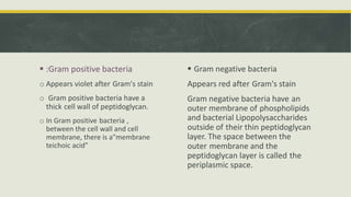
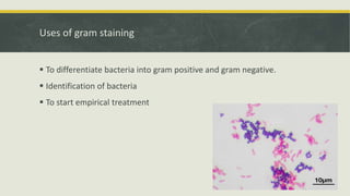
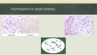
GRAM STAINING
Prepared by:Omar Arar
ID:22580680
 Gram staining is a method of differentiating bacterial species into two
large groups(Gram-positive and Gram-negative).
 The Gram staining is almost always the first step in the identification of
bacteria.
 Gram staining differentiates bacteria by the chemical and physical
properties of their cell walls.
INTRODUCTION: GRAM'S STAIN
 :Gram positive bacteria
o Appears violet after Gram's stain
o Gram positive bacteria have a
thick cell wall of peptidoglycan.
o In Gram positive bacteria ,
between the cell wall and cell
membrane, there is a"membrane
teichoic acid"
 Gram negative bacteria
Appears red after Gram's stain
Gram negative bacteria have an
outer membrane of phospholipids
and bacterial Lipopolysaccharides
outside of their thin peptidoglycan
layer. The space between the
outer membrane and the
peptidoglycan layer is called the
periplasmic space.
Uses of gram staining
 To differentiate bacteria into gram positive and gram negative.
 Identification of bacteria
 To start empirical treatment
FOUR BASIC STEPS OF GRAM STAINING
Applying a primary stain (Crystal Violet) to a heat-fixed smear of a bacterial
culture.
 The addition of Grams lodine , which binds to crystal violet and traps it in the
cell.
Decolourization with Alcohol or Acetone.
 Counter staining with Safranin.
PROCEDURE
 Prepare a heat fixed smear of the bacterial culture.
 Cover the smear with the Crystal Violet for 1 min.Add Grams lodine , which
washes the crystal violet stain.
 Rinse the slide in running water and add decolourizer(Alcohol).
 Again rinse the slide and cover the smear with the Safranin for 1 min.
 Wash off the safranin with water, air dry the slide and Observe under oil
immersion lens.
PHOTOGRAPHS OF GRAM STAINING:
Thank you

Omar Arar ( Gram's stain).pptx

  • 1.
  • 2.
     Gram stainingis a method of differentiating bacterial species into two large groups(Gram-positive and Gram-negative).  The Gram staining is almost always the first step in the identification of bacteria.  Gram staining differentiates bacteria by the chemical and physical properties of their cell walls. INTRODUCTION: GRAM'S STAIN
  • 3.
     :Gram positivebacteria o Appears violet after Gram's stain o Gram positive bacteria have a thick cell wall of peptidoglycan. o In Gram positive bacteria , between the cell wall and cell membrane, there is a"membrane teichoic acid"  Gram negative bacteria Appears red after Gram's stain Gram negative bacteria have an outer membrane of phospholipids and bacterial Lipopolysaccharides outside of their thin peptidoglycan layer. The space between the outer membrane and the peptidoglycan layer is called the periplasmic space.
  • 5.
    Uses of gramstaining  To differentiate bacteria into gram positive and gram negative.  Identification of bacteria  To start empirical treatment
  • 6.
    FOUR BASIC STEPSOF GRAM STAINING Applying a primary stain (Crystal Violet) to a heat-fixed smear of a bacterial culture.  The addition of Grams lodine , which binds to crystal violet and traps it in the cell. Decolourization with Alcohol or Acetone.  Counter staining with Safranin.
  • 7.
    PROCEDURE  Prepare aheat fixed smear of the bacterial culture.  Cover the smear with the Crystal Violet for 1 min.Add Grams lodine , which washes the crystal violet stain.  Rinse the slide in running water and add decolourizer(Alcohol).  Again rinse the slide and cover the smear with the Safranin for 1 min.  Wash off the safranin with water, air dry the slide and Observe under oil immersion lens.
  • 9.
  • 10.