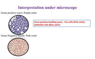
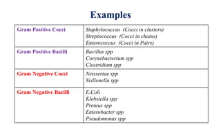
Gram Staining
Arun Kumar Parthasarathy Ph.d
Dept. of Microbiology
D.Y Patil Medical College, Kolhapur
Stain
• A stain, or dye, is a molecule that can bind to a cellular structure
and give it color.
• They have chromophore groups, with conjugated double bonds
that give the dye its color.
• They can bind with cells by ionic, covalent, or hydrophobic
bonding.
• Staining techniques make the microorganisms stand out against
their backgrounds.
• They are also used to help, examine the structural and chemical
differences in cellular structures, and look at the parts of the cell
Classification of dye (Stain)
Stains (Dyes)
Acidic dyes
(negatively charged),
attracted to any
positively charged cell
materials
Basic dyes(positively
charged),
dyes are attracted to
any negatively charged
cell
Neutral dyes
Acid dye is combine with
basic dye –Neutral dye is
formed
It contain both radicals it
gives colors to cytoplasm
and nucleus
simultaneously
Examples:- Methylene
blue, Crystal violet,
Safranin, and Malachite
green.
Examples:- Eosin and
Picric acid,
Example:- Leishman
Stain
Staining
Simple Staining
1. use of a single dye and
reveals basic cell shapes
and cell arrangements.
2. Methylene blue,
safranin, carbol fuchsin,
and crystal violet are
commonly used simple
stains.
Differential Staining
A differential stain
makes use of two or
more dyes and
distinguishes between
two kinds of
organisms
Example:- Gram Stain,
Acid Fast staining
Special staining
to study specific bacterial
structures with the light
microscope.
Example:-
1. Negative staining – capsule
2. Schaeffer-Fulton spore stain
3. Flagellar stain- Flagella
appear as dark lines with silver,
or red with carbol fuchsin
Gram Staining
The Gram stain was developed by Hans Christian Gram in 1884
and modified by Hucker in 1921.
The Gram stain separates bacteria into two groups:
(1) Gram-positive microorganisms that retain the primary dye
(Crystal violet) and
(2) Gram-negative microorganisms that take the color of the counter
stain (usually Safranin O).
Materials
Clean grease free slide
Microscope with oil immersion objective
Inoculation loop
Bacterial culture /suspension
Bunsen burner
Gram stain Reagents
a. Crystal Violet (Primary stain)
b. Grams Iodine (Mordant or Fixative )
c. Grams decolourizer (95% ethanol or Acetone)
d. Saffranin (Counter stain )
Smear Preparation
Take clean glass slide
Add one drop of normal saline or
distilled water with help of
inoculating loop
Open bacterial culture plate pick
one or 2 colonies with help of
inoculating loop and mix with
water
Heat fixation (10-15
secs )
Gram staining Protocol
1. Keep slide in staining rack
2. Add Crystal violet dye – wait for one minute
3. After one minute wash slide with slow running tap
water
4. After washing, add grams iodine – wait for one
minute
5. After one minute- wash slide with slow running tap
water
6. Add grams decolourizer (95% alcohol)- wait for 20-
25 secs
7. Wash slide with slow running tap water
8. Add saffranin (counter stain) wait for 30 seconds
9. Wash with slow running tap water
10. Allow to air dry
11. Observe under oil immersion (100X) objective
Interpretation under microscope
Gram positive cocci- Purple color
Gram Negative bacilli- Pink color
Gram positive budding yeast , Pus cells (Pink color),
Epithelial cells (Blue color)
Theories of Gram stain
1.PH Theory:-
Cytoplasm of gram positive bacteria is more acidic hence, can
retain basic dye (Crystal violet) for longer time.
Iodine acts as fixative (it combines with primary stain to form CV-I
complex which gets retained inside the cell
2. Magnesium Ribonucleate Theory:-
Magnesium ribonucleate and basic proteins are concentrated at the
cell membrane of gram positive bacteria.
Mg ribonucleate retain the basic dye (crystal violet) hence, gram
positive bacteria appear violet/purple in color
3. Cell wall Theory:-
Most important theory
• Gram Positive cell wall - Thick peptidoglycan layer (50-100 layers) with tight cross
linkages
•It acts as permeability barrier and prevent the loss of Crystal
violet dye
•Alcohol or acetone shrink the pores of the thick peptidoglycan
•Hence, large dye-iodine complex are not able to penetrate the
tightened peptidoglycan layer .
•Gram Positive bacteria – Crystal violet (Purple) in color
Gram Negative cell wall:- Thin peptidoglycan layer and not tightly cross linked
• Contain lipopolysaccharide layer, which gets disrupted easily
by the alcohol or acetone
• Forming larger pores that allows the dye-iodine complexes to
escape from cytoplasm
• Take only counter stain (Saffranin)
• Gram negative bacteria- Saffranin (Pink) in color
Examples
Gram Positive Cocci Staphylococcus (Cocci in clusters)
Streptococcus (Cocci in chains)
Enterococcus (Cocci in Pairs)
Gram Positive Bacilli Bacillus spp
Corynebacterium spp
Clostridium spp
Gram Negative Cocci Neisseriae spp
Veillonella spp
Gram Negative Bacilli E.Coli
Klebsiella spp
Proteus spp
Enterobacter spp
Pseudomonas spp
Uses of gram staining
1. To differentiate bacteria into gram positive and gram negative
2. Identification of bacteria
3. To start empirical treatment
Modification of Gram stain
Kopeloff and Beerman’s modification- primary stain and
counter stain are methyl blue and basic fuschin – anaerobic
bacterial identification
Jensen’s modification- use absolute alcohol as decolourizer and
neutral red as counter stain- useful for meningococci and
gonococci (Gram negative cocci)
.
Gram staining

Gram staining

  • 1.
    Gram Staining Arun KumarParthasarathy Ph.d Dept. of Microbiology D.Y Patil Medical College, Kolhapur
  • 2.
    Stain • A stain,or dye, is a molecule that can bind to a cellular structure and give it color. • They have chromophore groups, with conjugated double bonds that give the dye its color. • They can bind with cells by ionic, covalent, or hydrophobic bonding. • Staining techniques make the microorganisms stand out against their backgrounds. • They are also used to help, examine the structural and chemical differences in cellular structures, and look at the parts of the cell
  • 3.
    Classification of dye(Stain) Stains (Dyes) Acidic dyes (negatively charged), attracted to any positively charged cell materials Basic dyes(positively charged), dyes are attracted to any negatively charged cell Neutral dyes Acid dye is combine with basic dye –Neutral dye is formed It contain both radicals it gives colors to cytoplasm and nucleus simultaneously Examples:- Methylene blue, Crystal violet, Safranin, and Malachite green. Examples:- Eosin and Picric acid, Example:- Leishman Stain
  • 4.
    Staining Simple Staining 1. useof a single dye and reveals basic cell shapes and cell arrangements. 2. Methylene blue, safranin, carbol fuchsin, and crystal violet are commonly used simple stains. Differential Staining A differential stain makes use of two or more dyes and distinguishes between two kinds of organisms Example:- Gram Stain, Acid Fast staining Special staining to study specific bacterial structures with the light microscope. Example:- 1. Negative staining – capsule 2. Schaeffer-Fulton spore stain 3. Flagellar stain- Flagella appear as dark lines with silver, or red with carbol fuchsin
  • 5.
    Gram Staining The Gramstain was developed by Hans Christian Gram in 1884 and modified by Hucker in 1921. The Gram stain separates bacteria into two groups: (1) Gram-positive microorganisms that retain the primary dye (Crystal violet) and (2) Gram-negative microorganisms that take the color of the counter stain (usually Safranin O).
  • 6.
    Materials Clean grease freeslide Microscope with oil immersion objective Inoculation loop Bacterial culture /suspension Bunsen burner Gram stain Reagents a. Crystal Violet (Primary stain) b. Grams Iodine (Mordant or Fixative ) c. Grams decolourizer (95% ethanol or Acetone) d. Saffranin (Counter stain )
  • 7.
    Smear Preparation Take cleanglass slide Add one drop of normal saline or distilled water with help of inoculating loop Open bacterial culture plate pick one or 2 colonies with help of inoculating loop and mix with water Heat fixation (10-15 secs )
  • 8.
    Gram staining Protocol 1.Keep slide in staining rack 2. Add Crystal violet dye – wait for one minute 3. After one minute wash slide with slow running tap water 4. After washing, add grams iodine – wait for one minute 5. After one minute- wash slide with slow running tap water 6. Add grams decolourizer (95% alcohol)- wait for 20- 25 secs 7. Wash slide with slow running tap water 8. Add saffranin (counter stain) wait for 30 seconds 9. Wash with slow running tap water 10. Allow to air dry 11. Observe under oil immersion (100X) objective
  • 9.
    Interpretation under microscope Grampositive cocci- Purple color Gram Negative bacilli- Pink color Gram positive budding yeast , Pus cells (Pink color), Epithelial cells (Blue color)
  • 10.
    Theories of Gramstain 1.PH Theory:- Cytoplasm of gram positive bacteria is more acidic hence, can retain basic dye (Crystal violet) for longer time. Iodine acts as fixative (it combines with primary stain to form CV-I complex which gets retained inside the cell 2. Magnesium Ribonucleate Theory:- Magnesium ribonucleate and basic proteins are concentrated at the cell membrane of gram positive bacteria. Mg ribonucleate retain the basic dye (crystal violet) hence, gram positive bacteria appear violet/purple in color
  • 11.
    3. Cell wallTheory:- Most important theory • Gram Positive cell wall - Thick peptidoglycan layer (50-100 layers) with tight cross linkages •It acts as permeability barrier and prevent the loss of Crystal violet dye •Alcohol or acetone shrink the pores of the thick peptidoglycan •Hence, large dye-iodine complex are not able to penetrate the tightened peptidoglycan layer . •Gram Positive bacteria – Crystal violet (Purple) in color Gram Negative cell wall:- Thin peptidoglycan layer and not tightly cross linked • Contain lipopolysaccharide layer, which gets disrupted easily by the alcohol or acetone • Forming larger pores that allows the dye-iodine complexes to escape from cytoplasm • Take only counter stain (Saffranin) • Gram negative bacteria- Saffranin (Pink) in color
  • 12.
    Examples Gram Positive CocciStaphylococcus (Cocci in clusters) Streptococcus (Cocci in chains) Enterococcus (Cocci in Pairs) Gram Positive Bacilli Bacillus spp Corynebacterium spp Clostridium spp Gram Negative Cocci Neisseriae spp Veillonella spp Gram Negative Bacilli E.Coli Klebsiella spp Proteus spp Enterobacter spp Pseudomonas spp
  • 13.
    Uses of gramstaining 1. To differentiate bacteria into gram positive and gram negative 2. Identification of bacteria 3. To start empirical treatment
  • 14.
    Modification of Gramstain Kopeloff and Beerman’s modification- primary stain and counter stain are methyl blue and basic fuschin – anaerobic bacterial identification Jensen’s modification- use absolute alcohol as decolourizer and neutral red as counter stain- useful for meningococci and gonococci (Gram negative cocci) .

Editor's Notes

  • #2 It is important experiment, already morphology of bac over. So u have little idea about different shapes of bacteria
  • #3 stain or cld as dye, to increase the colour contrast and visibility of seeing bacter under microscope we go for staining techinques.
  • #5 It is process of applying dye to bacterial cell make colour.