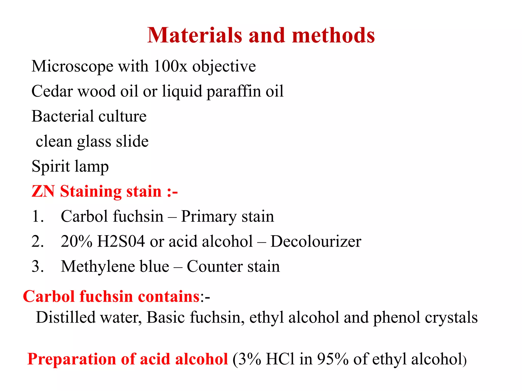
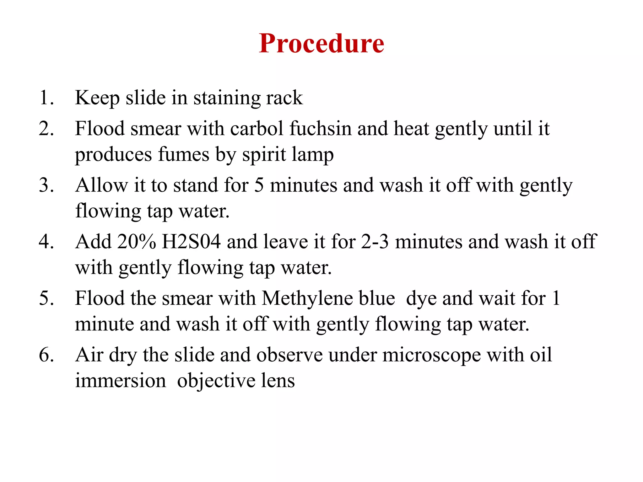
The document discusses the Ziehl-Neelsen staining technique, which is used to identify acid-fast bacteria such as Mycobacterium, Actinomycetes, and Nocardia. The staining method uses carbol fuchsin as the primary stain, along with heat to aid penetration. Acid-alcohol is used as a decolorizer, followed by methylene blue as the counterstain. Mycobacterium and other acid-fast bacteria will retain the pink carbol fuchsin stain due to their thick, waxy cell walls containing mycolic acid. This allows them to be differentiated from other non-acid fast bacteria that will take on the blue methylene blue counterstain
Acid fast staining is differential staining technique which differentiate bacteria into two group- acid fast bacteria and non acid bacteria. It used to identify acid-fast organisms such as members of the genus Mycobacterium .
It discusses laboratory tests involved in diagnosing meningitis with more emphasis on details of each test and findings, esp useful for microbiologists and medical students.
The PPT is mainly all about Mycobacterium Tuberculosis. Agents causing the disease Tuberculosis, pathogenesis, laboratory diagnosis, treatment and prophylaxis. It was made for both BSc and MSc students.
Erythrocyte Sedimentation Rate (ESR), a lecture for medical lab technicians at Baquba Technical Institute, Middle Technical University. All theoretical and practical notes about the test.
Smear preparation:
A drop of water is placed in the centre of a slide
One loopfuls of organisms is transferred to the centre of slide
Spread the organisms over the slide
The smear is allowed to dry
Slide is passed through flame several times to heat-kill and fix organisms
A bacterial stain is stained with crystal violet (fuchsin, methylene blue) 1 min
Observe under microscope
Basic Dyes
Methylene Blue
Crystal Violet
Carbol Fuchsin
Safranin
Malachite Green
Acidic Dyes
Picric Acid
Nigrosin
India Ink
Eosin
differrential statining
Two or more reagents Distinguish
Bacterial groups
Specific Structures
Example
Gram stain
Acid Fast Stain
Hereby you can get all about bacterial staining.
MICROBIAL STAINING
introduction
Microbial Staining - giving color to microbes. Because microbes are colorless and highly transparent structures.
Staining process in which microbes are getting color.
STAINES / DYES
Staines / dyes - organic compounds which carries either positive charges or negative charges or both .
Based on the charges
Basic stain / dyes :- stain with + ve charge .
Acidic stain / dyes - stain with -ve charge .
2. Based on function of stain :
Neutral stain / dyes - stain with both charges .
Simple staining only one dye is used differentiation among bacteria is impossible Eg . Simple Staining.
Differential staining- more than one dye is used- Differentiation among bacteria is possible- Eg. Gram's staining, Acid - fast staining.
Special staining - more than one dye used - Special structures are seen. Eg. Capsule staining , Spore staining .
This presentation mainly focuses on explanation about acid fast staining, their principle, reagents and procedure. And also about acid fast organisms. And a detailed explanation about the importance of Ziehl-neelson stain and errors occured during the procedure.
Slide 1: Title Slide
Extrachromosomal Inheritance
Slide 2: Introduction to Extrachromosomal Inheritance
Definition: Extrachromosomal inheritance refers to the transmission of genetic material that is not found within the nucleus.
Key Components: Involves genes located in mitochondria, chloroplasts, and plasmids.
Slide 3: Mitochondrial Inheritance
Mitochondria: Organelles responsible for energy production.
Mitochondrial DNA (mtDNA): Circular DNA molecule found in mitochondria.
Inheritance Pattern: Maternally inherited, meaning it is passed from mothers to all their offspring.
Diseases: Examples include Leber’s hereditary optic neuropathy (LHON) and mitochondrial myopathy.
Slide 4: Chloroplast Inheritance
Chloroplasts: Organelles responsible for photosynthesis in plants.
Chloroplast DNA (cpDNA): Circular DNA molecule found in chloroplasts.
Inheritance Pattern: Often maternally inherited in most plants, but can vary in some species.
Examples: Variegation in plants, where leaf color patterns are determined by chloroplast DNA.
Slide 5: Plasmid Inheritance
Plasmids: Small, circular DNA molecules found in bacteria and some eukaryotes.
Features: Can carry antibiotic resistance genes and can be transferred between cells through processes like conjugation.
Significance: Important in biotechnology for gene cloning and genetic engineering.
Slide 6: Mechanisms of Extrachromosomal Inheritance
Non-Mendelian Patterns: Do not follow Mendel’s laws of inheritance.
Cytoplasmic Segregation: During cell division, organelles like mitochondria and chloroplasts are randomly distributed to daughter cells.
Heteroplasmy: Presence of more than one type of organellar genome within a cell, leading to variation in expression.
Slide 7: Examples of Extrachromosomal Inheritance
Four O’clock Plant (Mirabilis jalapa): Shows variegated leaves due to different cpDNA in leaf cells.
Petite Mutants in Yeast: Result from mutations in mitochondrial DNA affecting respiration.
Slide 8: Importance of Extrachromosomal Inheritance
Evolution: Provides insight into the evolution of eukaryotic cells.
Medicine: Understanding mitochondrial inheritance helps in diagnosing and treating mitochondrial diseases.
Agriculture: Chloroplast inheritance can be used in plant breeding and genetic modification.
Slide 9: Recent Research and Advances
Gene Editing: Techniques like CRISPR-Cas9 are being used to edit mitochondrial and chloroplast DNA.
Therapies: Development of mitochondrial replacement therapy (MRT) for preventing mitochondrial diseases.
Slide 10: Conclusion
Summary: Extrachromosomal inheritance involves the transmission of genetic material outside the nucleus and plays a crucial role in genetics, medicine, and biotechnology.
Future Directions: Continued research and technological advancements hold promise for new treatments and applications.
Slide 11: Questions and Discussion
Invite Audience: Open the floor for any questions or further discussion on the topic.
Richard's aventures in two entangled wonderlandsRichard Gill
Since the loophole-free Bell experiments of 2020 and the Nobel prizes in physics of 2022, critics of Bell's work have retreated to the fortress of super-determinism. Now, super-determinism is a derogatory word - it just means "determinism". Palmer, Hance and Hossenfelder argue that quantum mechanics and determinism are not incompatible, using a sophisticated mathematical construction based on a subtle thinning of allowed states and measurements in quantum mechanics, such that what is left appears to make Bell's argument fail, without altering the empirical predictions of quantum mechanics. I think however that it is a smoke screen, and the slogan "lost in math" comes to my mind. I will discuss some other recent disproofs of Bell's theorem using the language of causality based on causal graphs. Causal thinking is also central to law and justice. I will mention surprising connections to my work on serial killer nurse cases, in particular the Dutch case of Lucia de Berk and the current UK case of Lucy Letby.
What is greenhouse gasses and how many gasses are there to affect the Earth.moosaasad1975
What are greenhouse gasses how they affect the earth and its environment what is the future of the environment and earth how the weather and the climate effects.
Seminar of U.V. Spectroscopy by SAMIR PANDASAMIR PANDA
Spectroscopy is a branch of science dealing the study of interaction of electromagnetic radiation with matter.
Ultraviolet-visible spectroscopy refers to absorption spectroscopy or reflect spectroscopy in the UV-VIS spectral region.
Ultraviolet-visible spectroscopy is an analytical method that can measure the amount of light received by the analyte.
Multi-source connectivity as the driver of solar wind variability in the heli...Sérgio Sacani
The ambient solar wind that flls the heliosphere originates from multiple
sources in the solar corona and is highly structured. It is often described
as high-speed, relatively homogeneous, plasma streams from coronal
holes and slow-speed, highly variable, streams whose source regions are
under debate. A key goal of ESA/NASA’s Solar Orbiter mission is to identify
solar wind sources and understand what drives the complexity seen in the
heliosphere. By combining magnetic feld modelling and spectroscopic
techniques with high-resolution observations and measurements, we show
that the solar wind variability detected in situ by Solar Orbiter in March
2022 is driven by spatio-temporal changes in the magnetic connectivity to
multiple sources in the solar atmosphere. The magnetic feld footpoints
connected to the spacecraft moved from the boundaries of a coronal hole
to one active region (12961) and then across to another region (12957). This
is refected in the in situ measurements, which show the transition from fast
to highly Alfvénic then to slow solar wind that is disrupted by the arrival of
a coronal mass ejection. Our results describe solar wind variability at 0.5 au
but are applicable to near-Earth observatories.
This pdf is about the Schizophrenia.
For more details visit on YouTube; @SELF-EXPLANATORY;
https://www.youtube.com/channel/UCAiarMZDNhe1A3Rnpr_WkzA/videos
Thanks...!
This presentation explores a brief idea about the structural and functional attributes of nucleotides, the structure and function of genetic materials along with the impact of UV rays and pH upon them.
2. Introduction
Ziehl-Neelsen staining (Acid Fast) technique is a
differential staining technique
It was initially developed by Ziehl and modified later
by Neelsen hence the name Ziehl-Neelsen stain
Neelsen used carbol fuchsin with heat and added a
decolorizing agent acid-alcohol and counter stain
methylene blue.
The use of acid-alcohol in the technique so it is called as
acid-fast stain
3. Where it is used?
Microorganisms that are not easily stained by basic
stains such as gram staining, negative staining
Examples:- Mycobacterium, Actinomycetes., Nocardia,
Cryptosporidium etc.
These microorganisms have thick cell wall made up of
lipodial complexes known as mycolic acid
4. Principle
The ziehl Neelsen stain uses basic fuchsin and phenol
compounds to stain the cell wall of mycobacterium spp.
Mycobacterium does not bind readily to simple stains
Use of heat along with carbol-fuchsin and phenol allows
the penetration through he bacterial cell.
Mycolic acid:- on its cell wall making it waxy,
hydrophobic and impermeable.
Mycolic acids are β-hydroxycarboxylic acids made up of
90 carbon atoms – defines the acid fastness of the
bacteria.
5. Materials and methods
Microscope with 100x objective
Cedar wood oil or liquid paraffin oil
Bacterial culture
clean glass slide
Spirit lamp
ZN Staining stain :-
1. Carbol fuchsin – Primary stain
2. 20% H2S04 or acid alcohol – Decolourizer
3. Methylene blue – Counter stain
Carbol fuchsin contains:-
Distilled water, Basic fuchsin, ethyl alcohol and phenol crystals
Preparation of acid alcohol (3% HCl in 95% of ethyl alcohol)
6. Procedure
1. Keep slide in staining rack
2. Flood smear with carbol fuchsin and heat gently until it
produces fumes by spirit lamp
3. Allow it to stand for 5 minutes and wash it off with gently
flowing tap water.
4. Add 20% H2S04 and leave it for 2-3 minutes and wash it off
with gently flowing tap water.
5. Flood the smear with Methylene blue dye and wait for 1
minute and wash it off with gently flowing tap water.
6. Air dry the slide and observe under microscope with oil
immersion objective lens
7.
8. Interpretation
Acid fast bacteria retain primary dye (Carbol fuchsin) –
Pink in colour E.x Mycobacterium, Cryptosporidium,
Nocardia, Isospora etc.
Non acid fast bacteria does not retain primary dye- Blue
in colour.
9. RNTCP grading
What is RNTCP?
Revised National Tuberculosis Control Program
Recently changed name, NTEP (National Tuberculosis
Elimination program)
RNTCP Grading : The number of bacilli seen in a smear reflects
disease severity and patient infectivity
It is given by WHO
It is mostly used in Pulmonary tuberculosis
10.
11. Modified acid fast stain
Cold Method with Gabbet’s methylene blue stain:-
The smear is flooded with basic fuschin-phenol
stain and allow to stand at RT for 10 mins and counter
stain Gabbet’s methylene blue stain for 2 mins.
Kinyoun’s Method:-
Nocardia and legionella resist decolourization by
1% cold sulphuric acid
Acid fast stain using by 5% sulphuric acid:-
M. leprae resist decolourization by 5% sulphuric
acid