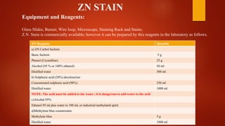
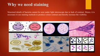
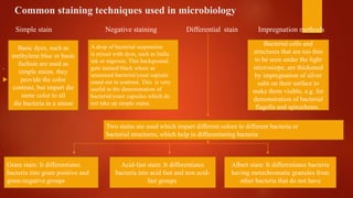
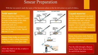
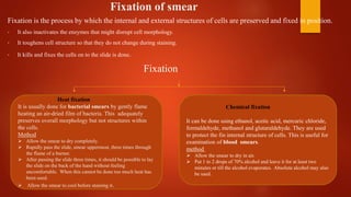
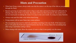
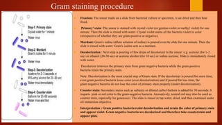
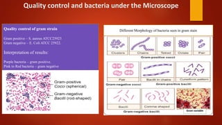
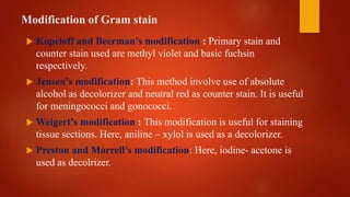
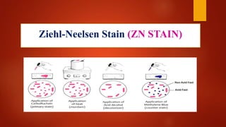
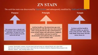
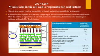
This document provides information about staining techniques used in microbiology. It discusses why staining is needed, as structural details of bacteria cannot be seen under a light microscope otherwise. It describes common staining methods like simple stains, negative stains, differential stains, and impregnation methods. Gram staining and Ziehl-Neelsen staining techniques are explained in detail, including the principles, procedures, and uses of each stain. Proper smear preparation and quality are also addressed.
this presentation involves a comprehensive outlines regarding the most common different methods used in diagnostic microbiology to stain bacteria and their structures
Capsule is an layer around the bacteria cell which gives bacteria the protection and pathogenicity. Staining such an layer is difficult with the normal stains so it is necessary to stain the background and the cell itself which makes the capsule appear colourless.
Hereby you can get all about bacterial staining.
MICROBIAL STAINING
introduction
Microbial Staining - giving color to microbes. Because microbes are colorless and highly transparent structures.
Staining process in which microbes are getting color.
STAINES / DYES
Staines / dyes - organic compounds which carries either positive charges or negative charges or both .
Based on the charges
Basic stain / dyes :- stain with + ve charge .
Acidic stain / dyes - stain with -ve charge .
2. Based on function of stain :
Neutral stain / dyes - stain with both charges .
Simple staining only one dye is used differentiation among bacteria is impossible Eg . Simple Staining.
Differential staining- more than one dye is used- Differentiation among bacteria is possible- Eg. Gram's staining, Acid - fast staining.
Special staining - more than one dye used - Special structures are seen. Eg. Capsule staining , Spore staining .
Acid fast staining is differential staining technique which differentiate bacteria into two group- acid fast bacteria and non acid bacteria. It used to identify acid-fast organisms such as members of the genus Mycobacterium .
Because microbial cytoplasm is usually transparent, it is necessary to stain microorganisms before they can be viewed with the light microscope. In some cases, staining is unnecessary, for example when microorganisms are very large or when motility is to be studied, and a drop of the microorganisms can be placed directly on the slide and observed.
Staining methods are helpful for presumptive identification of Microbes like Gram stain helps to identify Gram positive and Gram negative bacteria, similarly Z-N stain helps to identify acid fast bacilli and India ink preparation capsule observation of Cryptococcus neoformans.
Oxidase Test Microbiology - Principle, Procedure, Limitations, Results, QC - in lab #Oxidase Test
As the channel name suggests, our channel will be a perfect lounge for the malayali medicos..we wil be covering videos which will be like lecture classes related to the subjects biochemistry and microbiology in which we are specialised.. It will be a better learning experience for the students especially for those who are not able to understand and follow the normal classes in college..we assure the students that you will get a basic idea regarding the topic and extra reading can be done from the reference textbooks...
If you like my video
#like
#comment
#subscribe my channel
don't forget to subscribe my channel
Qualification
Maneesha M Joseph
MSc MLT (Microbiology)
Assistant Professor
Baby memorial college of allied Health science
Kozhikode
Our Partner Channel
Health & Voyage channel link - https://youtu.be/nzKqRVjlwc0
#Oxidase Test
#Medical
#Microbiology
# malayalam lecturer
#Mallu Medicos Lounge
#MalluMedicosLounge
#MLT
INTRODUCTION TO MICRO LAB, STAINING TECHNIQUES & MORPHOLOGY OF BACTERIADrBhavikapatel
This PPT is helpful to understand first practical to 2nd year MBBS student.
I have added 2 video in this PPT to understand staining techniques properly.
Reference: 1 Gram stain video: Dr.G Bhanu prakash animated medical videos
2. Zn stain video: sridhar Rao
This is a rundown of some staining techniques used in microbiology, including simple staining, negative staining, gram staining, acid-fast staining, endospore staining, and flagellar staining.
Title: Sense of Taste
Presenter: Dr. Faiza, Assistant Professor of Physiology
Qualifications:
MBBS (Best Graduate, AIMC Lahore)
FCPS Physiology
ICMT, CHPE, DHPE (STMU)
MPH (GC University, Faisalabad)
MBA (Virtual University of Pakistan)
Learning Objectives:
Describe the structure and function of taste buds.
Describe the relationship between the taste threshold and taste index of common substances.
Explain the chemical basis and signal transduction of taste perception for each type of primary taste sensation.
Recognize different abnormalities of taste perception and their causes.
Key Topics:
Significance of Taste Sensation:
Differentiation between pleasant and harmful food
Influence on behavior
Selection of food based on metabolic needs
Receptors of Taste:
Taste buds on the tongue
Influence of sense of smell, texture of food, and pain stimulation (e.g., by pepper)
Primary and Secondary Taste Sensations:
Primary taste sensations: Sweet, Sour, Salty, Bitter, Umami
Chemical basis and signal transduction mechanisms for each taste
Taste Threshold and Index:
Taste threshold values for Sweet (sucrose), Salty (NaCl), Sour (HCl), and Bitter (Quinine)
Taste index relationship: Inversely proportional to taste threshold
Taste Blindness:
Inability to taste certain substances, particularly thiourea compounds
Example: Phenylthiocarbamide
Structure and Function of Taste Buds:
Composition: Epithelial cells, Sustentacular/Supporting cells, Taste cells, Basal cells
Features: Taste pores, Taste hairs/microvilli, and Taste nerve fibers
Location of Taste Buds:
Found in papillae of the tongue (Fungiform, Circumvallate, Foliate)
Also present on the palate, tonsillar pillars, epiglottis, and proximal esophagus
Mechanism of Taste Stimulation:
Interaction of taste substances with receptors on microvilli
Signal transduction pathways for Umami, Sweet, Bitter, Sour, and Salty tastes
Taste Sensitivity and Adaptation:
Decrease in sensitivity with age
Rapid adaptation of taste sensation
Role of Saliva in Taste:
Dissolution of tastants to reach receptors
Washing away the stimulus
Taste Preferences and Aversions:
Mechanisms behind taste preference and aversion
Influence of receptors and neural pathways
Impact of Sensory Nerve Damage:
Degeneration of taste buds if the sensory nerve fiber is cut
Abnormalities of Taste Detection:
Conditions: Ageusia, Hypogeusia, Dysgeusia (parageusia)
Causes: Nerve damage, neurological disorders, infections, poor oral hygiene, adverse drug effects, deficiencies, aging, tobacco use, altered neurotransmitter levels
Neurotransmitters and Taste Threshold:
Effects of serotonin (5-HT) and norepinephrine (NE) on taste sensitivity
Supertasters:
25% of the population with heightened sensitivity to taste, especially bitterness
Increased number of fungiform papillae
Explore natural remedies for syphilis treatment in Singapore. Discover alternative therapies, herbal remedies, and lifestyle changes that may complement conventional treatments. Learn about holistic approaches to managing syphilis symptoms and supporting overall health.
Flu Vaccine Alert in Bangalore Karnatakaaddon Scans
As flu season approaches, health officials in Bangalore, Karnataka, are urging residents to get their flu vaccinations. The seasonal flu, while common, can lead to severe health complications, particularly for vulnerable populations such as young children, the elderly, and those with underlying health conditions.
Dr. Vidisha Kumari, a leading epidemiologist in Bangalore, emphasizes the importance of getting vaccinated. "The flu vaccine is our best defense against the influenza virus. It not only protects individuals but also helps prevent the spread of the virus in our communities," he says.
This year, the flu season is expected to coincide with a potential increase in other respiratory illnesses. The Karnataka Health Department has launched an awareness campaign highlighting the significance of flu vaccinations. They have set up multiple vaccination centers across Bangalore, making it convenient for residents to receive their shots.
To encourage widespread vaccination, the government is also collaborating with local schools, workplaces, and community centers to facilitate vaccination drives. Special attention is being given to ensuring that the vaccine is accessible to all, including marginalized communities who may have limited access to healthcare.
Residents are reminded that the flu vaccine is safe and effective. Common side effects are mild and may include soreness at the injection site, mild fever, or muscle aches. These side effects are generally short-lived and far less severe than the flu itself.
Healthcare providers are also stressing the importance of continuing COVID-19 precautions. Wearing masks, practicing good hand hygiene, and maintaining social distancing are still crucial, especially in crowded places.
Protect yourself and your loved ones by getting vaccinated. Together, we can help keep Bangalore healthy and safe this flu season. For more information on vaccination centers and schedules, residents can visit the Karnataka Health Department’s official website or follow their social media pages.
Stay informed, stay safe, and get your flu shot today!
Title: Sense of Smell
Presenter: Dr. Faiza, Assistant Professor of Physiology
Qualifications:
MBBS (Best Graduate, AIMC Lahore)
FCPS Physiology
ICMT, CHPE, DHPE (STMU)
MPH (GC University, Faisalabad)
MBA (Virtual University of Pakistan)
Learning Objectives:
Describe the primary categories of smells and the concept of odor blindness.
Explain the structure and location of the olfactory membrane and mucosa, including the types and roles of cells involved in olfaction.
Describe the pathway and mechanisms of olfactory signal transmission from the olfactory receptors to the brain.
Illustrate the biochemical cascade triggered by odorant binding to olfactory receptors, including the role of G-proteins and second messengers in generating an action potential.
Identify different types of olfactory disorders such as anosmia, hyposmia, hyperosmia, and dysosmia, including their potential causes.
Key Topics:
Olfactory Genes:
3% of the human genome accounts for olfactory genes.
400 genes for odorant receptors.
Olfactory Membrane:
Located in the superior part of the nasal cavity.
Medially: Folds downward along the superior septum.
Laterally: Folds over the superior turbinate and upper surface of the middle turbinate.
Total surface area: 5-10 square centimeters.
Olfactory Mucosa:
Olfactory Cells: Bipolar nerve cells derived from the CNS (100 million), with 4-25 olfactory cilia per cell.
Sustentacular Cells: Produce mucus and maintain ionic and molecular environment.
Basal Cells: Replace worn-out olfactory cells with an average lifespan of 1-2 months.
Bowman’s Gland: Secretes mucus.
Stimulation of Olfactory Cells:
Odorant dissolves in mucus and attaches to receptors on olfactory cilia.
Involves a cascade effect through G-proteins and second messengers, leading to depolarization and action potential generation in the olfactory nerve.
Quality of a Good Odorant:
Small (3-20 Carbon atoms), volatile, water-soluble, and lipid-soluble.
Facilitated by odorant-binding proteins in mucus.
Membrane Potential and Action Potential:
Resting membrane potential: -55mV.
Action potential frequency in the olfactory nerve increases with odorant strength.
Adaptation Towards the Sense of Smell:
Rapid adaptation within the first second, with further slow adaptation.
Psychological adaptation greater than receptor adaptation, involving feedback inhibition from the central nervous system.
Primary Sensations of Smell:
Camphoraceous, Musky, Floral, Pepperminty, Ethereal, Pungent, Putrid.
Odor Detection Threshold:
Examples: Hydrogen sulfide (0.0005 ppm), Methyl-mercaptan (0.002 ppm).
Some toxic substances are odorless at lethal concentrations.
Characteristics of Smell:
Odor blindness for single substances due to lack of appropriate receptor protein.
Behavioral and emotional influences of smell.
Transmission of Olfactory Signals:
From olfactory cells to glomeruli in the olfactory bulb, involving lateral inhibition.
Primitive, less old, and new olfactory systems with different path
Pulmonary Thromboembolism - etilogy, types, medical- Surgical and nursing man...VarunMahajani
Disruption of blood supply to lung alveoli due to blockage of one or more pulmonary blood vessels is called as Pulmonary thromboembolism. In this presentation we will discuss its causes, types and its management in depth.
The prostate is an exocrine gland of the male mammalian reproductive system
It is a walnut-sized gland that forms part of the male reproductive system and is located in front of the rectum and just below the urinary bladder
Function is to store and secrete a clear, slightly alkaline fluid that constitutes 10-30% of the volume of the seminal fluid that along with the spermatozoa, constitutes semen
A healthy human prostate measures (4cm-vertical, by 3cm-horizontal, 2cm ant-post ).
It surrounds the urethra just below the urinary bladder. It has anterior, median, posterior and two lateral lobes
It’s work is regulated by androgens which are responsible for male sex characteristics
Generalised disease of the prostate due to hormonal derangement which leads to non malignant enlargement of the gland (increase in the number of epithelial cells and stromal tissue)to cause compression of the urethra leading to symptoms (LUTS
micro teaching on communication m.sc nursing.pdfAnurag Sharma
Microteaching is a unique model of practice teaching. It is a viable instrument for the. desired change in the teaching behavior or the behavior potential which, in specified types of real. classroom situations, tends to facilitate the achievement of specified types of objectives.
Couples presenting to the infertility clinic- Do they really have infertility...Sujoy Dasgupta
Dr Sujoy Dasgupta presented the study on "Couples presenting to the infertility clinic- Do they really have infertility? – The unexplored stories of non-consummation" in the 13th Congress of the Asia Pacific Initiative on Reproduction (ASPIRE 2024) at Manila on 24 May, 2024.
New Drug Discovery and Development .....NEHA GUPTA
The "New Drug Discovery and Development" process involves the identification, design, testing, and manufacturing of novel pharmaceutical compounds with the aim of introducing new and improved treatments for various medical conditions. This comprehensive endeavor encompasses various stages, including target identification, preclinical studies, clinical trials, regulatory approval, and post-market surveillance. It involves multidisciplinary collaboration among scientists, researchers, clinicians, regulatory experts, and pharmaceutical companies to bring innovative therapies to market and address unmet medical needs.
Recomendações da OMS sobre cuidados maternos e neonatais para uma experiência pós-natal positiva.
Em consonância com os ODS – Objetivos do Desenvolvimento Sustentável e a Estratégia Global para a Saúde das Mulheres, Crianças e Adolescentes, e aplicando uma abordagem baseada nos direitos humanos, os esforços de cuidados pós-natais devem expandir-se para além da cobertura e da simples sobrevivência, de modo a incluir cuidados de qualidade.
Estas diretrizes visam melhorar a qualidade dos cuidados pós-natais essenciais e de rotina prestados às mulheres e aos recém-nascidos, com o objetivo final de melhorar a saúde e o bem-estar materno e neonatal.
Uma “experiência pós-natal positiva” é um resultado importante para todas as mulheres que dão à luz e para os seus recém-nascidos, estabelecendo as bases para a melhoria da saúde e do bem-estar a curto e longo prazo. Uma experiência pós-natal positiva é definida como aquela em que as mulheres, pessoas que gestam, os recém-nascidos, os casais, os pais, os cuidadores e as famílias recebem informação consistente, garantia e apoio de profissionais de saúde motivados; e onde um sistema de saúde flexível e com recursos reconheça as necessidades das mulheres e dos bebês e respeite o seu contexto cultural.
Estas diretrizes consolidadas apresentam algumas recomendações novas e já bem fundamentadas sobre cuidados pós-natais de rotina para mulheres e neonatos que recebem cuidados no pós-parto em unidades de saúde ou na comunidade, independentemente dos recursos disponíveis.
É fornecido um conjunto abrangente de recomendações para cuidados durante o período puerperal, com ênfase nos cuidados essenciais que todas as mulheres e recém-nascidos devem receber, e com a devida atenção à qualidade dos cuidados; isto é, a entrega e a experiência do cuidado recebido. Estas diretrizes atualizam e ampliam as recomendações da OMS de 2014 sobre cuidados pós-natais da mãe e do recém-nascido e complementam as atuais diretrizes da OMS sobre a gestão de complicações pós-natais.
O estabelecimento da amamentação e o manejo das principais intercorrências é contemplada.
Recomendamos muito.
Vamos discutir essas recomendações no nosso curso de pós-graduação em Aleitamento no Instituto Ciclos.
Esta publicação só está disponível em inglês até o momento.
Prof. Marcus Renato de Carvalho
www.agostodourado.com
Ozempic: Preoperative Management of Patients on GLP-1 Receptor Agonists Saeid Safari
Preoperative Management of Patients on GLP-1 Receptor Agonists like Ozempic and Semiglutide
ASA GUIDELINE
NYSORA Guideline
2 Case Reports of Gastric Ultrasound
Knee anatomy and clinical tests 2024.pdfvimalpl1234
This includes all relevant anatomy and clinical tests compiled from standard textbooks, Campbell,netter etc..It is comprehensive and best suited for orthopaedicians and orthopaedic residents.
NVBDCP.pptx Nation vector borne disease control programSapna Thakur
NVBDCP was launched in 2003-2004 . Vector-Borne Disease: Disease that results from an infection transmitted to humans and other animals by blood-feeding arthropods, such as mosquitoes, ticks, and fleas. Examples of vector-borne diseases include Dengue fever, West Nile Virus, Lyme disease, and malaria.
TEST BANK for Operations Management, 14th Edition by William J. Stevenson, Ve...kevinkariuki227
TEST BANK for Operations Management, 14th Edition by William J. Stevenson, Verified Chapters 1 - 19, Complete Newest Version.pdf
2. Why we need staining
Structural details of bacteria cannot be seen under light microscope due to lack of contrast. Hence, it is
necessary to use staining methods to produce colour contrast and thereby increase the visibility.
3. Aseptic technique
Any staining procedure should be done by using Aseptic technique.
Aseptic technique refers to the reduction of contamination from the
environment and other areas as the scientist is transferring bacteria
from one medium to another.
Aseptic techniques procedure
1. Wipe your work area before and after you are done working
2. If you ever spill something, the area needs to disinfected again with
disinfectant.
3. When dealing with microorganisms, you need to maintain sterile
conditions
4. Common staining techniques used in microbiology
Simple stain Negative staining Differential stain Impregnation methods
.
Basic dyes, such as
methylene blue or basic
fuchsin are used as
simple stains. they
provide the color
contrast, but impart die
same color to all
die bacteria in a smear
A drop of bacterial suspension
is mixed with dyes, such as India
ink or nigrosin. This background
gets stained black where as
unstained bacterial/yeast capsule
stand out in contrast. This is very
useful in the demonstration of
bacterial/yeast capsules which do
not take up simple stains.
Bacterial cells and
structures that are too thin
to be seen under the light
microscope, are thickened
by impregnation of silver
salts on their surface to
make them visible, e.g. for
demonstration of bacterial
flagella and spirochetes.
Two stains are used which impart different colors to different bacteria or
bacterial structures, which help in differentiating bacteria
Gram stain: It differentiates
bacteria into gram positive and
gram-negative groups
Acid-fast stain: It differentiates
bacteria into acid fast and non acid-
fast groups
Albert stain: It differentiates bacteria
having metachromatic granules from
other bacteria that do not have
5. SMEAR PREPARATION
A bacterial smear is a dried preparation of bacterial cells on a glass slide. Smear
preparation are made on 3× 1 glass slides .
QUALITY OF SMEAR
It is essential that the slides be perfectly clean and free from grease, otherwise film will be uneven.
The bacteria are evenly spread out on the slide in such a concentration that they are adequately separated
from one another, Smears should not be too thin or too thick.
The bacteria are not washed off the slide during staining.
Bacterial form is not distorted.
The smear should not be more than 2-3 cm in length and should not touch the edges of the slide.
Cleaning slides
Wipe the slide with dry cotton cloth and then holding its end with forceps, roast it free from grease by
passing it 6-12 times through a blue Bunsen flame.
Moisten the finger with water ,rub it on surface of some fine sand soap, and then smear the surface of the
slide , after removing the soapy film with clean cloth the surface is clean and free from grease
6. Smear Preparation
With the wax pencil, mark the name of the bacterial culture in the far left corner on each of slides.
Liquid material
For the liquid material e.g. broth
culture, urine, sputum , pus shake
the culture tube and, with an
inoculating loop, aseptically
transfer 1 to 2 loopfuls of bacteria
to the center of the slide. Spread the
bacteria over a large area.
Solid material
When preparing a smear from a
slant or culture plate, place a loopful
of water in the center of the slide.
With the inoculating needle,
aseptically pick up a
very small amount of culture and
mix into the drop of water. Spread
this out 2-3 cm.
Allow the slide to air dry, or place it
on a slide Warmer
Pass the slide through a Bunsen
burner flame three times to heat-
fix and kill the bacteria
Smears from swabs are prepared by
rubbing it with a rolling movement
at the center of a clean glass slide
Smear from urine, body fluids or
any other fluid are prepared from
centrifuged deposits.
7. Fixation of smear
Fixation is the process by which the internal and external structures of cells are preserved and fixed in position.
• It also inactivates the enzymes that might disrupt cell morphology.
• It toughens cell structure so that they do not change during staining.
• It kills and fixes the cells on to the slide is done.
Fixation
Heat fixation
It is usually done for bacterial smears by gently flame
heating an air-dried film of bacteria. This adequately
preserves overall morphology but not structures within
the cells.
Method
Allow the smear to dry completely.
Rapidly pass the slide, smear uppermost, three times through
the flame of a burner.
After passing the slide three times, it should be possible to lay
the slide on the back of the hand without feeling
uncomfortable. When this cannot be done too much heat has
been used.
Allow the smear to cool before staining it.
Chemical fixation
It can be done using ethanol, acetic acid, mercuric chloride,
formaldehyde, methanol and glutaraldehyde. They are used
to protect the fin internal structure of cells. This is useful for
examination of blood smears.
method
Allow the smear to dry in air.
Put 1 to 2 drops of 70% alcohol and leave it for at least two
minutes or till the alcohol evaporates. Absolute alcohol may also
be used.
8. Hints and Precaution
When heat-fixing a smear, always make sure that the smear is on the top of the slide as you pass it
through the flame.
Bacteria growing on solid media tend to cling to each other and must be dispersed sufficiently by
diluting with water. If this is not done, the smear will be too thick and uneven. Be careful not to
use too much paste in making the smear. It is easy to ruin your results by using too many bacteria.
Always wait until the slide is dry before heat-fixing.
Fixing smears with an open flame may create artifacts.
The inoculating loop must be relatively cool before inserting it into any broth. If the loop is too
hot, it will spatter the broth and suspend bacteria into the air. Always flame the inoculating loop
after using it and before setting it down.
When rinsing with water, direct the stream of water so that it runs gently over the smear.
9. Gram stains
This staining technique was originally developed by Hans Christian Gram (1884). Later modification gives
better results.
Purpose Principle of the method Type of primary sample
Visualization of microbes
in clinical samples or from
culture plates and their
classification as either gram
positive , gram negative
and few species gram
variable
Gram-positive bacteria retain the primary
stain (crystal violet) by resisting
decolorization appear purple. Gram-negative
bacteria on the action with acetone thus other
hand get completely decolorized and take up
the colour of the counter stain safranin and
appear light red or pink.
Pus, Throat Swab, Body
fluids, Culture Isolates,
Sputum, Wound Swab,
Eye Swab etc.
11. Gram stain
A Crystal violet or methyl violet (0.5-2%)
Absolute Alcohol (100% ethanol)
Distilled water
10 g
100 ml
1000 ml
B Grams Iodine:
Iodine Crystal
Potassium Iodine
Distilled Water
10 g
20 gm
1000 ml
C Decolourizer :
Acetone or absolute alcohol or acetone alcohol or Iodine
acetone
D Counter Stain
Safranin containing 99% dye
Distilled Water
5 g
1000 ml
Equipments and reagents:
Glass Slides, Burner, Wire loop, Microscope, Staining Rack and Stains.
Gram stain is commercially available ; however it can be prepared in the laboratory as follows..
12. Gram staining procedure
Fixation: The smear made on a slide from bacterial culture or specimen, is air dried and then heat
fixed.
Primary' stain: The smear is stained with crystal violet (or gentian violet or methyl violet) for one
minute. Then the slide is rinsed with water. Crystal violet stains all the bacteria violet in color
(irrespective of whether they are gram-positive or negative).
Mordant: Gram's iodine (dilute solution of iodine) is poured oven he slide for one minute. Then the
slide is rinsed with water. Gram's iodine acts as a mordant.
Decolorization : Next step is pouring of few drops of decolorizer to the smear: e.g. acetone (for 1-2
sec) or ethanol (20-30 sec) or acetone alcohol (for 10 sec) or iodine acetone. Slide is immediately rinsed
with water.
o Decolorizer removes the primary stain from gram negative bacteria while the gram-positive
bacteria retain the primary stain.
o Note: Decolorization is the most crucial step of Gram stain. If the decolorizer is poured for more time,
even gram positive bacteria loose color (over decolorization) and if poured for less time, the
gram·negative bacteria do not lose the color of primary stain properly (under decolorization).
Counter stain: Secondary stains such as safranin or diluted carbol fuchsin is added for 30 seconds. It
imparts pink or red color to the gram-negative bacteria. Aternatively, neutral red may also be used as
counter stain, especially for gonococci. The slide is rinsed in tap water, dried, and then examined under
oil immersion objective.
Interpretation : Gram positive bacteria resist decolorization and retain the color of primary stain
and appear violet. Gram negative bacteria are decolorized and therefore take counterstain and
appear pink.
13. Mechanism of Gram stain and Structure of bacterial cell wall
The violet dye and iodine combine to form an insoluble, dark
purple compound in the bacterial protoplasm and cell wall.
This compound is dissociable in the dicolouizer, which
dissolves and removes its two components from the cell.
But the removal is much slower from Gram positive than
Gram negative bacteria, so that by correct timing the former
stay dark purple while the letter become colourless.
The difference between the two types of bacteria is that gram
positive have thicker and denser peptidoglycan layers in their
cell walls, which makes them less permeable to the strain than
those of the Gram negative bacteria.
The iodine has a critical role in enhancing this difference. It
seems to bind temporarily to the peptidoglycan and make it
even less permeable to the dye.
14. Quality control and bacteria under the Microscope
.
Quality control of gram strain
Gram positive – S. aureus ATCC25923
Gram negative – E. Coli ATCC 25922.
Interpretation of results:
Purple bacteria – gram positive.
Pink to Red bacteria – gram negative
Gram variable
Different Morphology of bacteria seen in gram stain
15. Modification of Gram stain
Kopeloff and Beerman’s modification : Primary stain and
counter stain used are methyl violet and basic fuchsin
respectively.
Jensen’s modification: This method involve use of absolute
alcohol as decolorizer and neutral red as counter stain. It is useful
for meningococci and gonococci.
Weigert’s modification : This modification is useful for staining
tissue sections. Here, aniline – xylol is used as a decolorizer.
Preston and Morrell’s modification: Here, iodine- acetone is
used as decolrizer.
16. Automated Gram Stainer
A market-leading automated Gram Stainer
PREVI® COLOR sprays
•Standardization – slides are all stained the same
way
•Up to 30 slides ready in 5 minutes
•Eco-friendly system
•Staining adjustable to your practices/habits
17. Uses of Gram Stain
Differentiation of bacteria into gram-positive and gram-negative:
For identification: Gram staining from bacterial culture gives an idea to put the
corresponding biochemical test for further identification of bacteria
To start empircal treatment: Gram stain from specimen gives a preliminary
clue about the bacteria present (based on the shape and Gram staining property
of the bacteria) so that the empirical treatment with broad spectrum antib iotics
can be started early b efore the culture report is available.
For fastidious organisms, such as Haemophilus which takes time to grow in
culture; Gram stain helps in early presumptive identification .
Anaerobic organisms, such as Clostridium, which do not grow in routine
culture. Hence, Gram stain gives a preliminary clue to put anaerobic culture.
Yeasts: In addition to stain the bacteria, Gram stain is useful for staining
certain fungi such as Candida and Cryptococcus (appear gram-positive).
19. ZN STAIN
The acid-fast stain was discovered by Paul Ehrlich and subsequently modified by Ziehl and Neelsen.
Purpose Principle Sample
This staining is done co identify acid-
fast organisms, such as Mycobacterium
tuberculosis , M. lepare etc. Acid
fastness is due to presence of mycolic
acid in the cell wall.
Acid fast bacilli e.g. Mycobacterium spp resist
decolorisation by acid after they have been stained
with a strong phenolic dye like carbol fuchsin by
virtue of their unique cell wall stricture. Sulphuric
or hydrochloric acid may be used for
decolorisation and methylene blue or malachite
green as counter stain
To detect acid fast bacilli in
different samples e.g. sputum,
urine, CSF, pus, BAL fluid, other
body fluids, nasal smear, slit skin
smear, tissue etc.
Container: Sterile plastic container. (slit skin smears and nasal smears are collected directly onto a glass slide).
Specimens that cannot be processed within 1 hour of the time of collection should be refrigerated during transport to the
laboratory. Sputum – Early morning sample is best.
20. ZN STAIN
Mycolic acid in the cell wall is responsible for acid fastness
Mycolic acid confers very low permeability to the cell wall and is responsible for acid fastness.
Concentration of sulphuric acid may vary depending on the acid fastness of the structure to be demonstrated .
More the content of mycolic acid in cell wall, more is the acid fastness, hence more is the percentage of
sulphuric acid needed for decolorization.
Cell wall of acid fast bacteria
21. ZN STAIN
Equipment and Reagents:
Glass Slides, Burner, Wire loop, Microscope, Staining Rack and Stains.
Z.N. Stain is commercially available; however it can be prepared by this reagents in the laboratory as follows.
ZN Reagents Quantity
a) ZN Carbol fuchsin
Basic fuchsin 5 g
Phenol (Crystalline) 25 g
Alcohol (95 % or 100% ethanol) 50 ml
Distilled water 500 ml
b) Sulphuric acid (20%) decolourizer
Concentrated sulphuric acid (98%) 250 ml
Distilled water 1000 ml
NOTE: The acid must be added to the water ; it is dangerous to add water to the acid
c)Alcohal 95%
Ethanol 95 ml plus water to 100 ml, or industrial methylated spirit
d)Methylene blue counterstain
Methylene blue 5 g
Distilled water 1000 ml
22. Ziehl-Neelsen Technique (Hot Method)
Preparation of smear
Smears (2x3 cm size) are prepared from thick mucopurulent part of sputum
Smears from swabs are prepared by rubbing it with a rolling movement at the center of a clean glass slide.
The smear should not be more than 2-3 cm in length and should not touch the edges of the slide.
Smear from urine, body fluids or any other fluid are prepared from centrifuged deposits. Smears should not be too thin or too thick.
fixation
The smear is heat fixed.
Primary stain
Cover the slide with ZN carbol fuchsin filtered through Whatman No. 1 paper. for 5 minutes with intermittent heating by flaming the
underneath of the slide until the fumes appear. Heating helps in better penetration of the strain. Do not allow the strain to dry on the
slide. Heating act as mordant here.
Decolorization
Cover the slide with 20% sulfuric acid after wash with water. Decolourization generally requires contact with sulpuric
acid for a total time of at least 10 min. When it is complete, the film, after washing , is only very faintly pink.
Counterstain
It is done by adding methylene blue for 1 minute after wash with water. Slide is rinsed
in tap water, dried, and then examined under oil immersion objective.
Interpretation
Acid fast bacilli are stained bright red/pink coloured acid-fast while other non-acid fast organisms present in
the smear and the background take up the counter stain and appear blue
23. Acid fast bacteria under microscope
GRADING OF POSTIVE SMEARS :
RNTCP GRADING
Mycobacterium tuberculosis
appers long, slender,beaded,
red coloured acid fast
bacillilus
Grading for smear of
Mycobacterium leprae red
acid bacilli , arranged
singly or in groups(cigar
like bundles/ globi
No. of bacilli per oil immersion field (OIF) Grading
1-10 bacilli in 100 OI 1+
1-10 bacilli in 10 OIF 2+
1-10 bacilli per OIF 3+
10-100 bacilli per OIF 4+
100-1000 bacilli per OIF 5+
>1000 bacilli or bacilli in clumbs and globi in each OIF 6+
24. Modifications of Acid Fast Staining
Modifications are as follows:
Cold method (Kinyoun's method): It is differs from ZN staining in that
It is modification, where the intermittent heating is not required.
Phenol concentration in carbol fuchsin is increased,
Duration of carbol fuchsin staining is more.
Example : Nocardia spp.
Acid-alcohol can be used as decolorizer alternatively.
Acid alcohol (3% hydrochloric acid+ 95% ethyl alcohol): It can be used to differentiate M. tuberculosis (acid and
alcohol fast) from M. smegmatis(acid fast but not alcohol fast) in urine sample.
Nocardia SPP: Nocardiae are partially acid fast
and appear as branching and filamentous red
colored acid fast bacilli 1% sulfuric acid use as
decolorizer
25. Auramine Phenol Technique
It is a florescent staining technique, uses auramine phenol as primary stain, acid alcohol as decolorizer
and potassium permanganate as counter stain.
- The bacilli appear bright brilliant green against dark background.
- Smears screened by using 20 x objective , hence can be screened faster (2 min for 100 fields)
- It is widely used bt RNTCP in laboratory having higher sample load
Tubercle bacilli seen under fluorescence microscope
27. ALBERT STAIN
Albert stain is used to demonstrate the metachromatic granules of Corynebacterium diphtheriae.
Procedure
Fixation: The smear is heat fixed.
Smear is covered with Albert 1 (Albert's stain) for 5 minutes, then is washed in water, and blotted
dry.
Albert ll (iodine solution) is added for 1 minute.
Slide is washed in water, blotted dry and examined under oil immersion field.
Interpretation
C. diphtheriae appears as green colored bacilli arranged in Chinese
letter or cuneiform pattern, with bluish black metachromatic
granules at polar ends. These can be differentiated from
diphtheroids which do not show granules and are arranged in
palisade pattern.
28. Microscopy of bacteria in living state
Unstained (Wet) Preparations :
Unstained preparations are examined mainly for checking bacterial motility (e.g. hanging drop and
wet mount preparations) and for demonstration of spirochetes (e.g. dark field or phase contrast
microscopy).
Vital Stains :
Vital stains are capable of differentiating the living cells from dead cells. The live cells are capable of
excluding the dye and stain negatively whereas the dead cells stain positively as they cannot exlude
the dye. the viability can be assessed by counting the percentage of total cells that stain negatively.
Vital stains have greater applications in some diagnostic and surgical techniques.
In supravital staining, living cells that have been removed from an organism are stained (in
vitro), whereas the intravital staining is done by injecting stain into the body (in vivo).
Examples of vital stains are eosin, propidium iodide, trypan blue, erythrosine and neutral red.
30. Staining use for fungal disease
1) Potassium hydroxide (KOH) preparation: Keratinized tissue specimens such as skin scrapings and plucked
hair samples are treated with 10% KOH which digests die keratin material so that the fungal hyphae will be
clearly seen under the microscope.
Concentration of KOH
10% KOH – most commomly use
20-40% KOH – specimens takes longer times to dissolve example. Nail scraping, biopsy tissues.
Glycerol (10%) is added to prevent drying
DMSO ( dimethyl sulfoxide) is added which helps in tissue digestion.
2) Gram stain: lt is useful in identifying the yeasts (e.g. Cryptococcus ) and yeast like fungi (e.g. Candida). They
appear as gram-positive budding yeast cells.
KOH mount Gram stain cryptococoocus Gram stain candida
31. Staining use for fungal disease
3) India ink and nigrosin stains: They are used as negative stains for demonstration of capsule of Cryptococcus neoformans.
4) Lactophenol cotton blue (LPCB): It is used to Study the microscopic appearance of the fungal isolates grown in
culture.
It contains:
• Phenol acts as disinfectant.
• Lactic acid preserves the morphology of fungi.
• Glycerol prevents drying.
• Cotton blue stains the fungal elements blue.
5) Calcofluor white stain: lt is more sensitive than other stains; binds to cellulose and chitin of fungal cell wall and fluoresce
under UV light.
India ink nigrosin stain cryptococcus
32. Staining use for fungal disease
6) Histopathological stains: They are useful for demonstrating fungal elements from biopsy tissues. This is useful for
detecting fungi causing deep mycoses.
a) Periodic acid Schiff (PAS) stain: lt is the recommended stain for detecting fungi. PAS positive fungi appear magenta/deep
pink, whereas die nuclei stain blue.
b) Gomori methenamine silver (CMS) stain: 1t is used as an alternative to PAS for detecting fungi. lt stains both live and dead
fungi, as compared to PAS which stains only the live fungi. GMS stains the polysaccharide component of !he cell wall. Fungi
appear black whereas the background tissue takes pale green color.
c) Mucicarmine stain: lt is used for staining die carminophilic cell wall of Cryptococcus and Rhinosporidium.
d) Masson fontana stain: lt is used for pigmemed (or pheoid) fungi.
PAS stain shows septate fungal hyphae
34. Stains use for Parasitology
1) Direct Wet Mount(Saline and Iodine Mount): Drops of saline and Lugol’s Iodine are placed on two corners
of slide. A small amount of feces is mixed by a stick to form a uniform smooth suspension. Cover slip is placed
on the mount and examined under low power objective(10 X) followed by high power objective (40 X).
Parasites Eggs/Larvae seen in wet preparation
35. Stains use for Parasitology
2) Parmanent Stained Smear: Permanent stained smears are required for accurate diagnosis of
intestinal parasites.
Iron hematoxyline stain
Trichrome stain
Modified acid fast stain
3) Romanowsky’s stain for Thick and thin blood smear
Water based stains- Geimsa stain, Jaswant singh bhattacharya (JSB) , Field stains
Methanol based stains- Leishman’s stain and wright’s stain.
Modified acid fast stain:Cryptosporidium spp Isospora