- The Gram stain technique was developed in 1884 by Danish physician Hans Christian Gram to classify bacteria based on differences in their cell walls.
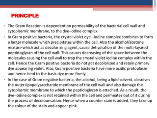
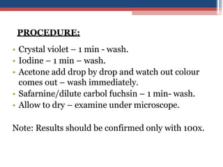
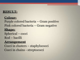
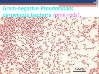
- Gram staining involves staining bacteria with crystal violet dye followed by treatment with iodine and decolorizing agent. Bacteria that retain the crystal violet stain are Gram-positive, while those that lose it and take up the counterstain are Gram-negative.
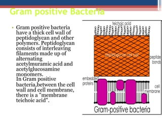
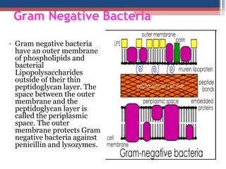
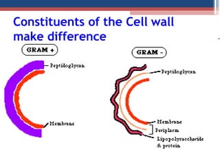
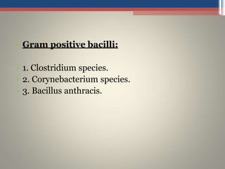
- The key difference between Gram-positive and Gram-negative bacteria is the thickness and composition of their cell walls, with Gram-positive having a thicker peptidoglycan layer that retains the crystal violet iodine complex.