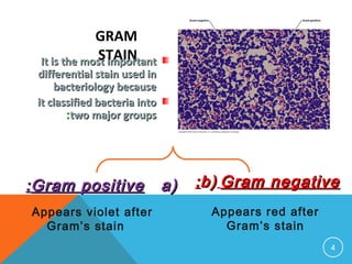
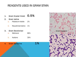
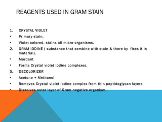
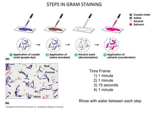
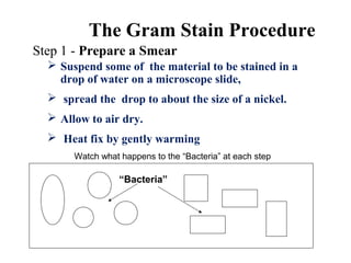
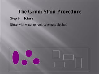
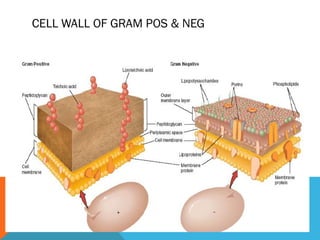
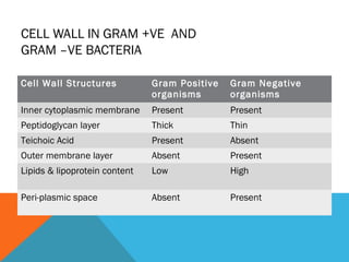
SARDAR VALLABHAI PATEL UNI
OF AGRICULTURE AND
TECHNOLOGY
PRESENTATION ON GRAM STAINING
OF BACTERIA
SUBMITTED BY:
ANUPAM KUMAR
SUBMITTED TO:-
PROF. KAMAL KHILADI
DEPTT- NEMATOLOGY
3
4
Gram StainGram Stain::
It is the most importantIt is the most important
differential stain used indifferential stain used in
bacteriology becausebacteriology because
it classified bacteria intoit classified bacteria into
two major groupstwo major groups::
a)a)Gram positiveGram positive::
Appears violet after
Gram’s stain
b)b) Gram negativeGram negative::
Appears red after
Gram’s stain
GRAM
STAIN
OBJECTIVES
 Describe reagents used in Gram stain &
purpose of these reagents.
 Color expected of Gram Pos & Gram Neg
after performing the procedure.
 Explain reason of differential stain by Gram
Pos & Gram Neg
 Describe cell wall structure of Gram Pos &
Negative bacteria.
REAGENTS USED IN GRAM STAIN
1. Gram Crystal Violet 0.5%
2. Gram Iodine
a. Potassium Iodide 2%
b. Resublimed Iodine 1%
3. Gram Decolorizer
a. Methanol 80%
b. Acetone 20%
4. Gram Safranine 1%
Gram negative
Gram positive
REAGENTS USED IN GRAM STAIN
1. CRYSTAL VIOLET
• Primary stain.
• Violet colored, stains all micro-organisms.
2. GRAM IODINE ( substance that combine with stain & there by fixes it in
material).
• Mordant
• Forms Crystal violet iodine complexes.
3. DECOLORIZER
• Acetone + Methanol
• Removes Crystal violet iodine complex from thin peptidoglycan layers
• Dissolves outer layer of Gram negative organism.
Time Frame
1) 1 minute
2) 1 minute
3) 15 seconds
4) 1 minute
Rinse with water between each step
STEPS IN GRAM STAINING
The Gram Stain Procedure
Step 1 - Prepare a Smear
Watch what happens to the “Bacteria” at each step
“Bacteria”
 Suspend some of the material to be stained in a
drop of water on a microscope slide,
 spread the drop to about the size of a nickel.
 Allow to air dry.
 Heat fix by gently warming
The Gram Stain Procedure
STEP 2- CRYSTAL VIOLETthe Primary
Stain
 Flood the Smear with Crystal Violet
 Allow to stand for 1 min.
 Rinse with water to remove excess stain.
The Gram Stain Procedure
Step 3 - Apply the Mordant
 Flood the Smear with Iodine solution.
 Allow to stand 2 mins.
The Gram Stain Procedure
Step 4 - Rinse
Rinse with water to remove excess Iodine
The Gram Stain Procedure
Step 5 - Decolorize
 Drip Decolorizer (80% Methanol +20% Acetone)
across the slide about 5 sec
 The effluent should appear pale or clear
The Gram Stain Procedure
Step 6 - Rinse
Rinse with water to remove excess alcohol
The Gram Stain Procedure
Step 7 - Counterstain
 Flood the slide with Safranin solution
 Let stand for 2 minutes
The Gram Stain
Step 8 - Rinse, Dry and Observe
Gram-Positive Gram-Negative
 Rinse with water to remove excess stain.
 Blot dry
 Observe under Oil Immersion
CELL WALL OF GRAM POS & NEG
CELL WALL IN GRAM +VE AND
GRAM –VE BACTERIA
Cell Wall Structures Gram Positive
organisms
Gram Negative
organisms
Inner cytoplasmic membrane Present Present
Peptidoglycan layer Thick Thin
Teichoic Acid Present Absent
Outer membrane layer Absent Present
Lipids & lipoprotein content Low High
Peri-plasmic space Absent Present
Gram's staining

Gram's staining

  • 2.
    SARDAR VALLABHAI PATELUNI OF AGRICULTURE AND TECHNOLOGY PRESENTATION ON GRAM STAINING OF BACTERIA SUBMITTED BY: ANUPAM KUMAR SUBMITTED TO:- PROF. KAMAL KHILADI DEPTT- NEMATOLOGY
  • 3.
  • 4.
    4 Gram StainGram Stain:: Itis the most importantIt is the most important differential stain used indifferential stain used in bacteriology becausebacteriology because it classified bacteria intoit classified bacteria into two major groupstwo major groups:: a)a)Gram positiveGram positive:: Appears violet after Gram’s stain b)b) Gram negativeGram negative:: Appears red after Gram’s stain GRAM STAIN
  • 5.
    OBJECTIVES  Describe reagentsused in Gram stain & purpose of these reagents.  Color expected of Gram Pos & Gram Neg after performing the procedure.  Explain reason of differential stain by Gram Pos & Gram Neg  Describe cell wall structure of Gram Pos & Negative bacteria.
  • 6.
    REAGENTS USED INGRAM STAIN 1. Gram Crystal Violet 0.5% 2. Gram Iodine a. Potassium Iodide 2% b. Resublimed Iodine 1% 3. Gram Decolorizer a. Methanol 80% b. Acetone 20% 4. Gram Safranine 1% Gram negative Gram positive
  • 7.
    REAGENTS USED INGRAM STAIN 1. CRYSTAL VIOLET • Primary stain. • Violet colored, stains all micro-organisms. 2. GRAM IODINE ( substance that combine with stain & there by fixes it in material). • Mordant • Forms Crystal violet iodine complexes. 3. DECOLORIZER • Acetone + Methanol • Removes Crystal violet iodine complex from thin peptidoglycan layers • Dissolves outer layer of Gram negative organism.
  • 8.
    Time Frame 1) 1minute 2) 1 minute 3) 15 seconds 4) 1 minute Rinse with water between each step STEPS IN GRAM STAINING
  • 9.
    The Gram StainProcedure Step 1 - Prepare a Smear Watch what happens to the “Bacteria” at each step “Bacteria”  Suspend some of the material to be stained in a drop of water on a microscope slide,  spread the drop to about the size of a nickel.  Allow to air dry.  Heat fix by gently warming
  • 10.
    The Gram StainProcedure STEP 2- CRYSTAL VIOLETthe Primary Stain  Flood the Smear with Crystal Violet  Allow to stand for 1 min.  Rinse with water to remove excess stain.
  • 11.
    The Gram StainProcedure Step 3 - Apply the Mordant  Flood the Smear with Iodine solution.  Allow to stand 2 mins.
  • 12.
    The Gram StainProcedure Step 4 - Rinse Rinse with water to remove excess Iodine
  • 13.
    The Gram StainProcedure Step 5 - Decolorize  Drip Decolorizer (80% Methanol +20% Acetone) across the slide about 5 sec  The effluent should appear pale or clear
  • 14.
    The Gram StainProcedure Step 6 - Rinse Rinse with water to remove excess alcohol
  • 15.
    The Gram StainProcedure Step 7 - Counterstain  Flood the slide with Safranin solution  Let stand for 2 minutes
  • 16.
    The Gram Stain Step8 - Rinse, Dry and Observe Gram-Positive Gram-Negative  Rinse with water to remove excess stain.  Blot dry  Observe under Oil Immersion
  • 17.
    CELL WALL OFGRAM POS & NEG
  • 18.
    CELL WALL INGRAM +VE AND GRAM –VE BACTERIA Cell Wall Structures Gram Positive organisms Gram Negative organisms Inner cytoplasmic membrane Present Present Peptidoglycan layer Thick Thin Teichoic Acid Present Absent Outer membrane layer Absent Present Lipids & lipoprotein content Low High Peri-plasmic space Absent Present