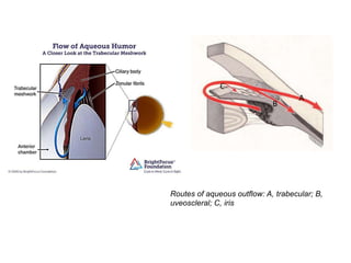
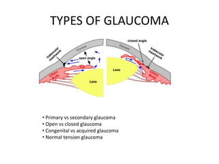
By Muhd Ariff Mahdzub
GLAUCOMA
GLAUCOMA
A group of eye conditions that
damage the optic nerve, often
caused by increase of IOP
AQUEOUS HUMOUR
• Maintains the intraocular pressure (IOP).
• Normal range of IOP varies between 10-21mmHg but there
is no absolute limit.
• It is secreted by ciliary body into posterior chamber which
the space between iris and lens
• Then, flows through the pupil into anterior chamber
between iris and cornea
• It drains to a sponge like structure located at base of iris
called trabecular meshwork and leave the eye
• There are two other route the fluid exits which are
uveoscleral drainage and iris.
• In healthy eyes, the rate of secretion balances the rate of
drainage
Routes of aqueous outflow: A, trabecular; B,
uveoscleral; C, iris
A
B
C
• For most types of glaucoma, optic neuropathy is
associated with a raised IOP
• This has given rise to the hypothesis of retinal
ganglion apoptosis, whose rate is influenced by
the hydrostatic pressure on the optic nerve head
and by compromise of the local microvasculature
• The resulting optic neuropathy gives rise to the
characteristic optic disc changes and visual field
loss.
TYPES OF GLAUCOMA
• Primary vs secondary glaucoma
• Open vs closed glaucoma
• Congenital vs acquired glaucoma
• Normal tension glaucoma
CLOSE ANGLE GLAUCOMA
• the normal drainage canals within the eye are
physically blocked.
• can be acute (sudden) or chronic (long-lasting).
• In acute angle-closure glaucoma, a sudden
increase in IOP occurs because of the build up of
aqueous humour.
• It is considered an emergency because
optic nerve damage and vision loss can occur
within hours of the onset of the problem.
• Chronic angle-closure glaucoma may cause vision
damage without symptoms.
ACUTE CLOSED ANGLE
– Symptom
• Painful red eye
(unilateral)
• Blurred vision
• Nausea and vomiting
• Unilateral headache
– Sign
• Conjunctival injection
• Hazy cornea
• Eclipse sign
• Non-reactive mid-dilated
pupil
CHRONIC CLOSED ANGLE
GLAUCOMA
– Symptom
• Often asymptomatic
• Hx of previous episode of
painful red aye with blurring
vision
• Hx of painful red eye
following pupil dilation
– Sign
• Elevated IOP
• Optic disc examination
shows glaucomatous
changes
• Visual field abnormalities
are seen on perimetry
OPEN ANGLE GLAUCOMA
• Also known as chronic simple glaucoma.
• The most common type of glaucoma affected people in worldwide.
• Caused by slow clogging of drainage canals due to wider angle btwn
iris and the cornea, thus increase the IOP
• Pathogenesis
– Direct mechanical damage to the retinal nerve fibre at the optic nerves
perhaps as they pass through the lamina cribrosa
– ischemic damage- possibly due to compression of blood vessels
supplying optic nerve head
These mechanism might lead to:
-reduction in axoplasmic flow
-interference with delivery of nutrients or removal of metabolic products
-deprivation of neuronal growth factor
-oxidative injury
-Initiation of immune-mediated damage
Greater than six-tenths is generally considered to be suspicious for glaucoma.
Asymmetry between two eyes of 0.2 is also significant
NORMAL TENSION GLAUCOMA
• Normal-tension glaucoma (NTG), also known as
low tension or normal pressure glaucoma, is a
form of glaucoma in which damage occurs to the
optic nerve without eye pressure exceeding the
normal range.
• Signs of optic nerve damage(glaucomatose
pattern)
• Visual field loss as damage progress
• No features of secondary glaucoma or a non
glaucomatose cause for neuropathy
NEOVASCULAR GLAUCOMA
• Secondary glaucoma.
• Retinal ischemia  release of VEGF  new
vascular structure over the meshwork in AC 
obstruction of meshwork  open/closed
angle glaucoma.
• Delayed management  loss of vision
• Manage by reduce the IOP, treat the causes.
MANAGEMENT
• Suppress of aqueous humor formation :
• Beta-blocker such as timolol, betaxolol and carteolol but it
contraindicated with patient asthmatic and bronchitis
• Carbonic anhydrase inhibitors by inhibiting beta-adrenoceptors on
the ciliary body such as dorzolamide and brinzolamide.
Contraindicated with metabolite impairment
• Alpha-adrenergic agonist such as apraclonidine and brimonidine.
• Enhancement of aqueous humor drainage :
• Prostaglandin analogues such as latanoprost, travoprost and
bimatoprost by increase outflow uveoscleral route but
contraindicated with pregnancy and breast feeding
• Cholinergic such as pilocarpine
MANAGEMENTCont.
-Argon laser trabeculoplasty (ALT) - lasering to the trabecular meshwork in
the iridocorneal angle, so enhancing aqueous outflow. It has the benefit of
reducing (or stopping) the need for drops whilst not having the
complications of surgery.
-Selective laser trabeculoplasty (SLT) - uses a laser at very low intensity,
treating specific areas and leaving parts of the trabecular meshwork intact. It
therefore, unlike ALT, preserves the trabecular meshwork architecture and
can be repeated
-Cyclodiode laser trabeculoplasty - similar principle as above, using a higher
laser power.This is often used in refractory cases and for relief of pain in end-
stage glaucoma
-YAG laser iridotomy - usually reserved for angle-closure glaucoma, a small
hole is made in the iris in patients with angle-closure glaucoma, to enhance
aqueous outflow.
-Diode laser cycloablation - part of the secretory component of the ciliary
body is destroyed, so reducing aqueous secretion. This is used in intractable
end-stage glaucoma.
MANAGEMENTCont.
• Trabeculectomy. This procedure creates a fistula
between the anterior chamber of the eye and
the sub-Tenon space (immediately around the
globe), so allowing aqueous outflow.
• Trabeculotomy for open-angle glaucoma aims to
reduce IOP by removing a portion of the
trabecular meshwork to improve drainage. It
avoids creation of the subconjunctival bleb
associated with traditional trabeculectomy
MANAGEMENTCont.
• Peripheral iridotomy (PI) - this refers to (usually two) holes
made in each iris with a laser, usually at around the 11 and
2 o'clock positions. This is to provide a free-flow transit
passage for the aqueous. Both eyes are treated, as the
fellow eye will be predisposed to an AAC attack too
• Surgical iridectomy - this is carried out where PI is not
possible. It is a less favoured option, as it is more invasive
and therefore more prone to complications
• Treatment aims is to slow down progression but cannot
reverse it. However, if treatment is timely, appropriate and
maintained, useful vision can be expected to be maintained
throughout the patient's lifetime

Glaucoma (opthalmology)

  • 1.
    By Muhd AriffMahdzub GLAUCOMA
  • 2.
    GLAUCOMA A group ofeye conditions that damage the optic nerve, often caused by increase of IOP
  • 3.
    AQUEOUS HUMOUR • Maintainsthe intraocular pressure (IOP). • Normal range of IOP varies between 10-21mmHg but there is no absolute limit. • It is secreted by ciliary body into posterior chamber which the space between iris and lens • Then, flows through the pupil into anterior chamber between iris and cornea • It drains to a sponge like structure located at base of iris called trabecular meshwork and leave the eye • There are two other route the fluid exits which are uveoscleral drainage and iris. • In healthy eyes, the rate of secretion balances the rate of drainage
  • 4.
    Routes of aqueousoutflow: A, trabecular; B, uveoscleral; C, iris A B C
  • 5.
    • For mosttypes of glaucoma, optic neuropathy is associated with a raised IOP • This has given rise to the hypothesis of retinal ganglion apoptosis, whose rate is influenced by the hydrostatic pressure on the optic nerve head and by compromise of the local microvasculature • The resulting optic neuropathy gives rise to the characteristic optic disc changes and visual field loss.
  • 6.
    TYPES OF GLAUCOMA •Primary vs secondary glaucoma • Open vs closed glaucoma • Congenital vs acquired glaucoma • Normal tension glaucoma
  • 7.
    CLOSE ANGLE GLAUCOMA •the normal drainage canals within the eye are physically blocked. • can be acute (sudden) or chronic (long-lasting). • In acute angle-closure glaucoma, a sudden increase in IOP occurs because of the build up of aqueous humour. • It is considered an emergency because optic nerve damage and vision loss can occur within hours of the onset of the problem. • Chronic angle-closure glaucoma may cause vision damage without symptoms.
  • 8.
    ACUTE CLOSED ANGLE –Symptom • Painful red eye (unilateral) • Blurred vision • Nausea and vomiting • Unilateral headache – Sign • Conjunctival injection • Hazy cornea • Eclipse sign • Non-reactive mid-dilated pupil CHRONIC CLOSED ANGLE GLAUCOMA – Symptom • Often asymptomatic • Hx of previous episode of painful red aye with blurring vision • Hx of painful red eye following pupil dilation – Sign • Elevated IOP • Optic disc examination shows glaucomatous changes • Visual field abnormalities are seen on perimetry
  • 9.
    OPEN ANGLE GLAUCOMA •Also known as chronic simple glaucoma. • The most common type of glaucoma affected people in worldwide. • Caused by slow clogging of drainage canals due to wider angle btwn iris and the cornea, thus increase the IOP • Pathogenesis – Direct mechanical damage to the retinal nerve fibre at the optic nerves perhaps as they pass through the lamina cribrosa – ischemic damage- possibly due to compression of blood vessels supplying optic nerve head These mechanism might lead to: -reduction in axoplasmic flow -interference with delivery of nutrients or removal of metabolic products -deprivation of neuronal growth factor -oxidative injury -Initiation of immune-mediated damage
  • 10.
    Greater than six-tenthsis generally considered to be suspicious for glaucoma. Asymmetry between two eyes of 0.2 is also significant
  • 11.
    NORMAL TENSION GLAUCOMA •Normal-tension glaucoma (NTG), also known as low tension or normal pressure glaucoma, is a form of glaucoma in which damage occurs to the optic nerve without eye pressure exceeding the normal range. • Signs of optic nerve damage(glaucomatose pattern) • Visual field loss as damage progress • No features of secondary glaucoma or a non glaucomatose cause for neuropathy
  • 12.
    NEOVASCULAR GLAUCOMA • Secondaryglaucoma. • Retinal ischemia  release of VEGF  new vascular structure over the meshwork in AC  obstruction of meshwork  open/closed angle glaucoma. • Delayed management  loss of vision • Manage by reduce the IOP, treat the causes.
  • 13.
    MANAGEMENT • Suppress ofaqueous humor formation : • Beta-blocker such as timolol, betaxolol and carteolol but it contraindicated with patient asthmatic and bronchitis • Carbonic anhydrase inhibitors by inhibiting beta-adrenoceptors on the ciliary body such as dorzolamide and brinzolamide. Contraindicated with metabolite impairment • Alpha-adrenergic agonist such as apraclonidine and brimonidine. • Enhancement of aqueous humor drainage : • Prostaglandin analogues such as latanoprost, travoprost and bimatoprost by increase outflow uveoscleral route but contraindicated with pregnancy and breast feeding • Cholinergic such as pilocarpine
  • 14.
    MANAGEMENTCont. -Argon laser trabeculoplasty(ALT) - lasering to the trabecular meshwork in the iridocorneal angle, so enhancing aqueous outflow. It has the benefit of reducing (or stopping) the need for drops whilst not having the complications of surgery. -Selective laser trabeculoplasty (SLT) - uses a laser at very low intensity, treating specific areas and leaving parts of the trabecular meshwork intact. It therefore, unlike ALT, preserves the trabecular meshwork architecture and can be repeated -Cyclodiode laser trabeculoplasty - similar principle as above, using a higher laser power.This is often used in refractory cases and for relief of pain in end- stage glaucoma -YAG laser iridotomy - usually reserved for angle-closure glaucoma, a small hole is made in the iris in patients with angle-closure glaucoma, to enhance aqueous outflow. -Diode laser cycloablation - part of the secretory component of the ciliary body is destroyed, so reducing aqueous secretion. This is used in intractable end-stage glaucoma.
  • 15.
    MANAGEMENTCont. • Trabeculectomy. Thisprocedure creates a fistula between the anterior chamber of the eye and the sub-Tenon space (immediately around the globe), so allowing aqueous outflow. • Trabeculotomy for open-angle glaucoma aims to reduce IOP by removing a portion of the trabecular meshwork to improve drainage. It avoids creation of the subconjunctival bleb associated with traditional trabeculectomy
  • 16.
    MANAGEMENTCont. • Peripheral iridotomy(PI) - this refers to (usually two) holes made in each iris with a laser, usually at around the 11 and 2 o'clock positions. This is to provide a free-flow transit passage for the aqueous. Both eyes are treated, as the fellow eye will be predisposed to an AAC attack too • Surgical iridectomy - this is carried out where PI is not possible. It is a less favoured option, as it is more invasive and therefore more prone to complications • Treatment aims is to slow down progression but cannot reverse it. However, if treatment is timely, appropriate and maintained, useful vision can be expected to be maintained throughout the patient's lifetime