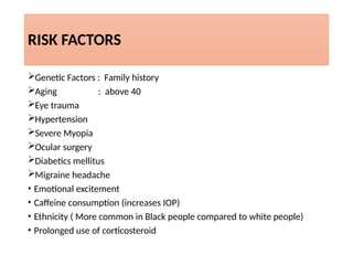
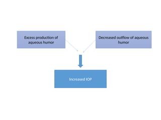
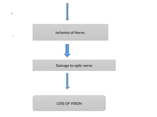
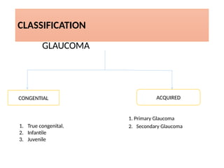
.
GLAUCOMA
PRESENTED BY
Sinead Malama
INTRODUCTION
The term Glaucoma is a term used to refer group of ocular condition
characterized by the optic nerve damage. Glaucoma occurs as a result
of increased intraocular pressure (IOP) caused by a malformation or
malfunction of the eyes drainage system. Normal IOP is 19 – 21 inches
of mercury. The increased pressure causes compression of the retina
and the optic nerve, and causes progressive , permanent loss of
eyesight if left untreated.
DEFINITION
Glaucoma is a group of disorder characterized by an abnormally high
intraocular pressure , optic nerve dystrophy, and peripheral filed loss.
Glaucoma is a symptomatic condition of the eye where the
intraocular pressure is more than normal.
.
• Normal IOP is 16 to 23 mmHg.
INCIDENCE
Globally 6 to 67 million.
More common in peoples older than 40 years.
ETIOLOGY
Increased intraocular pressure.( more than 24 mmhg)
Optic nerve dystrophy.
RISK FACTORS
Genetic Factors : Family history
Aging : above 40
Eye trauma
Hypertension
Severe Myopia
Ocular surgery
Diabetics mellitus
Migraine headache
• Emotional excitement
• Caffeine consumption (increases IOP)
• Ethnicity ( More common in Black people compared to white people)
• Prolonged use of corticosteroid
Excess production of
aqueous humor
Decreased outflow of aqueous
humor
Increased IOP
.
. Ischemia of Nerve.
Damage to optic nerve
LOSS OF VISION
CLASSIFICATION
GLAUCOMA
1. Primary Glaucoma
2. Secondary Glaucoma
CONGENTIAL ACQUIRED
1. True congenital.
2. Infantile
3. Juvenile
CONGENTIAL GLUCOMA
 Rare disease.
Occurs when a congenital defect in the angle of the anterior chamber
obstructs the outflow of aqueous humor.
If remains untreated causes damage to optic nerve damage and
blindness.
1. True Congenital Glaucoma
occur when IOP increases during intrauterine period.
Child is born with ocular enlargement.
occur in 40 % of cases
.
• INFANTILE GLAUCOMA
occurs during third birthday
occurs about 10% of cases.
• JUVENILE GLAUCOMA
Occurs during 3 to 16 years of life.
occurs in about 10 % of casas
Clinical features of congenital glaucoma
• Lacrimation
• Photophobia
• Corneal oedema
• Raised IOP
• Eyes Become MYOPIC
• Corneal diameter more than 13mmhg
ACQUIRED GLAUCOMA
• PRIMARY GLUCOMA.
o Primary Open angle glaucoma.
o Primary angle closure Glaucoma
o Chronic angle closure glaucoma.
• SECONDARY GLAUCOMA
o Lens induced glaucoma
o Glaucoma due to uveitis
o Neurovascular Glaucoma
o Glaucoma associated with intraocular tumor
o Steroid induced glaucoma
.
• PRIMARY GLAUCOMA.
Primary Glaucoma is a progressive condition and is most common
cause of irreversible blindness across world wide.
a. Primary Open Angle Glaucoma
 Also Called as open angle Glaucoma or chronic simple Glaucoma or
simple complex Glaucoma.
Results from the overproduction of aqueous humour through
trabecular Mesh work results in increased IOP and Damage to optic
nerve, results in loss of vision.
.
• The clinical features of Primary Open angle glaucoma includes.
 Mild ache in the eyes
Headache
Increased IOP ( more than24 mmhg)
Loss of Peripheral vision
Reduced visual acquity at night.
Corneal edema
Visual field deficit.
b. Primary angle closure glaucoma
• It is also called as Primary closed angle glaucoma, Narrow angle
glaucoma, Pupil block glaucoma and acute
congestive glaucoma.
•Onset : rapid, ophthalmic emergency. Unless
treated promptly the causes blindness in 3 or 5
days.
•This is due to the abnormality of structure infront of
the eyes. This result from the obstruction to the
outflow of aqueous humor.
Clinical features of primary angle closure
glaucoma
• Pain and redness in eyes
• Increased IOP
• Blurred vision
• Headache
• Nausea
• Vomiting
• Oedematous cornea
• Decreased visual acuity
• Moderate Pupillary dilation.
c. Chronic angle closure Glaucoma
• Chronic angle closure Glaucoma may develop as the sequelae to an
attack of acute angle glaucoma.
• Clinical features include
Increased IOP.
visual field defect.
Decreased visual
acquity.
2. Secondary glaucoma
• Secondary glaucoma occurs as a result of either diseases within the
eyes such as uveitis, Inflammation , Trauma, intra ocular
haemorrhage, previous surgeries, diabetics and steroid medication
etc
• The major types include
.
• TYPES
 LENS INDUCED GLAUCOMA
 GLAUCOMA DUE TO UVEITIS
 NEUROVASULAR GLAUCOMA
 GLAUCOMA ASSOCIATED WITH INTRA OCULAR
TUMOR.
 STEROID INDUCED GLAUCOMA
.
1. LENS INDUCED GLAUCOMA
o It occur due to trabecular blockage.
o it occur due to clogging of trabeculae by
inflammatory material.
2. GLAUCOMA DUE TO UVEITIS
o IOP is raised due to clogging by inflammatory material & associated
trabeculitis.
3. Neurovascular glaucoma
• Uncommon type of glaucoma
• Difficult to treat
• Caused by proliferative diabetic retinopathy.
• Individual with poor blood flow to the eyes are highly at risk for this
condition.
4. Glaucoma associated with intra ocular
Tumor.
Intraocular tumor such as retinoblastoma & Malignant melanoma may
rise IOP.
5. Steroid induced Glaucoma
• Developed due to sensitivity to steroid.
• Sudden rise in IOP may occur, this can be prevented by judicious use
of steroid.
6. Pigmentary Glaucoma
• Rare Condition
• This is caused by pigment cells sloughing off from the back of the iris
& floating around the aqueous humor.
DIAGNOSTIC MEASURES
oHistory collection
oPatient ocular & Medical history.
oTonometry ( to measure IOP)
oOphthalmoscopy ( To show the cupping of the optic disc )
oGonioscopy (To determine the angle of the eyes anterior chamber)
oPerimetry or visual field test.( To detect loss of peripheral vision)
.
oSlit Lamp Examination.
oFundus Photography ( To monitor the Disc for changes)
oPachymetry
oNerve fiber analysis ( to asses the thickness of nerve fiber layer)
MANAGEMENT
• Goal of Management measures include
 To reduce IOP
To prevent the damage of optic nerve.
Management measures mainly include
 Medical Management
Surgical management
 Nursing Management
MEDICAL MANAGEMENT
• Beta adrenergic blockers : decreases aqueous humor production , eg
Timolol, betaxolol.
• Cholinergic ( Miotics) : Reduces IOP by facilitating the outflow of
aqueous humor.
eg Pilocarpine, Carbacol.
• Carbonic anhydrase inhibitor : Decreases the formation & secretion
of aqueous humor.
.
• Prostaglandin Analogs : Reduces IOP by increasing uveoscleral Flow.
• Osmotic Agents : Iv mannitol 20% or oral glycerine 50% is used to
reduce IOP by creating an osmotic pressure between blood and
intraocular fluid
SURGICAL MANAGEMENT
• Argon Laser Trabeculoplasty :
Used to treat open angle glaucoma.
Thermal Argon laser burns are applied to the inner surface of
trabecular Meshwork to open intra trabecular spaces , thus reduces
outflow of aqueous humor and decreases IOP.
• Laser Iridotomy : In this surgical procedure,
an opening is made by the laser beam in the
iris to eliminate pupillary block.
• Relive the pressure & preserve the vision by promoting outflow of
the aqueous humor.
• Cyclocryotherapy
Application of a freezing probe to the sclera over the Cillary body that
destroy some of the cillary processes , results in the reduction of the
amount of the aqueous humor
.
• Cyclodialysis
Through a small incision in the sclera a spatula type instrument is passed
into the anterior chamber, creating an opening in the angle.
• Filtering Procedure
For Chronic Glaucoma filtering procedure are used to create an
opening or fistula in the trabecular meshwork to drain aqueous humor.
This allow the aqueous humor to flow & exit different route.
.
• Trabeculotomy
In this procedure a partial thickness incision is made in the sclera.
Section of sclera is removed to produce an opening for outflow of
aqueous humor.
• Drainage implants and shunts
 Used to Shunt the aqueous humor in the Conjunctival space.
Implants and shunts are the open tubes implanted in the anterior
chamber through sclerotomy
.
General Physical examination
• Assess for Sudden severe pain in eyes, Halo light, blurred vision etc.
Check for diagnostic Measures
Assess the patient Understanding and emotional response to the
condition.
Complications
• Complete loss of vision
• Choroidal detachment
• Retinal detachment
• Chronic hypotony
• Anterior Segment necrosis
Research studies
Safety & efficiency of Biomatoprost sustained – release in patients
with open angle glaucoma or Ocular hypertension.
CONCLUSION
• Glaucoma is a condition that causes damage to the eyes optic nerve&
gets worse over the time. Without treatment the glaucoma can cause
permanent blindness within few years. So the treatment should be
given at right time to prevent complications.
.

glaucoma Final PowerPoint done my Malama

  • 1.
  • 2.
    INTRODUCTION The term Glaucomais a term used to refer group of ocular condition characterized by the optic nerve damage. Glaucoma occurs as a result of increased intraocular pressure (IOP) caused by a malformation or malfunction of the eyes drainage system. Normal IOP is 19 – 21 inches of mercury. The increased pressure causes compression of the retina and the optic nerve, and causes progressive , permanent loss of eyesight if left untreated.
  • 3.
    DEFINITION Glaucoma is agroup of disorder characterized by an abnormally high intraocular pressure , optic nerve dystrophy, and peripheral filed loss. Glaucoma is a symptomatic condition of the eye where the intraocular pressure is more than normal.
  • 4.
    . • Normal IOPis 16 to 23 mmHg. INCIDENCE Globally 6 to 67 million. More common in peoples older than 40 years.
  • 5.
    ETIOLOGY Increased intraocular pressure.(more than 24 mmhg) Optic nerve dystrophy.
  • 6.
    RISK FACTORS Genetic Factors: Family history Aging : above 40 Eye trauma Hypertension Severe Myopia Ocular surgery Diabetics mellitus Migraine headache • Emotional excitement • Caffeine consumption (increases IOP) • Ethnicity ( More common in Black people compared to white people) • Prolonged use of corticosteroid
  • 8.
    Excess production of aqueoushumor Decreased outflow of aqueous humor Increased IOP
  • 9.
    . . Ischemia ofNerve. Damage to optic nerve LOSS OF VISION
  • 10.
    CLASSIFICATION GLAUCOMA 1. Primary Glaucoma 2.Secondary Glaucoma CONGENTIAL ACQUIRED 1. True congenital. 2. Infantile 3. Juvenile
  • 11.
    CONGENTIAL GLUCOMA  Raredisease. Occurs when a congenital defect in the angle of the anterior chamber obstructs the outflow of aqueous humor. If remains untreated causes damage to optic nerve damage and blindness. 1. True Congenital Glaucoma occur when IOP increases during intrauterine period. Child is born with ocular enlargement. occur in 40 % of cases
  • 12.
    . • INFANTILE GLAUCOMA occursduring third birthday occurs about 10% of cases. • JUVENILE GLAUCOMA Occurs during 3 to 16 years of life. occurs in about 10 % of casas
  • 13.
    Clinical features ofcongenital glaucoma • Lacrimation • Photophobia • Corneal oedema • Raised IOP • Eyes Become MYOPIC • Corneal diameter more than 13mmhg
  • 14.
    ACQUIRED GLAUCOMA • PRIMARYGLUCOMA. o Primary Open angle glaucoma. o Primary angle closure Glaucoma o Chronic angle closure glaucoma. • SECONDARY GLAUCOMA o Lens induced glaucoma o Glaucoma due to uveitis o Neurovascular Glaucoma o Glaucoma associated with intraocular tumor o Steroid induced glaucoma
  • 15.
    . • PRIMARY GLAUCOMA. PrimaryGlaucoma is a progressive condition and is most common cause of irreversible blindness across world wide. a. Primary Open Angle Glaucoma  Also Called as open angle Glaucoma or chronic simple Glaucoma or simple complex Glaucoma. Results from the overproduction of aqueous humour through trabecular Mesh work results in increased IOP and Damage to optic nerve, results in loss of vision.
  • 16.
    . • The clinicalfeatures of Primary Open angle glaucoma includes.  Mild ache in the eyes Headache Increased IOP ( more than24 mmhg) Loss of Peripheral vision Reduced visual acquity at night. Corneal edema Visual field deficit.
  • 17.
    b. Primary angleclosure glaucoma • It is also called as Primary closed angle glaucoma, Narrow angle glaucoma, Pupil block glaucoma and acute congestive glaucoma. •Onset : rapid, ophthalmic emergency. Unless treated promptly the causes blindness in 3 or 5 days. •This is due to the abnormality of structure infront of the eyes. This result from the obstruction to the outflow of aqueous humor.
  • 18.
    Clinical features ofprimary angle closure glaucoma • Pain and redness in eyes • Increased IOP • Blurred vision • Headache • Nausea • Vomiting • Oedematous cornea • Decreased visual acuity • Moderate Pupillary dilation.
  • 19.
    c. Chronic angleclosure Glaucoma • Chronic angle closure Glaucoma may develop as the sequelae to an attack of acute angle glaucoma. • Clinical features include Increased IOP. visual field defect. Decreased visual acquity.
  • 20.
    2. Secondary glaucoma •Secondary glaucoma occurs as a result of either diseases within the eyes such as uveitis, Inflammation , Trauma, intra ocular haemorrhage, previous surgeries, diabetics and steroid medication etc • The major types include
  • 21.
    . • TYPES  LENSINDUCED GLAUCOMA  GLAUCOMA DUE TO UVEITIS  NEUROVASULAR GLAUCOMA  GLAUCOMA ASSOCIATED WITH INTRA OCULAR TUMOR.  STEROID INDUCED GLAUCOMA
  • 22.
    . 1. LENS INDUCEDGLAUCOMA o It occur due to trabecular blockage. o it occur due to clogging of trabeculae by inflammatory material. 2. GLAUCOMA DUE TO UVEITIS o IOP is raised due to clogging by inflammatory material & associated trabeculitis.
  • 23.
    3. Neurovascular glaucoma •Uncommon type of glaucoma • Difficult to treat • Caused by proliferative diabetic retinopathy. • Individual with poor blood flow to the eyes are highly at risk for this condition. 4. Glaucoma associated with intra ocular Tumor. Intraocular tumor such as retinoblastoma & Malignant melanoma may rise IOP.
  • 24.
    5. Steroid inducedGlaucoma • Developed due to sensitivity to steroid. • Sudden rise in IOP may occur, this can be prevented by judicious use of steroid. 6. Pigmentary Glaucoma • Rare Condition • This is caused by pigment cells sloughing off from the back of the iris & floating around the aqueous humor.
  • 25.
    DIAGNOSTIC MEASURES oHistory collection oPatientocular & Medical history. oTonometry ( to measure IOP) oOphthalmoscopy ( To show the cupping of the optic disc ) oGonioscopy (To determine the angle of the eyes anterior chamber) oPerimetry or visual field test.( To detect loss of peripheral vision)
  • 26.
    . oSlit Lamp Examination. oFundusPhotography ( To monitor the Disc for changes) oPachymetry oNerve fiber analysis ( to asses the thickness of nerve fiber layer)
  • 27.
    MANAGEMENT • Goal ofManagement measures include  To reduce IOP To prevent the damage of optic nerve. Management measures mainly include  Medical Management Surgical management  Nursing Management
  • 28.
    MEDICAL MANAGEMENT • Betaadrenergic blockers : decreases aqueous humor production , eg Timolol, betaxolol. • Cholinergic ( Miotics) : Reduces IOP by facilitating the outflow of aqueous humor. eg Pilocarpine, Carbacol. • Carbonic anhydrase inhibitor : Decreases the formation & secretion of aqueous humor.
  • 29.
    . • Prostaglandin Analogs: Reduces IOP by increasing uveoscleral Flow. • Osmotic Agents : Iv mannitol 20% or oral glycerine 50% is used to reduce IOP by creating an osmotic pressure between blood and intraocular fluid
  • 30.
    SURGICAL MANAGEMENT • ArgonLaser Trabeculoplasty : Used to treat open angle glaucoma. Thermal Argon laser burns are applied to the inner surface of trabecular Meshwork to open intra trabecular spaces , thus reduces outflow of aqueous humor and decreases IOP.
  • 31.
    • Laser Iridotomy: In this surgical procedure, an opening is made by the laser beam in the iris to eliminate pupillary block. • Relive the pressure & preserve the vision by promoting outflow of the aqueous humor. • Cyclocryotherapy Application of a freezing probe to the sclera over the Cillary body that destroy some of the cillary processes , results in the reduction of the amount of the aqueous humor
  • 32.
    . • Cyclodialysis Through asmall incision in the sclera a spatula type instrument is passed into the anterior chamber, creating an opening in the angle. • Filtering Procedure For Chronic Glaucoma filtering procedure are used to create an opening or fistula in the trabecular meshwork to drain aqueous humor. This allow the aqueous humor to flow & exit different route.
  • 33.
    . • Trabeculotomy In thisprocedure a partial thickness incision is made in the sclera. Section of sclera is removed to produce an opening for outflow of aqueous humor. • Drainage implants and shunts  Used to Shunt the aqueous humor in the Conjunctival space. Implants and shunts are the open tubes implanted in the anterior chamber through sclerotomy
  • 34.
    . General Physical examination •Assess for Sudden severe pain in eyes, Halo light, blurred vision etc. Check for diagnostic Measures Assess the patient Understanding and emotional response to the condition.
  • 35.
    Complications • Complete lossof vision • Choroidal detachment • Retinal detachment • Chronic hypotony • Anterior Segment necrosis
  • 36.
    Research studies Safety &efficiency of Biomatoprost sustained – release in patients with open angle glaucoma or Ocular hypertension.
  • 37.
    CONCLUSION • Glaucoma isa condition that causes damage to the eyes optic nerve& gets worse over the time. Without treatment the glaucoma can cause permanent blindness within few years. So the treatment should be given at right time to prevent complications.
  • 38.