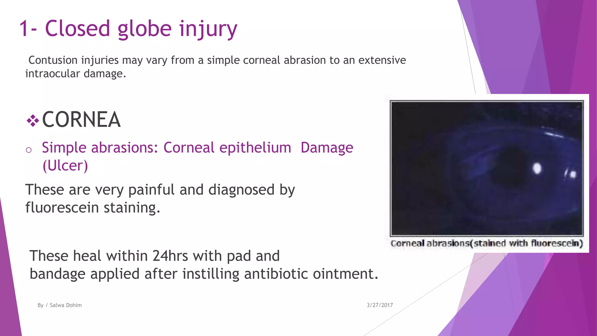
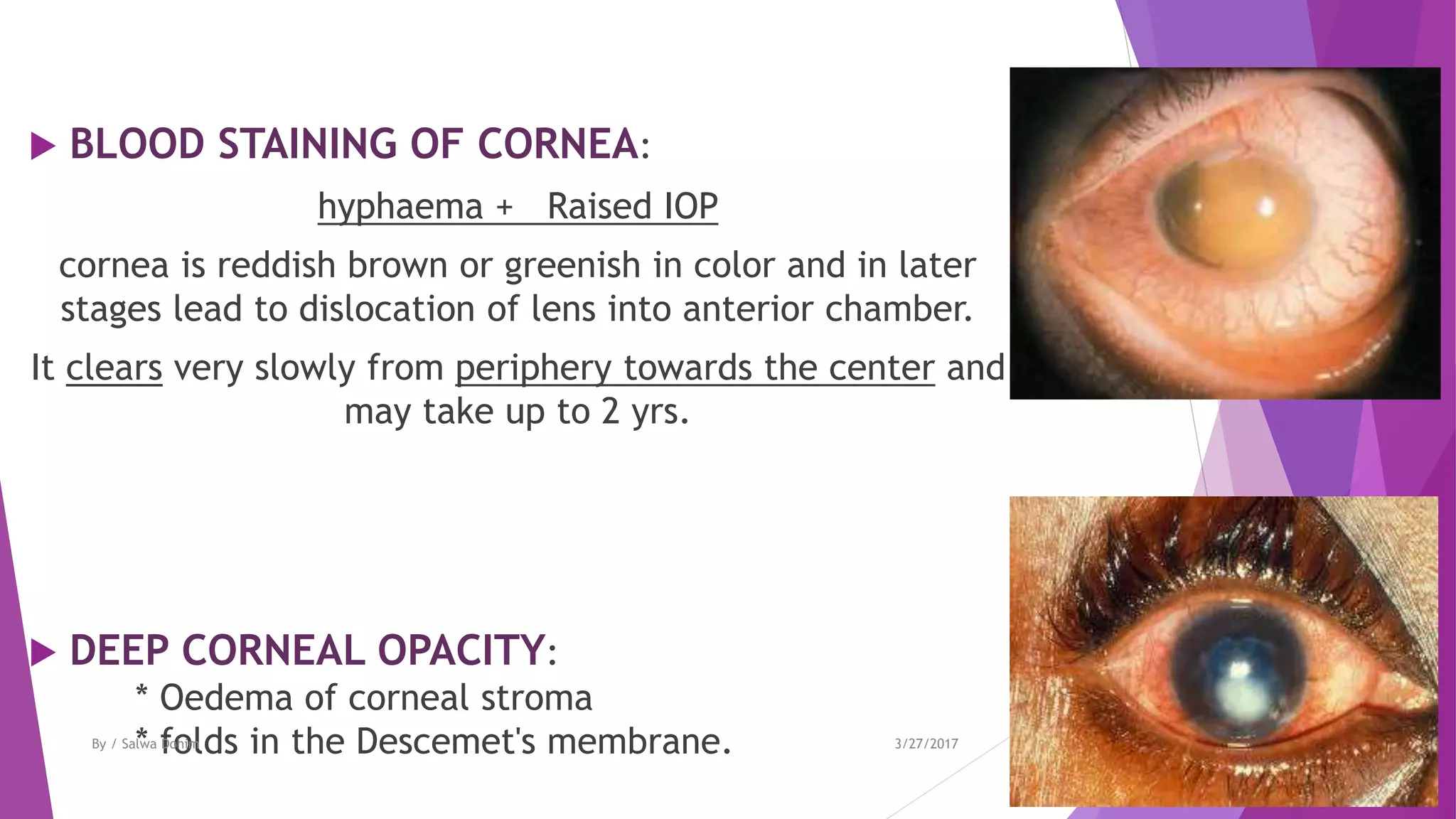
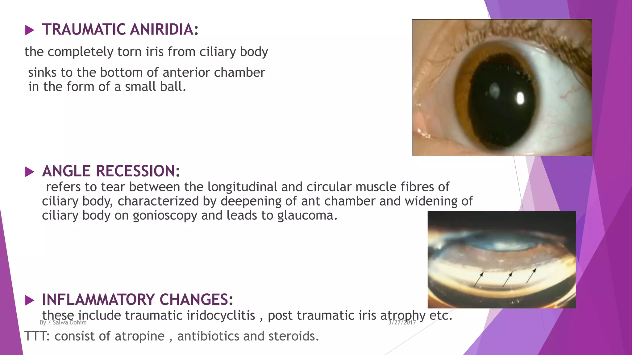
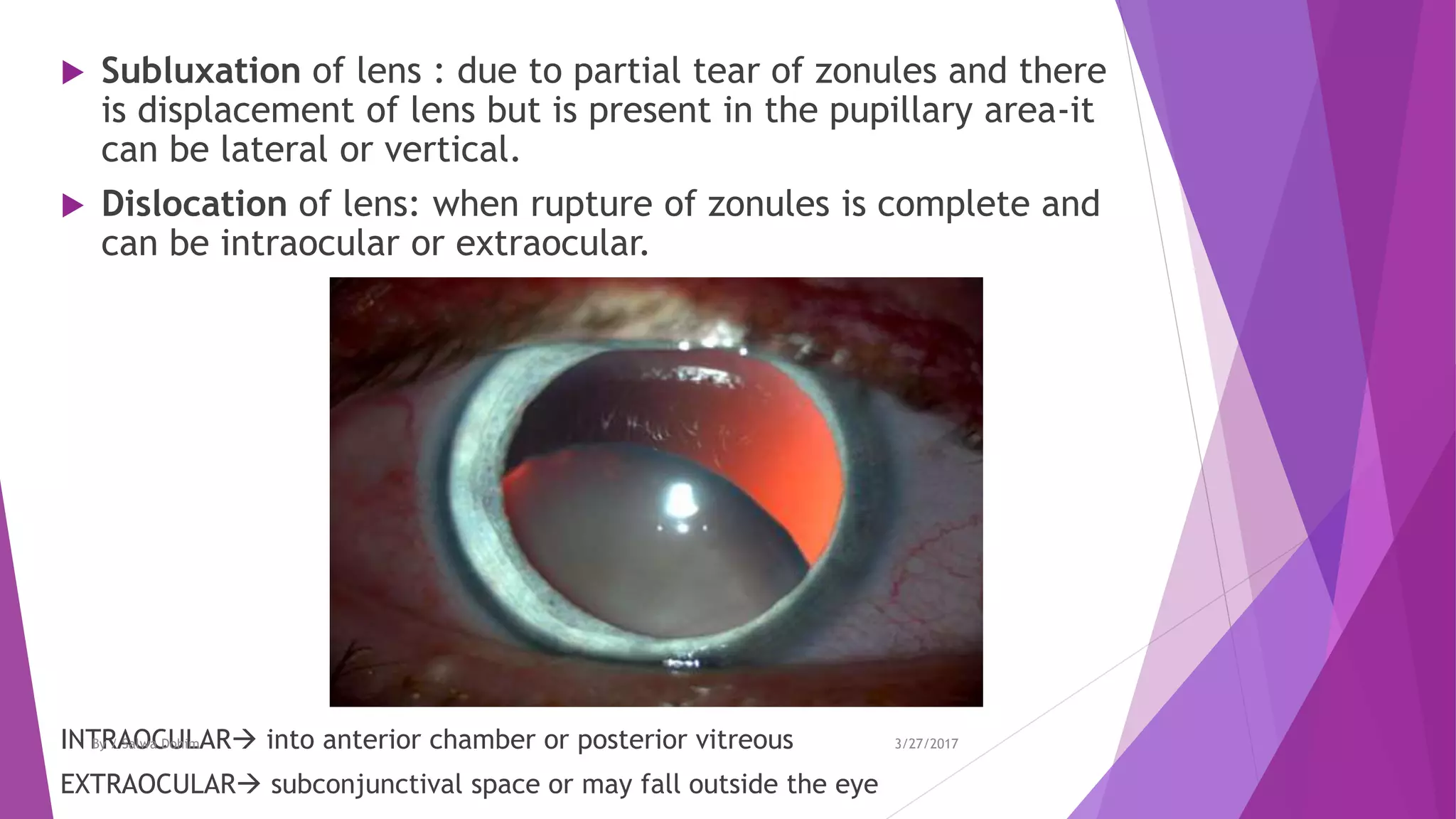
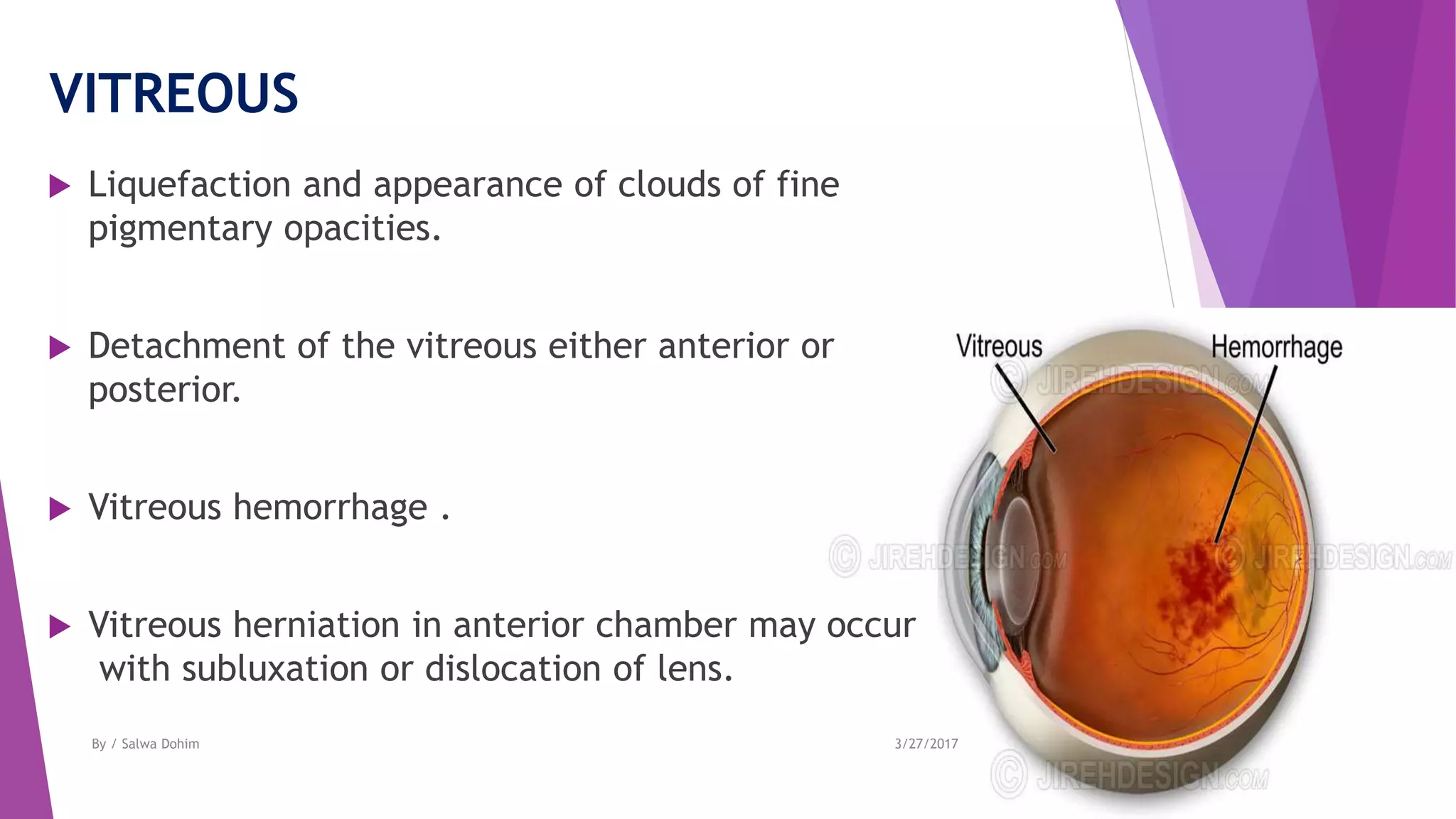
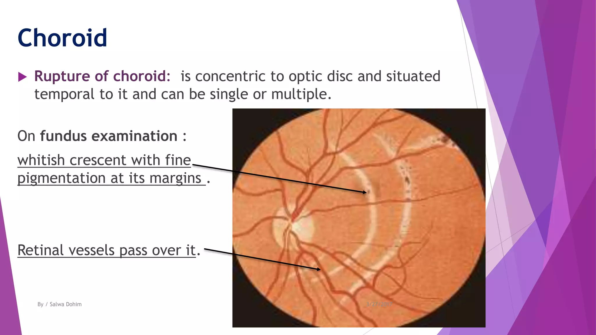
Blunt trauma
of the eye
Presented By:
Salwa Ahmed Elkelany
5th year – Ophthalmology department
Faculty of medicine –Suez Canal university
3/27/2017By / Salwa Dohim
Types of Ocular injuries :
1. Blunt
2. Perforating
3. Foreign Body
4. Radiation
5. Chemical
6. Thermal
3/27/2017By / Salwa Dohim
Injuries occur during blunt
trauma
Coup or direct
Eg: corneal
abrasions
Antero-posterior
compression &
Horizontal
expansion
Eg: rupture globe
Countercoup due to
transmitted pressure
waves to the post.
Pole
Eg : commotio retina
3/27/2017By / Salwa Dohim
Modes of injury
- Blunt trauma may occur by :
Direct blow to the eye ball by fist, ball or blunt instruments like sticks, and
big stones.
- Accidental blunt trauma to eye ball
may also occur in accidents, injuries by instruments , fall upon the projecting
blunt objects.
3/27/2017By / Salwa Dohim
Mechanism of blunt trauma to Eye Ball
3/27/2017By / Salwa Dohim
Traumatic lesions of blunt trauma
1. CLOSED GLOBE INJURY
2. GLOBE RUPTURE
3. EXTRAOCULAR LESIONS
3/27/2017By / Salwa Dohim
1- Closed globe injury
Contusion injuries may vary from a simple corneal abrasion to an extensive
intraocular damage.
CORNEA
o Simple abrasions: Corneal epithelium Damage
(Ulcer)
These are very painful and diagnosed by
fluorescein staining.
These heal within 24hrs with pad and
bandage applied after instilling antibiotic ointment.
3/27/2017By / Salwa Dohim
○ Recurrent corneal erosions
(recurrent keractalgia)
Caused by fingernail trauma. Patient presents with recurrent
attacks of acute pain and lacrimation on opening eye in the
morning and is due to abnormally loose attachment of epithelium
to Bowman’s membrane.
○ Partial corneal tears ( lamellar corneal lacerations )
3/27/2017By / Salwa Dohim
 BLOOD STAINING OF CORNEA:
hyphaema + Raised IOP
cornea is reddish brown or greenish in color and in later
stages lead to dislocation of lens into anterior chamber.
It clears very slowly from periphery towards the center and
may take up to 2 yrs.
 DEEP CORNEAL OPACITY:
* Oedema of corneal stroma
* folds in the Descemet's membrane. 3/27/2017By / Salwa Dohim
Sclera
 Partial thickness scleral wounds occur alone or
with other lesions of closed globe type.
3/27/2017By / Salwa Dohim
Anterior chamber
 TRAUMATIC HYPHAEMA or blood in anterior chamber which
occurs due to injury to iris or anterior ciliary vessels.
 EXUDATES: these collect here following traumatic uveitis
3/27/2017By / Salwa Dohim
Iris , pupil and ciliary body
 TRAUMATIC MIOSIS:
occurs initially due to irritation of ciliary nerves or with spasm of
accommodation.
 TRAUMATIC MYDRIASIS(iridoplegia):
It is permanent and is associated with traumatic cycloplegia.
 RUPTURE OF PUPILARY MARGIN
 RADIATING TEARS IN THE IRIS STROMA
 IRIDODIALYSIS:
detachment of iris from its root at ciliary body
which results in ‘D’ shaped pupil
* Anti-flexion * Retro-flexion
3/27/2017By / Salwa Dohim
 TRAUMATIC ANIRIDIA:
the completely torn iris from ciliary body
sinks to the bottom of anterior chamber
in the form of a small ball.
 ANGLE RECESSION:
refers to tear between the longitudinal and circular muscle fibres of
ciliary body, characterized by deepening of ant chamber and widening of
ciliary body on gonioscopy and leads to glaucoma.
 INFLAMMATORY CHANGES:
these include traumatic iridocyclitis , post traumatic iris atrophy etc.
TTT: consist of atropine , antibiotics and steroids.
3/27/2017By / Salwa Dohim
lens
 VOSSIUS RING :
It is a circular ring of brown pigment seen on the anterior capsule .
It occurs due to striking of contracted
pupillary margin against the lens.
 CONCUSSION CATARACT:
due to imbibition of aqueous and due to direct mechanical effects
of the injury on lens fibers
rosette cataract
3/27/2017By / Salwa Dohim
 Subluxation of lens : due to partial tear of zonules and there
is displacement of lens but is present in the pupillary area-it
can be lateral or vertical.
 Dislocation of lens: when rupture of zonules is complete and
can be intraocular or extraocular.
INTRAOCULAR into anterior chamber or posterior vitreous
EXTRAOCULAR subconjunctival space or may fall outside the eye
3/27/2017By / Salwa Dohim
VITREOUS
 Liquefaction and appearance of clouds of fine
pigmentary opacities.
 Detachment of the vitreous either anterior or
posterior.
 Vitreous hemorrhage .
 Vitreous herniation in anterior chamber may occur
with subluxation or dislocation of lens.
3/27/2017By / Salwa Dohim
Choroid
 Rupture of choroid: is concentric to optic disc and situated
temporal to it and can be single or multiple.
On fundus examination :
whitish crescent with fine
pigmentation at its margins .
Retinal vessels pass over it.
3/27/2017By / Salwa Dohim
- Choroidal hemorrhage may occur under retina
or may even enter the vitreous if retina is torn.
- Choroidal detachment
- Traumatic choroiditis :
seen on fundus examination as patches
of pigmentation and discoloration after eye becomes silent.
3/27/2017By / Salwa Dohim
Retina
 COMMOTIO RETINAE(Berlin’s oedema):
It manifests as milky white cloudiness involving of posterior pole
with a cherry- red spot in the foveal region .
disappear after some days pigmentary changes.
 RETINAL HAEMORRHAGES:
eg: flame shaped and preretinal D shaped haemorrhage may be
associated with traumatic retinopathy.
 RETINAL TEARS:
these follow a contusion in eyes suffering from myopia or senile
degenerations.
3/27/2017By / Salwa Dohim
 TRAUMATIC PROLIFERATIVE RETINOPATHY :
occur secondary to vitreous haemorrhage.
 RETINAL DETACHMENT:
follows retinal tears or vitreo-retinal tractional bands.
 CONCUSSION CHANGES AT MACULA: traumatic macular oedema
followed by pigmentary degeneration.
Sometimes, a macular cyst is formed , which on rupture is
converted to a lamellar or full thickness macular hole.
3/27/2017By / Salwa Dohim
IOP changes in closed globe injury
 TRAUMATIC GLAUCOMA
 TRAUMATIC HYPOTONY
it may follow damage to the ciliary body.
3/27/2017By / Salwa Dohim
Traumatic changes in refraction
 Myopia may follow ciliary spasm or rupture of zonules or
anterior shift of lens
 Hypermetropia and loss of accommodation may result
from damage to the ciliary body(cycloplegia).
3/27/2017By / Salwa Dohim
2- GLOBE RUPTURE
It is a full-thickness wound of eye-wall caused by blunt object and
can occur in 2 ways:
 DIRECT RUPTURE: at the site of injury.
 INDIRECT RUPTURE : occurs because of compression force .The
impact results in momentary increase in IOP and inside out injury at the
weakest part of the eye wall
TREATMENT:
A badly damaged globe should be enucleated .
In less severe cases it can be repaired under general anesthesia.
3/27/2017By / Salwa Dohim
Eye lids lesion Conjunctival Injury Orbital injury Optic n. Injury
- Laceration and avulsion of
lids may occur .
subconjunctival
hemorrhage
-fracture of orbital walls.
-Orbital hemorrhage
sudden proptosis
-Orbital emphysema may
occur following ethmoidal
sinus rupture
associated with
fracture of base
of skull
3- Extraocular lesions
3/27/2017By / Salwa Dohim
You can refer to :
 http://eyewiki.aao.org/Ocular_Trauma%3A_Acute_Evaluation,_Cataract,_G
laucoma
 http://www.medscape.com/viewarticle/813605
3/27/2017By / Salwa Dohim
Merci Bien
Take care of your Eyes ..
3/27/2017By / Salwa Dohim

Blunt trauma of the eye

  • 1.
    Blunt trauma of theeye Presented By: Salwa Ahmed Elkelany 5th year – Ophthalmology department Faculty of medicine –Suez Canal university 3/27/2017By / Salwa Dohim
  • 2.
    Types of Ocularinjuries : 1. Blunt 2. Perforating 3. Foreign Body 4. Radiation 5. Chemical 6. Thermal 3/27/2017By / Salwa Dohim
  • 3.
  • 4.
    Injuries occur duringblunt trauma Coup or direct Eg: corneal abrasions Antero-posterior compression & Horizontal expansion Eg: rupture globe Countercoup due to transmitted pressure waves to the post. Pole Eg : commotio retina 3/27/2017By / Salwa Dohim
  • 5.
    Modes of injury -Blunt trauma may occur by : Direct blow to the eye ball by fist, ball or blunt instruments like sticks, and big stones. - Accidental blunt trauma to eye ball may also occur in accidents, injuries by instruments , fall upon the projecting blunt objects. 3/27/2017By / Salwa Dohim
  • 6.
    Mechanism of blunttrauma to Eye Ball 3/27/2017By / Salwa Dohim
  • 7.
    Traumatic lesions ofblunt trauma 1. CLOSED GLOBE INJURY 2. GLOBE RUPTURE 3. EXTRAOCULAR LESIONS 3/27/2017By / Salwa Dohim
  • 8.
    1- Closed globeinjury Contusion injuries may vary from a simple corneal abrasion to an extensive intraocular damage. CORNEA o Simple abrasions: Corneal epithelium Damage (Ulcer) These are very painful and diagnosed by fluorescein staining. These heal within 24hrs with pad and bandage applied after instilling antibiotic ointment. 3/27/2017By / Salwa Dohim
  • 9.
    ○ Recurrent cornealerosions (recurrent keractalgia) Caused by fingernail trauma. Patient presents with recurrent attacks of acute pain and lacrimation on opening eye in the morning and is due to abnormally loose attachment of epithelium to Bowman’s membrane. ○ Partial corneal tears ( lamellar corneal lacerations ) 3/27/2017By / Salwa Dohim
  • 10.
     BLOOD STAININGOF CORNEA: hyphaema + Raised IOP cornea is reddish brown or greenish in color and in later stages lead to dislocation of lens into anterior chamber. It clears very slowly from periphery towards the center and may take up to 2 yrs.  DEEP CORNEAL OPACITY: * Oedema of corneal stroma * folds in the Descemet's membrane. 3/27/2017By / Salwa Dohim
  • 11.
    Sclera  Partial thicknessscleral wounds occur alone or with other lesions of closed globe type. 3/27/2017By / Salwa Dohim
  • 12.
    Anterior chamber  TRAUMATICHYPHAEMA or blood in anterior chamber which occurs due to injury to iris or anterior ciliary vessels.  EXUDATES: these collect here following traumatic uveitis 3/27/2017By / Salwa Dohim
  • 13.
    Iris , pupiland ciliary body  TRAUMATIC MIOSIS: occurs initially due to irritation of ciliary nerves or with spasm of accommodation.  TRAUMATIC MYDRIASIS(iridoplegia): It is permanent and is associated with traumatic cycloplegia.  RUPTURE OF PUPILARY MARGIN  RADIATING TEARS IN THE IRIS STROMA  IRIDODIALYSIS: detachment of iris from its root at ciliary body which results in ‘D’ shaped pupil * Anti-flexion * Retro-flexion 3/27/2017By / Salwa Dohim
  • 14.
     TRAUMATIC ANIRIDIA: thecompletely torn iris from ciliary body sinks to the bottom of anterior chamber in the form of a small ball.  ANGLE RECESSION: refers to tear between the longitudinal and circular muscle fibres of ciliary body, characterized by deepening of ant chamber and widening of ciliary body on gonioscopy and leads to glaucoma.  INFLAMMATORY CHANGES: these include traumatic iridocyclitis , post traumatic iris atrophy etc. TTT: consist of atropine , antibiotics and steroids. 3/27/2017By / Salwa Dohim
  • 15.
    lens  VOSSIUS RING: It is a circular ring of brown pigment seen on the anterior capsule . It occurs due to striking of contracted pupillary margin against the lens.  CONCUSSION CATARACT: due to imbibition of aqueous and due to direct mechanical effects of the injury on lens fibers rosette cataract 3/27/2017By / Salwa Dohim
  • 16.
     Subluxation oflens : due to partial tear of zonules and there is displacement of lens but is present in the pupillary area-it can be lateral or vertical.  Dislocation of lens: when rupture of zonules is complete and can be intraocular or extraocular. INTRAOCULAR into anterior chamber or posterior vitreous EXTRAOCULAR subconjunctival space or may fall outside the eye 3/27/2017By / Salwa Dohim
  • 17.
  • 18.
    VITREOUS  Liquefaction andappearance of clouds of fine pigmentary opacities.  Detachment of the vitreous either anterior or posterior.  Vitreous hemorrhage .  Vitreous herniation in anterior chamber may occur with subluxation or dislocation of lens. 3/27/2017By / Salwa Dohim
  • 19.
    Choroid  Rupture ofchoroid: is concentric to optic disc and situated temporal to it and can be single or multiple. On fundus examination : whitish crescent with fine pigmentation at its margins . Retinal vessels pass over it. 3/27/2017By / Salwa Dohim
  • 20.
    - Choroidal hemorrhagemay occur under retina or may even enter the vitreous if retina is torn. - Choroidal detachment - Traumatic choroiditis : seen on fundus examination as patches of pigmentation and discoloration after eye becomes silent. 3/27/2017By / Salwa Dohim
  • 21.
    Retina  COMMOTIO RETINAE(Berlin’soedema): It manifests as milky white cloudiness involving of posterior pole with a cherry- red spot in the foveal region . disappear after some days pigmentary changes.  RETINAL HAEMORRHAGES: eg: flame shaped and preretinal D shaped haemorrhage may be associated with traumatic retinopathy.  RETINAL TEARS: these follow a contusion in eyes suffering from myopia or senile degenerations. 3/27/2017By / Salwa Dohim
  • 22.
     TRAUMATIC PROLIFERATIVERETINOPATHY : occur secondary to vitreous haemorrhage.  RETINAL DETACHMENT: follows retinal tears or vitreo-retinal tractional bands.  CONCUSSION CHANGES AT MACULA: traumatic macular oedema followed by pigmentary degeneration. Sometimes, a macular cyst is formed , which on rupture is converted to a lamellar or full thickness macular hole. 3/27/2017By / Salwa Dohim
  • 23.
    IOP changes inclosed globe injury  TRAUMATIC GLAUCOMA  TRAUMATIC HYPOTONY it may follow damage to the ciliary body. 3/27/2017By / Salwa Dohim
  • 24.
    Traumatic changes inrefraction  Myopia may follow ciliary spasm or rupture of zonules or anterior shift of lens  Hypermetropia and loss of accommodation may result from damage to the ciliary body(cycloplegia). 3/27/2017By / Salwa Dohim
  • 25.
    2- GLOBE RUPTURE Itis a full-thickness wound of eye-wall caused by blunt object and can occur in 2 ways:  DIRECT RUPTURE: at the site of injury.  INDIRECT RUPTURE : occurs because of compression force .The impact results in momentary increase in IOP and inside out injury at the weakest part of the eye wall TREATMENT: A badly damaged globe should be enucleated . In less severe cases it can be repaired under general anesthesia. 3/27/2017By / Salwa Dohim
  • 26.
    Eye lids lesionConjunctival Injury Orbital injury Optic n. Injury - Laceration and avulsion of lids may occur . subconjunctival hemorrhage -fracture of orbital walls. -Orbital hemorrhage sudden proptosis -Orbital emphysema may occur following ethmoidal sinus rupture associated with fracture of base of skull 3- Extraocular lesions 3/27/2017By / Salwa Dohim
  • 27.
  • 28.
  • 29.
  • 30.
    You can referto :  http://eyewiki.aao.org/Ocular_Trauma%3A_Acute_Evaluation,_Cataract,_G laucoma  http://www.medscape.com/viewarticle/813605 3/27/2017By / Salwa Dohim
  • 31.
    Merci Bien Take careof your Eyes .. 3/27/2017By / Salwa Dohim