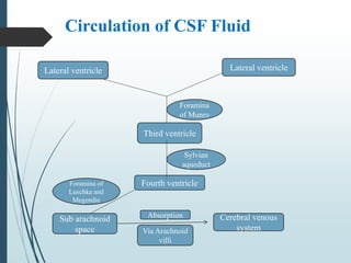
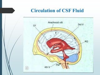
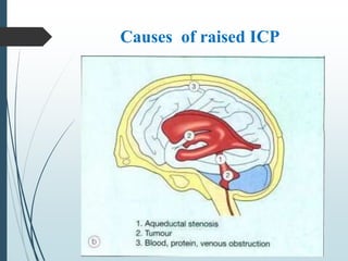
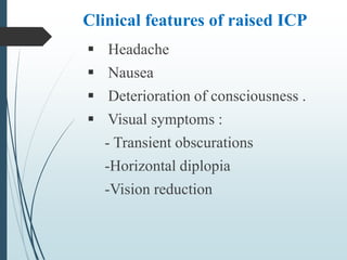
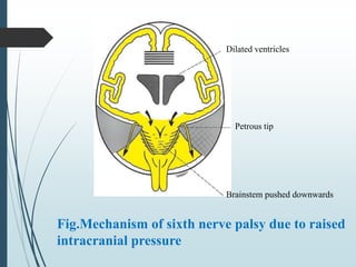
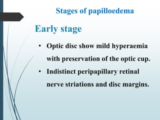
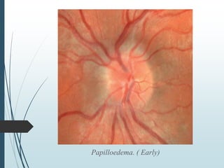
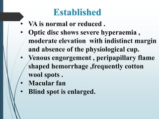
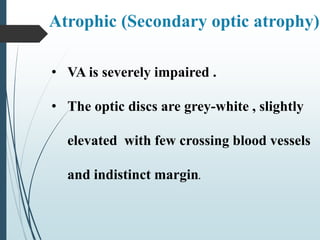
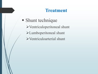
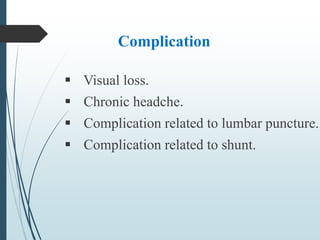
PAPILLOEDEMA
Papilloedema
 It is swelling of optic nerve head due to
raised ICP .
 It is nearly always bilateral.
 All patient with papilloedema should be
suspected of having intracranial mass.
Production of CSF
CSF is formed by Choroid plexus in
the ventricles of the brain .
Normal CSF pressure 10-18 cm H2O
Circulation of CSF Fluid
Lateral ventricle Lateral ventricle
Foramina
of Munro
Third ventricle
Fourth ventricle
Sylvian
aqueduct
Sub arachnoid
space
Foramina of
Luschka and
Megendie
Cerebral venous
system
Absorption
Via Arachnoid
villi
Circulation of CSF Fluid
Causes of raised ICP
 Idiopathic intracranial HTN.
 Obstruction of ventricular system.
 Space occupying intracranial lesions.
 Impairment of CSF absorption.
Cerebral venous sinus thrombosis .
 Cerebral oedema from blunt head
trauma .
Severe systemic HTN.
Hypersecretion of CSF
Causes of raised ICP
Causes of optic disc elevation
1.Papilloedema
2. Accelerated hypertension
3. Anterior optic neuropathy
- Ischaemia
- Inflammatory
- Infiltrative
- Compressive
4. Pseudopapilloedema
- Optic disc drusen
- Tilted optic disc
- Peripapillary myelinated nerve fibres
- Crowded disc in hypermetropia
5. Mitochondrial optic neuropathy
- Leber hereditary optic neuropathy
- Methanol poisoning
6. Intra ocular disease
- Central retinal vein occlusion
- uveitis
- Posterior scleritis
- Hypotony
Clinical features of raised ICP
 Headache
 Nausea
 Deterioration of consciousness .
 Visual symptoms :
- Transient obscurations
-Horizontal diplopia
-Vision reduction
Fig.Mechanism of sixth nerve palsy due to raised
intracranial pressure
Dilated ventricles
Petrous tip
Brainstem pushed downwards
Stages of papilloedema
• Optic disc show mild hyperaemia
with preservation of the optic cup.
• Indistinct peripapillary retinal
nerve striations and disc margins.
Early stage
Papilloedema. ( Early)
• VA is normal or reduced .
• Optic disc shows severe hyperaemia ,
moderate elevation with indistinct margin
and absence of the physiological cup.
• Venous engorgement , peripapillary flame
shaped hemorrhage ,frequently cotton
wool spots .
• Macular fan
• Blind spot is enlarged.
Established
Papilloedema.- acute established
• VA is variable and visual fields begin to
constrict
• Optic disc is not elevated
• Cotton wool spots and hemorrhages are absent
• Opto-cilliary shunt and drusen like crystalline
deposits may be present on the disc surface .
Chronic
Papilloedema.-chronic
• VA is severely impaired .
• The optic discs are grey-white , slightly
elevated with few crossing blood vessels
and indistinct margin.
Atrophic (Secondary optic atrophy)
Papilloedema- atrophic
Investigations
 B-scan ultrasonography
 CT scan of brain
 MRI of Brain
 Venography
 Lumbar puncture
Treatment
 According to cause .
 Neuro-surgical consultation .
 Method of reducing ICP
Mannitol infusion
CSF withdrawal
Sedative –propofol, barbiturate
Control hyperventilation
Decompressive craniotomy
Removal of mass lesion if present
Hypothermia
Steriods
Treatment
 Shunt technique
Ventriculoperitoneal shunt
Lumboperitoneal shunt
Ventriculoarterial shunt
Complication
 Visual loss.
 Chronic headche.
 Complication related to lumbar puncture.
 Complication related to shunt.
Thanks to all

Papilloedema presentation1

  • 1.
  • 2.
    Papilloedema  It isswelling of optic nerve head due to raised ICP .  It is nearly always bilateral.  All patient with papilloedema should be suspected of having intracranial mass.
  • 3.
    Production of CSF CSFis formed by Choroid plexus in the ventricles of the brain . Normal CSF pressure 10-18 cm H2O
  • 4.
    Circulation of CSFFluid Lateral ventricle Lateral ventricle Foramina of Munro Third ventricle Fourth ventricle Sylvian aqueduct Sub arachnoid space Foramina of Luschka and Megendie Cerebral venous system Absorption Via Arachnoid villi
  • 5.
  • 6.
    Causes of raisedICP  Idiopathic intracranial HTN.  Obstruction of ventricular system.  Space occupying intracranial lesions.  Impairment of CSF absorption.
  • 7.
    Cerebral venous sinusthrombosis .  Cerebral oedema from blunt head trauma . Severe systemic HTN. Hypersecretion of CSF Causes of raised ICP
  • 8.
  • 9.
    Causes of opticdisc elevation 1.Papilloedema 2. Accelerated hypertension 3. Anterior optic neuropathy - Ischaemia - Inflammatory - Infiltrative - Compressive
  • 10.
    4. Pseudopapilloedema - Opticdisc drusen - Tilted optic disc - Peripapillary myelinated nerve fibres - Crowded disc in hypermetropia 5. Mitochondrial optic neuropathy - Leber hereditary optic neuropathy - Methanol poisoning
  • 11.
    6. Intra oculardisease - Central retinal vein occlusion - uveitis - Posterior scleritis - Hypotony
  • 12.
    Clinical features ofraised ICP  Headache  Nausea  Deterioration of consciousness .  Visual symptoms : - Transient obscurations -Horizontal diplopia -Vision reduction
  • 13.
    Fig.Mechanism of sixthnerve palsy due to raised intracranial pressure Dilated ventricles Petrous tip Brainstem pushed downwards
  • 14.
    Stages of papilloedema •Optic disc show mild hyperaemia with preservation of the optic cup. • Indistinct peripapillary retinal nerve striations and disc margins. Early stage
  • 15.
  • 16.
    • VA isnormal or reduced . • Optic disc shows severe hyperaemia , moderate elevation with indistinct margin and absence of the physiological cup. • Venous engorgement , peripapillary flame shaped hemorrhage ,frequently cotton wool spots . • Macular fan • Blind spot is enlarged. Established
  • 17.
  • 18.
    • VA isvariable and visual fields begin to constrict • Optic disc is not elevated • Cotton wool spots and hemorrhages are absent • Opto-cilliary shunt and drusen like crystalline deposits may be present on the disc surface . Chronic
  • 19.
  • 20.
    • VA isseverely impaired . • The optic discs are grey-white , slightly elevated with few crossing blood vessels and indistinct margin. Atrophic (Secondary optic atrophy)
  • 21.
  • 22.
    Investigations  B-scan ultrasonography CT scan of brain  MRI of Brain  Venography  Lumbar puncture
  • 23.
    Treatment  According tocause .  Neuro-surgical consultation .  Method of reducing ICP Mannitol infusion CSF withdrawal Sedative –propofol, barbiturate Control hyperventilation Decompressive craniotomy Removal of mass lesion if present Hypothermia Steriods
  • 24.
    Treatment  Shunt technique Ventriculoperitonealshunt Lumboperitoneal shunt Ventriculoarterial shunt
  • 25.
    Complication  Visual loss. Chronic headche.  Complication related to lumbar puncture.  Complication related to shunt.
  • 26.