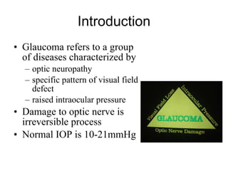
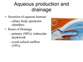
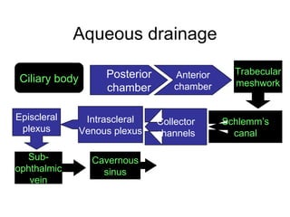
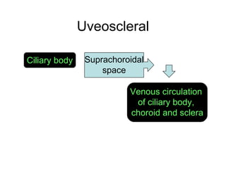
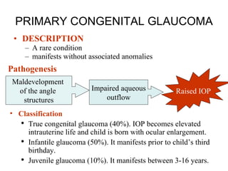
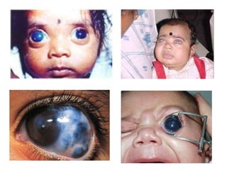
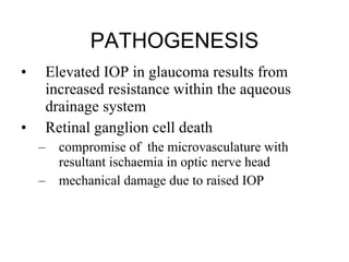
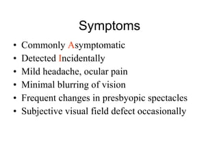
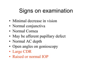
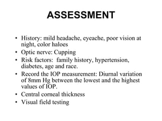
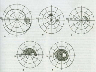
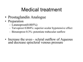
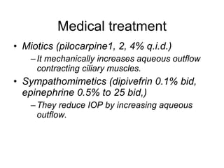
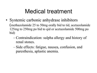
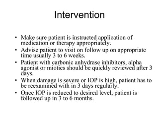
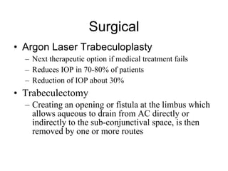
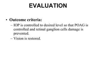
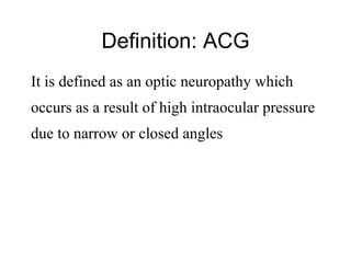
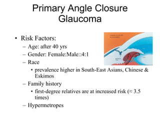
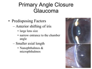
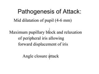
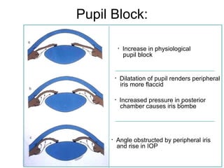
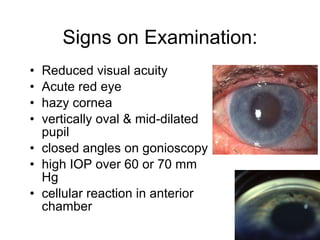
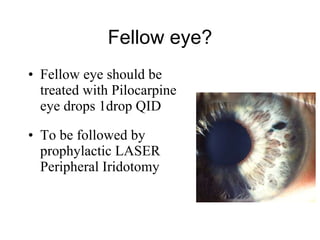
Glaucoma Gauri S. Shrestha, M.Optom, FIACLE
Introduction Glaucoma refers to a group of diseases characterized by  optic neuropathy  specific pattern of visual field defect  raised intraocular pressure Damage to optic nerve is irreversible process Normal IOP is 10-21mmHg
Aqueous production and drainage Secretion of aqueous humour ciliary body (posterior chamber ) Route of Drainage primary (90%): trabecular meshwork uveal-scleral outflow (10%)
Aqueous drainage Ciliary body Posterior chamber Anterior chamber Trabecular meshwork Schlemm’s canal Collector channels Intrascleral Venous plexus Episcleral plexus Sub- ophthalmic vein Cavernous sinus
Uveoscleral Ciliary body Suprachoroidal space Venous circulation  of ciliary body,  choroid and sclera
Classification Congenital and developmental glaucoma Primary congenital glaucoma  Developmental glaucoma  With  associated anomalies  such as Rieger syndrome, peter’s anomaly, aniridian, ectopia lentis, Sturge-Weber syndrome, nanophthalmos, congenital microcornea etc., Primary glaucoma Primary open angle glaucoma (POAG) Primary angle closure glaucoma (PACG)
Classification Secondary glaucoma  Phacomorphic glaucoma Phacolytic glaucoma Glaucoma due to uveitis Pigmentary glaucoma Neovascular glaucoma Glaucoma associated with intraocular tumours Steroid induced glaucoma Secondary glaucoma   Traumatic glaucoma Ciliary block glaucoma Glaucoma associated with intraocular haemorrhage Glaucoma associated with iridocorneal endothelial syndrome Glaucoma due to pseudoexfoliation syndrome
PRIMARY CONGENITAL GLAUCOMA DESCRIPTION A rare condition  manifests without associated anomalies Pathogenesis Classification True congenital glaucoma (40%). IOP becomes elevated intrauterine life and child is born with ocular enlargement. Infantile glaucoma (50%). It manifests prior to child’s third birthday.  Juvenile glaucoma (10%). It manifests between 3-16 years. Maldevelopment  of the angle  structures Impaired aqueous outflow Raised IOP
 
Symptoms and signs Symptoms Photophobia, Lacrimation, blepharospasm, enlarged eyeball,  Signs Corneal  edema, corneal enlargement more than 13mm diameter. Sclera   become thin and appers blue Iris   may show iridodonesis and atrophic patches in late stage Lens   becomes flat or subluxated Optic disc   shows increased cup/disc ratio and atrophy specially after third year. IOP  is invariably high.
NURSING ASSESSMENT  Child may need examination under anesthesia History on lacrimation, photophobia, and blepharospasm Assess visual acuity and perform refraction to find out loss of vision Examine cornea for edema and opacity Measure IOP with hand held perkin’s applanation tonometer Measure corneal diameter by calipers. Perform a dilated fundus examination to evaluate the optic disc and retina
NURSING DIAGNOSIS Altered visual perception secondary to increased intraocular pressure and manifested as Profound lacrimation, photophobia, corneal haze, and buphthalmos  Loss of vision  EXPECTED OUTCOME Intraocular pressure will be controlled and bring down to normal. Lacrimation will be controlled. Photophobia and corneal haze will be eliminated Prevent eye from loss of vision.
Intervention Counsel the child’s parents for urgent need of surgery Prepare them psychologically Check the ophthalmologic order of management plan Topical beta-blocker  (timolol 0.25% to 0.50% b.i.d.) Goniotomy  is the first choice of surgery (Clear cornea) Trabeculotomy   corneal clouding prevents visualization of the angle  Failed repeated goniotomy Monitor IOP, optic disc, and corneal diameter on regular follow up
EVALUATION Outcome criteria Cornea should be transparent; and IOP should be maintained with in normal range
PRIMARY OPEN ANGLE GLAUCOMA Usually bilateral with asymmetry in onset.  Slow progressive rise in IOP (above 21mmHg),  Glaucomatous optic nerve damage, Visual field loss
Risk-factors Ocular risk Factors . IOP . Myopia . Increased cup/disc ratio . Asymmetric cupping . Disc hemorrhage
Risk-factors Non ocular risk factors . Age . Race . Family history . Diabetes and Systemic hypertension . Migraine and peripheral vasospasm . Alcohol consumption . Cigarette smoking
PATHOGENESIS Elevated IOP in glaucoma results from increased resistance within the aqueous drainage system Retinal ganglion cell death  compromise of  the microvasculature with resultant ischaemia in optic nerve head mechanical damage due to raised IOP
Symptoms Commonly  A symptomatic Detected  I ncidentally Mild headache, ocular pain Minimal blurring of vision Frequent changes in presbyopic spectacles Subjective visual field defect occasionally
Signs on examination Minimal decrease in vision Normal conjunctiva Normal Cornea May be afferent pupillary defect Normal AC depth Open angles on gonioscopy Large CDR Raised or normal IOP
ASSESSMENT History: mild headache, eyeache, poor vision at night, color haloes  Optic nerve: Cupping Risk factors:  family history, hypertension, diabetes, age and race.  Record the IOP measurement: Diurnal variation of 8mm Hg between the lowest and the highest values of IOP. Central corneal thickness  Visual field testing
NURSING DIAGNOSIS Progressive loss of vision EXPECTED OUTCOME Stop progression of POAG and damage to retinal ganglion cells. Prevent permanent loss of vision.
 
 
Glaucomatous optic atrophy
 
Intervention Counseling: tests, retest, and examination Instruct the patient to avoid conditions that will increase IOP such as emotional stress, heavy exertion, wearing tight clothes around neck. Institute proper treatment advice
Medical treatment B-blocker: 1 st  drug of choice for initial therapy Mechanism of action: Lowers IOP by reducing the aqueous secretion by effect on beta -receptor in ciliary processes Preparation:  Timolol Maleate(0.25, 0.5%) BD- non selective Levobunolol (0.25,0.5%) BD- nonselective Betaxolol (0.25%) BD- cardioselective Contraindication : bronchial asthma, chronic obstructive pulmonary disease, heart-block, congestive hearth failure, depression.
Medical treatment Carbonic anhydrase inhibitor Preparation Dorzolamide 2% t.i.d Inhibits enzyme carbonic anhydrase -thus reduces aqueous humor formation
Medical treatment Prostaglandin Analogue Preparation Latanoprost(0.005%)  Tarvoprost 0.004%: superior ocular hypotensive effect Bimatoprost 0.3%: potentiate trabecular outflow Increase the uveo - scleral outflow of Aqueous and decrease episcleral venous pressure
Medical treatment Miotics (pilocarpine1, 2, 4% q.i.d.) It mechanically increases aqueous outflow contracting ciliary muscles.  Sympathomimetics (dipivefrin 0.1% bid, epinephrine 0.5% to 25 bid,) They reduce IOP by increasing aqueous outflow.
Medical treatment Systemic carbonic anhydrase inhibitors ( methazolamide 25 to 50mg orally bid to tid, acetazolamide 125mg to 250mg po bid to qid or acetazolamide 500mg po bid) Contraindication: sulpha allergy and history of renal stones.  Side effects: fatigue, nausea, confusion, and paresthesia, aplastic anemia.
Intervention Make sure patient is instructed application of medication or therapy appropriately.  Advise patient to visit on follow up on appropriate time usually 3 to 6 weeks.  Patient with carbonic anhydrase inhibitors, alpha agonist or miotics should be quickly reviewed after 3 days. When damage is severe or IOP is high, patient has to be reexamined with in 3 days regularly. Once IOP is reduced to desired level, patient is followed up in 3 to 6 months.
Surgical Argon Laser Trabeculoplasty   Next therapeutic option if medical treatment fails Reduces IOP in 70-80% of patients Reduction of IOP about 30% Trabeculectomy Creating an opening or fistula at the limbus which allows aqueous to drain from AC directly or indirectly to the sub-conjunctival space, is then removed by one or more routes
EVALUATION Outcome criteria:  IOP is controlled to desired level so that POAG is controlled and retinal ganglion cells damage is prevented.  Vision is restored.
Definition: ACG It is defined as an optic neuropathy which  occurs as a result of high intraocular pressure due to narrow or closed angles
Primary Angle Closure Glaucoma Risk Factors: Age: after 40 yrs Gender: Female:Male::4:1 Race prevalence higher in South-East Asians, Chinese & Eskimos Family history first-degree relatives are at increased risk (≈ 3.5 times) Hypermetropes
Predisposing Factors Anterior shifting of iris large lens size narrow entrance to the chamber angle Smaller axial length Nanophthalmos & microphthalmos Primary Angle Closure Glaucoma
Precipitating Factors Dim illumination Emotional Stress Trauma/illness Intense concentration Pharmacological pupil dilatation Primary Angle Closure Glaucoma
Pathogenesis of Attack: Mid dilatation of pupil (4-6 mm) Maximum pupillary block and relaxation  of peripheral iris allowing  forward displacement of iris Angle closure attack
Pupil Block: Increase in physiological pupil block Dilatation of pupil renders peripheral iris more flaccid Increased pressure in posterior chamber causes iris bombe Angle obstructed by peripheral iris and rise in IOP
Symptom presents at  emergency  unit sudden onset of acute pain in eye / head diminution of vision colour halos around the bulb lacrimation / lid oedema anxiety and fatigue - nausea & vomiting
Signs on Examination: Reduced visual acuity Acute red eye hazy cornea vertically oval & mid-dilated pupil closed angles on gonioscopy high IOP over 60 or 70 mm Hg cellular reaction in anterior chamber
Management: Principle of therapy is to bring down the IOP as quicker as possible Admit the patient Counsel the patient to make her anxiety free as far as possible Give IOP lowering drugs Reduce inflammation  LASER therapy
Medical Therapy: IV Acetazolamide (Inj Diamox, 500mg IV) followed by (250 mg tablet TID) Beta-blockers Miotics Steroid Eye drops
Medical Therapy: If IOP is still more than 50mmHg  Hyperosmotic agents: Mannitol 20% 1-2 gm/kg given I/V within 30-45 minutes Oral Glycerol 50%-----1-1.5 gm/kg Raise serum osmotic pressure and withdraw fluid from the eye specially from vitreous Vitreous dehydration allows the lens to fall back deepening the AC
Fellow eye? Fellow eye should be treated with Pilocarpine eye drops 1drop QID To be followed by prophylactic LASER Peripheral Iridotomy
Nursing management of glaucoma Measures taken to relieve pain Reassure patient that pain and other signs and symptoms should subside with reduction of IOP Provide reassurance and calm to reduce anxiety and fear Provide preoperative care Post operative care
THANK YOU !

Glaucoma

  • 1.
    Glaucoma Gauri S.Shrestha, M.Optom, FIACLE
  • 2.
    Introduction Glaucoma refersto a group of diseases characterized by optic neuropathy specific pattern of visual field defect raised intraocular pressure Damage to optic nerve is irreversible process Normal IOP is 10-21mmHg
  • 3.
    Aqueous production anddrainage Secretion of aqueous humour ciliary body (posterior chamber ) Route of Drainage primary (90%): trabecular meshwork uveal-scleral outflow (10%)
  • 4.
    Aqueous drainage Ciliarybody Posterior chamber Anterior chamber Trabecular meshwork Schlemm’s canal Collector channels Intrascleral Venous plexus Episcleral plexus Sub- ophthalmic vein Cavernous sinus
  • 5.
    Uveoscleral Ciliary bodySuprachoroidal space Venous circulation of ciliary body, choroid and sclera
  • 6.
    Classification Congenital anddevelopmental glaucoma Primary congenital glaucoma Developmental glaucoma With associated anomalies such as Rieger syndrome, peter’s anomaly, aniridian, ectopia lentis, Sturge-Weber syndrome, nanophthalmos, congenital microcornea etc., Primary glaucoma Primary open angle glaucoma (POAG) Primary angle closure glaucoma (PACG)
  • 7.
    Classification Secondary glaucoma Phacomorphic glaucoma Phacolytic glaucoma Glaucoma due to uveitis Pigmentary glaucoma Neovascular glaucoma Glaucoma associated with intraocular tumours Steroid induced glaucoma Secondary glaucoma Traumatic glaucoma Ciliary block glaucoma Glaucoma associated with intraocular haemorrhage Glaucoma associated with iridocorneal endothelial syndrome Glaucoma due to pseudoexfoliation syndrome
  • 8.
    PRIMARY CONGENITAL GLAUCOMADESCRIPTION A rare condition manifests without associated anomalies Pathogenesis Classification True congenital glaucoma (40%). IOP becomes elevated intrauterine life and child is born with ocular enlargement. Infantile glaucoma (50%). It manifests prior to child’s third birthday. Juvenile glaucoma (10%). It manifests between 3-16 years. Maldevelopment of the angle structures Impaired aqueous outflow Raised IOP
  • 9.
  • 10.
    Symptoms and signsSymptoms Photophobia, Lacrimation, blepharospasm, enlarged eyeball, Signs Corneal edema, corneal enlargement more than 13mm diameter. Sclera become thin and appers blue Iris may show iridodonesis and atrophic patches in late stage Lens becomes flat or subluxated Optic disc shows increased cup/disc ratio and atrophy specially after third year. IOP is invariably high.
  • 11.
    NURSING ASSESSMENT Child may need examination under anesthesia History on lacrimation, photophobia, and blepharospasm Assess visual acuity and perform refraction to find out loss of vision Examine cornea for edema and opacity Measure IOP with hand held perkin’s applanation tonometer Measure corneal diameter by calipers. Perform a dilated fundus examination to evaluate the optic disc and retina
  • 12.
    NURSING DIAGNOSIS Alteredvisual perception secondary to increased intraocular pressure and manifested as Profound lacrimation, photophobia, corneal haze, and buphthalmos Loss of vision EXPECTED OUTCOME Intraocular pressure will be controlled and bring down to normal. Lacrimation will be controlled. Photophobia and corneal haze will be eliminated Prevent eye from loss of vision.
  • 13.
    Intervention Counsel thechild’s parents for urgent need of surgery Prepare them psychologically Check the ophthalmologic order of management plan Topical beta-blocker (timolol 0.25% to 0.50% b.i.d.) Goniotomy is the first choice of surgery (Clear cornea) Trabeculotomy corneal clouding prevents visualization of the angle Failed repeated goniotomy Monitor IOP, optic disc, and corneal diameter on regular follow up
  • 14.
    EVALUATION Outcome criteriaCornea should be transparent; and IOP should be maintained with in normal range
  • 15.
    PRIMARY OPEN ANGLEGLAUCOMA Usually bilateral with asymmetry in onset. Slow progressive rise in IOP (above 21mmHg), Glaucomatous optic nerve damage, Visual field loss
  • 16.
    Risk-factors Ocular riskFactors . IOP . Myopia . Increased cup/disc ratio . Asymmetric cupping . Disc hemorrhage
  • 17.
    Risk-factors Non ocularrisk factors . Age . Race . Family history . Diabetes and Systemic hypertension . Migraine and peripheral vasospasm . Alcohol consumption . Cigarette smoking
  • 18.
    PATHOGENESIS Elevated IOPin glaucoma results from increased resistance within the aqueous drainage system Retinal ganglion cell death compromise of the microvasculature with resultant ischaemia in optic nerve head mechanical damage due to raised IOP
  • 19.
    Symptoms Commonly A symptomatic Detected I ncidentally Mild headache, ocular pain Minimal blurring of vision Frequent changes in presbyopic spectacles Subjective visual field defect occasionally
  • 20.
    Signs on examinationMinimal decrease in vision Normal conjunctiva Normal Cornea May be afferent pupillary defect Normal AC depth Open angles on gonioscopy Large CDR Raised or normal IOP
  • 21.
    ASSESSMENT History: mildheadache, eyeache, poor vision at night, color haloes Optic nerve: Cupping Risk factors: family history, hypertension, diabetes, age and race. Record the IOP measurement: Diurnal variation of 8mm Hg between the lowest and the highest values of IOP. Central corneal thickness Visual field testing
  • 22.
    NURSING DIAGNOSIS Progressiveloss of vision EXPECTED OUTCOME Stop progression of POAG and damage to retinal ganglion cells. Prevent permanent loss of vision.
  • 23.
  • 24.
  • 25.
  • 26.
  • 27.
    Intervention Counseling: tests,retest, and examination Instruct the patient to avoid conditions that will increase IOP such as emotional stress, heavy exertion, wearing tight clothes around neck. Institute proper treatment advice
  • 28.
    Medical treatment B-blocker:1 st drug of choice for initial therapy Mechanism of action: Lowers IOP by reducing the aqueous secretion by effect on beta -receptor in ciliary processes Preparation: Timolol Maleate(0.25, 0.5%) BD- non selective Levobunolol (0.25,0.5%) BD- nonselective Betaxolol (0.25%) BD- cardioselective Contraindication : bronchial asthma, chronic obstructive pulmonary disease, heart-block, congestive hearth failure, depression.
  • 29.
    Medical treatment Carbonicanhydrase inhibitor Preparation Dorzolamide 2% t.i.d Inhibits enzyme carbonic anhydrase -thus reduces aqueous humor formation
  • 30.
    Medical treatment ProstaglandinAnalogue Preparation Latanoprost(0.005%) Tarvoprost 0.004%: superior ocular hypotensive effect Bimatoprost 0.3%: potentiate trabecular outflow Increase the uveo - scleral outflow of Aqueous and decrease episcleral venous pressure
  • 31.
    Medical treatment Miotics(pilocarpine1, 2, 4% q.i.d.) It mechanically increases aqueous outflow contracting ciliary muscles. Sympathomimetics (dipivefrin 0.1% bid, epinephrine 0.5% to 25 bid,) They reduce IOP by increasing aqueous outflow.
  • 32.
    Medical treatment Systemiccarbonic anhydrase inhibitors ( methazolamide 25 to 50mg orally bid to tid, acetazolamide 125mg to 250mg po bid to qid or acetazolamide 500mg po bid) Contraindication: sulpha allergy and history of renal stones. Side effects: fatigue, nausea, confusion, and paresthesia, aplastic anemia.
  • 33.
    Intervention Make surepatient is instructed application of medication or therapy appropriately. Advise patient to visit on follow up on appropriate time usually 3 to 6 weeks. Patient with carbonic anhydrase inhibitors, alpha agonist or miotics should be quickly reviewed after 3 days. When damage is severe or IOP is high, patient has to be reexamined with in 3 days regularly. Once IOP is reduced to desired level, patient is followed up in 3 to 6 months.
  • 34.
    Surgical Argon LaserTrabeculoplasty Next therapeutic option if medical treatment fails Reduces IOP in 70-80% of patients Reduction of IOP about 30% Trabeculectomy Creating an opening or fistula at the limbus which allows aqueous to drain from AC directly or indirectly to the sub-conjunctival space, is then removed by one or more routes
  • 35.
    EVALUATION Outcome criteria: IOP is controlled to desired level so that POAG is controlled and retinal ganglion cells damage is prevented. Vision is restored.
  • 36.
    Definition: ACG Itis defined as an optic neuropathy which occurs as a result of high intraocular pressure due to narrow or closed angles
  • 37.
    Primary Angle ClosureGlaucoma Risk Factors: Age: after 40 yrs Gender: Female:Male::4:1 Race prevalence higher in South-East Asians, Chinese & Eskimos Family history first-degree relatives are at increased risk (≈ 3.5 times) Hypermetropes
  • 38.
    Predisposing Factors Anteriorshifting of iris large lens size narrow entrance to the chamber angle Smaller axial length Nanophthalmos & microphthalmos Primary Angle Closure Glaucoma
  • 39.
    Precipitating Factors Dimillumination Emotional Stress Trauma/illness Intense concentration Pharmacological pupil dilatation Primary Angle Closure Glaucoma
  • 40.
    Pathogenesis of Attack:Mid dilatation of pupil (4-6 mm) Maximum pupillary block and relaxation of peripheral iris allowing forward displacement of iris Angle closure attack
  • 41.
    Pupil Block: Increasein physiological pupil block Dilatation of pupil renders peripheral iris more flaccid Increased pressure in posterior chamber causes iris bombe Angle obstructed by peripheral iris and rise in IOP
  • 42.
    Symptom presents at emergency unit sudden onset of acute pain in eye / head diminution of vision colour halos around the bulb lacrimation / lid oedema anxiety and fatigue - nausea & vomiting
  • 43.
    Signs on Examination:Reduced visual acuity Acute red eye hazy cornea vertically oval & mid-dilated pupil closed angles on gonioscopy high IOP over 60 or 70 mm Hg cellular reaction in anterior chamber
  • 44.
    Management: Principle oftherapy is to bring down the IOP as quicker as possible Admit the patient Counsel the patient to make her anxiety free as far as possible Give IOP lowering drugs Reduce inflammation LASER therapy
  • 45.
    Medical Therapy: IVAcetazolamide (Inj Diamox, 500mg IV) followed by (250 mg tablet TID) Beta-blockers Miotics Steroid Eye drops
  • 46.
    Medical Therapy: IfIOP is still more than 50mmHg Hyperosmotic agents: Mannitol 20% 1-2 gm/kg given I/V within 30-45 minutes Oral Glycerol 50%-----1-1.5 gm/kg Raise serum osmotic pressure and withdraw fluid from the eye specially from vitreous Vitreous dehydration allows the lens to fall back deepening the AC
  • 47.
    Fellow eye? Felloweye should be treated with Pilocarpine eye drops 1drop QID To be followed by prophylactic LASER Peripheral Iridotomy
  • 48.
    Nursing management ofglaucoma Measures taken to relieve pain Reassure patient that pain and other signs and symptoms should subside with reduction of IOP Provide reassurance and calm to reduce anxiety and fear Provide preoperative care Post operative care
  • 49.