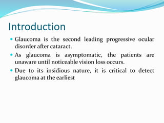
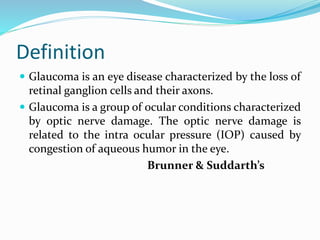
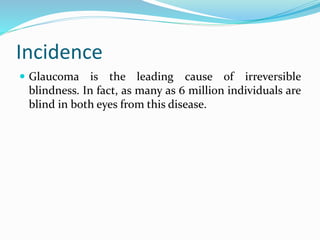
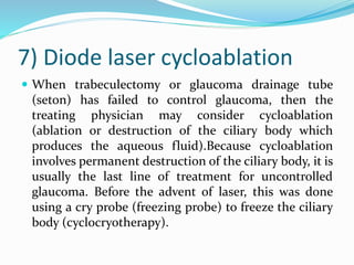
Introduction
 Glaucoma is the second leading progressive ocular
disorder after cataract.
 As glaucoma is asymptomatic, the patients are
unaware until noticeable vision loss occurs.
 Due to its insidious nature, it is critical to detect
glaucoma at the earliest
Definition
 Glaucoma is an eye disease characterized by the loss of
retinal ganglion cells and their axons.
 Glaucoma is a group of ocular conditions characterized
by optic nerve damage. The optic nerve damage is
related to the intra ocular pressure (IOP) caused by
congestion of aqueous humor in the eye.
Brunner & Suddarth’s
Incidence
 Glaucoma is the leading cause of irreversible
blindness. In fact, as many as 6 million individuals are
blind in both eyes from this disease.
Types Glaucoma
1. Congenitalinfantile/Buphthalmos/Hydrophthalmos
2. Acquired glaucoma
a. Primary glaucoma
a. Open angle/wide angle/ chronic
b. Angle closure/narrow angle/closed angle
b. Secondary glaucoma
Congenital infantile buphthalmos
hydrophthalmos
 It is characterized by elevation of intra ocular pressure
(IOP) associated with developmental abnormalities of
the angle of anterior chamber depending upon the age
of onset.
Congenital infantile buphthalmos
hydrophthalmos
 This developmental glaucoma’s are termed as:
1. True or primary congenital glaucoma (IOP is raised
during intrauterine life)
2. Infantile glaucoma’s (Diseases manifests prior to the
child)
3. Juvenile glaucoma (Children develop pressure rise
between 3-16 yrs of life).
OPEN ANGLE GLAUCOMA
 It is the most common form of the disease and
generally does not affect people until they are in their
40s.
1) Primary chronic open angle
glaucoma (COAG)
 It is the most common type of the glaucoma. Its
frequency increases greatly with age. The aqueous
fluid does not drain from the eye properly. The
pressure within the eye, therefore, builds up painlessly
and without symptoms.
2) Normal tension (pressure)
glaucoma or low tension
glaucoma
This type of glaucoma is thought to be due to decreased
blood flow to the optic nerve. This condition is
characterized by progressive optic-nerve damage and
loss of peripheral vision (visual field) despite
intraocular pressures in the normal range or even
below normal
3) Childhood glaucoma
 Childhood glaucoma is an uncommon pediatric
condition often associated with significant visual loss.
It may most commonly be caused by trauma, surgery
or other acquired or secondary causes or abnormal
increase intra ocular pressure.
4) Secondary open angle
glaucoma
 It can result from an eye (ocular) injury, inflammation
in the iris (iritis), rétinal vein blockage etc.
5) Pigmentary glaucoma
 In this granules of pigment detach from the iris, which
is the colored part of the eye. These granules then may
block the trabecular meshwork, is a key element in the
drainage system of the eye. Finally the blocked
drainage system leads to elevated intraocular pressure
which results in damage to the optic nerve.
6) Exfoliative glaucoma
 This type of glaucoma is characterized by deposits of
flaky material on the front surface of the lens (anterior
capsule) & in the angle of the eye. The accumulation of
this material in the angle is believed to block the
drainage system of the eye and raise the eye pressure.
ANGLE CLOSURE GLAUCOMA
 Angle-closure glaucoma may be acute or chronic. The
common element in both is that the entire drainage
angle becomes anatomically closed, so that the
aqueous fluid within the eye cannot even reach all or
part of the trabecular meshwork.
1) Acute angle closure glaucoma
 When the drainage angle of the eye suddenly becomes
completely blocked, pressure builds up rapidly, and
this is called acute angle-closure glaucoma. The
symptoms include severe eye pain, blurred vision,
headache, nausea & vomiting.
2) Chronic angle closure glaucoma
 When the drainage angle of the eye gradually becomes
completely blocked, pressure builds up gradually, and
this is called chronic angle-closure glaucoma. The
drainage tissues gradually start to scar. This condition
is generally silent, and severe glaucoma damage can
occur without the person's knowledge.
Etiology
1) Eye injury
2) Eye surgery
3) Eye tumors
4) Diabetes
5) Cataract
6) Trauma
7) Steroid use
8) Emotional stress
9) Anti-histamine use
10) Hypothyroidism
11) Sleep apnea
12) Leukemia
13) Sickle cell anemia
Risk factors
1) Age over 45 years
2) Family history of glaucoma
3) Diabetes
4) History of elevated intra-ocular pressure
5) Near-sightedness (Myopia)
6) Use of steroids
7) Thin cornea
8) A history of severe anemia or shock
9) Cardiovascular disease
10) Eye trauma
11) Race
12) Abnormally high intra-ocular pressure
13) Peripheral vision is decreased.
14) Provision eye injury
15) Not seeing a rainbow.
Clinical manifestation
1) Severe eye pain
2) Eye redness
3) Blurred vision
4) Severe headache
5) Nausea
6) Vomiting
7) Dry eyes with itching or burning
8) Dark spot at the center of viewing
9) Excess tearing or watery eyes
10) Difficulty focusing on near or distant object
Pathophysiology
1. Initiating Events: - Precipitating factors include
illness, emotional stress, congenital narrow angles,
long term use of corticosteroids & mydriatics
(medications causing papillary dilation).These
events lead to second stage.
2. Structural alterations in the aqueous outflow
system: - Tissue & cellular changes caused by factors
that affect aqueous humor dynamics lead to structural
alterations & to the third stage
Pathophysiology
3. Functional alterations: - Conditions such as
increased IOP or impaired blood flow create
functional changes that lead to fourth stage.
4. Optic nerve damage: - Atrophy of the optic nerve is
characterized by loss of nerve fibers & blood supply &
this fourth stage inevitably progresses to the fifth
stage.
5. Vision loss: - Progressive loss of vision is
characterized by visual field defects.
Diagnostic evaluation
1) Ocular & medical history
2) Tonometry
3) Ophthalmoscope
4) Gonioscopy
5) Perimeter
6) Pachymetry
7) Dilated pupil examination
Medical management
 Cholinergic(Miotics): Pilocarpine, Carbachol
 Adrenergic agonists : Dipivefrin, Epinephrine
 Beta blockers : Betaxolol, Timolol
 Alpha adrenergic agonists : Apraclonidin,Brimonidine
 Carbonic anhydrase inhibitors:
Acetazolamide,methazolamide
Surgical management :-
1) Laser trabeculoplasty:
 In this laser burns are applied to the inner surface of
the trabecular meshwork to open the intra trabecular
spaces.Thereby promoting outflow of aqueous humor
& decreasing IOP.The procedure is indicated when
IOP is inadequately controlled by medication’s serious
complication is a transient rise in IOP (usually 2
hrs.after surgery).
2) Laser iridotomy :- ( for papillary
block glaucoma)
 In this an opening is made in the iris to eliminate the
papillary block. This procedure is contraindicated in
pts with corneal edema.
 Potential complication is burns to the cornea, lens or
retina, transient elevated IOP.
3) Filtering procedures
 These are used to create an opening or fistula in the
trabecular meshwork to drain aqueous humor from
the anterior chamber to the subconjunctival space,
thereby bypassing the usual drainage structures. This
allows the aqueous humor to flow & exit by different
routes.
4) Trabeculectomy
 It is the standard filtering technique used to remove
part of the trabecular meshwork. Complication
include hemorrhage, loe or elevated IOP, cataract etc.
5) Drainage implants or shunts
 These are an open tubes implanted in the anterior
chamber to shunt aqueous humor to an attached place
in the conjunctiva space. A fibrous capsule develops
around the episcleral plate & filters the aqueous
humor, thereby regulating the outflow & controlling
IOP.
6) Canaloplasty
 Canaloplasty utilizes a micro catheter or tube placed in
the Canal of Schlemm (the natural site of drainage for
healthy eyes) to enlarge the drainage canal, relieving
pressure inside the eye. Studies have been published
demonstrating longterm efficacy and safety
7) Diode laser cycloablation
 When trabeculectomy or glaucoma drainage tube
(seton) has failed to control glaucoma, then the
treating physician may consider cycloablation
(ablation or destruction of the ciliary body which
produces the aqueous fluid).Because cycloablation
involves permanent destruction of the ciliary body, it is
usually the last line of treatment for uncontrolled
glaucoma. Before the advent of laser, this was done
using a cry probe (freezing probe) to freeze the ciliary
body (cyclocryotherapy).
Nursing management :-
 Acute pain related to increased intra-ocular pressure
 Fear related to pain & potential loss of vision.
 Nausea & vomiting related to opioids & other
medications.
 Knowledge deficit related to disease

Glaucoma

  • 2.
    Introduction  Glaucoma isthe second leading progressive ocular disorder after cataract.  As glaucoma is asymptomatic, the patients are unaware until noticeable vision loss occurs.  Due to its insidious nature, it is critical to detect glaucoma at the earliest
  • 3.
    Definition  Glaucoma isan eye disease characterized by the loss of retinal ganglion cells and their axons.  Glaucoma is a group of ocular conditions characterized by optic nerve damage. The optic nerve damage is related to the intra ocular pressure (IOP) caused by congestion of aqueous humor in the eye. Brunner & Suddarth’s
  • 4.
    Incidence  Glaucoma isthe leading cause of irreversible blindness. In fact, as many as 6 million individuals are blind in both eyes from this disease.
  • 5.
    Types Glaucoma 1. Congenitalinfantile/Buphthalmos/Hydrophthalmos 2.Acquired glaucoma a. Primary glaucoma a. Open angle/wide angle/ chronic b. Angle closure/narrow angle/closed angle b. Secondary glaucoma
  • 6.
    Congenital infantile buphthalmos hydrophthalmos It is characterized by elevation of intra ocular pressure (IOP) associated with developmental abnormalities of the angle of anterior chamber depending upon the age of onset.
  • 7.
    Congenital infantile buphthalmos hydrophthalmos This developmental glaucoma’s are termed as: 1. True or primary congenital glaucoma (IOP is raised during intrauterine life) 2. Infantile glaucoma’s (Diseases manifests prior to the child) 3. Juvenile glaucoma (Children develop pressure rise between 3-16 yrs of life).
  • 8.
    OPEN ANGLE GLAUCOMA It is the most common form of the disease and generally does not affect people until they are in their 40s.
  • 9.
    1) Primary chronicopen angle glaucoma (COAG)  It is the most common type of the glaucoma. Its frequency increases greatly with age. The aqueous fluid does not drain from the eye properly. The pressure within the eye, therefore, builds up painlessly and without symptoms.
  • 10.
    2) Normal tension(pressure) glaucoma or low tension glaucoma This type of glaucoma is thought to be due to decreased blood flow to the optic nerve. This condition is characterized by progressive optic-nerve damage and loss of peripheral vision (visual field) despite intraocular pressures in the normal range or even below normal
  • 11.
    3) Childhood glaucoma Childhood glaucoma is an uncommon pediatric condition often associated with significant visual loss. It may most commonly be caused by trauma, surgery or other acquired or secondary causes or abnormal increase intra ocular pressure.
  • 12.
    4) Secondary openangle glaucoma  It can result from an eye (ocular) injury, inflammation in the iris (iritis), rétinal vein blockage etc.
  • 13.
    5) Pigmentary glaucoma In this granules of pigment detach from the iris, which is the colored part of the eye. These granules then may block the trabecular meshwork, is a key element in the drainage system of the eye. Finally the blocked drainage system leads to elevated intraocular pressure which results in damage to the optic nerve.
  • 14.
    6) Exfoliative glaucoma This type of glaucoma is characterized by deposits of flaky material on the front surface of the lens (anterior capsule) & in the angle of the eye. The accumulation of this material in the angle is believed to block the drainage system of the eye and raise the eye pressure.
  • 15.
    ANGLE CLOSURE GLAUCOMA Angle-closure glaucoma may be acute or chronic. The common element in both is that the entire drainage angle becomes anatomically closed, so that the aqueous fluid within the eye cannot even reach all or part of the trabecular meshwork.
  • 16.
    1) Acute angleclosure glaucoma  When the drainage angle of the eye suddenly becomes completely blocked, pressure builds up rapidly, and this is called acute angle-closure glaucoma. The symptoms include severe eye pain, blurred vision, headache, nausea & vomiting.
  • 17.
    2) Chronic angleclosure glaucoma  When the drainage angle of the eye gradually becomes completely blocked, pressure builds up gradually, and this is called chronic angle-closure glaucoma. The drainage tissues gradually start to scar. This condition is generally silent, and severe glaucoma damage can occur without the person's knowledge.
  • 18.
    Etiology 1) Eye injury 2)Eye surgery 3) Eye tumors 4) Diabetes 5) Cataract 6) Trauma 7) Steroid use 8) Emotional stress 9) Anti-histamine use 10) Hypothyroidism 11) Sleep apnea 12) Leukemia 13) Sickle cell anemia
  • 19.
    Risk factors 1) Ageover 45 years 2) Family history of glaucoma 3) Diabetes 4) History of elevated intra-ocular pressure 5) Near-sightedness (Myopia) 6) Use of steroids 7) Thin cornea 8) A history of severe anemia or shock 9) Cardiovascular disease 10) Eye trauma 11) Race 12) Abnormally high intra-ocular pressure 13) Peripheral vision is decreased. 14) Provision eye injury 15) Not seeing a rainbow.
  • 20.
    Clinical manifestation 1) Severeeye pain 2) Eye redness 3) Blurred vision 4) Severe headache 5) Nausea 6) Vomiting 7) Dry eyes with itching or burning 8) Dark spot at the center of viewing 9) Excess tearing or watery eyes 10) Difficulty focusing on near or distant object
  • 21.
    Pathophysiology 1. Initiating Events:- Precipitating factors include illness, emotional stress, congenital narrow angles, long term use of corticosteroids & mydriatics (medications causing papillary dilation).These events lead to second stage. 2. Structural alterations in the aqueous outflow system: - Tissue & cellular changes caused by factors that affect aqueous humor dynamics lead to structural alterations & to the third stage
  • 22.
    Pathophysiology 3. Functional alterations:- Conditions such as increased IOP or impaired blood flow create functional changes that lead to fourth stage. 4. Optic nerve damage: - Atrophy of the optic nerve is characterized by loss of nerve fibers & blood supply & this fourth stage inevitably progresses to the fifth stage. 5. Vision loss: - Progressive loss of vision is characterized by visual field defects.
  • 23.
    Diagnostic evaluation 1) Ocular& medical history 2) Tonometry 3) Ophthalmoscope 4) Gonioscopy 5) Perimeter 6) Pachymetry 7) Dilated pupil examination
  • 24.
    Medical management  Cholinergic(Miotics):Pilocarpine, Carbachol  Adrenergic agonists : Dipivefrin, Epinephrine  Beta blockers : Betaxolol, Timolol  Alpha adrenergic agonists : Apraclonidin,Brimonidine  Carbonic anhydrase inhibitors: Acetazolamide,methazolamide
  • 25.
    Surgical management :- 1)Laser trabeculoplasty:  In this laser burns are applied to the inner surface of the trabecular meshwork to open the intra trabecular spaces.Thereby promoting outflow of aqueous humor & decreasing IOP.The procedure is indicated when IOP is inadequately controlled by medication’s serious complication is a transient rise in IOP (usually 2 hrs.after surgery).
  • 26.
    2) Laser iridotomy:- ( for papillary block glaucoma)  In this an opening is made in the iris to eliminate the papillary block. This procedure is contraindicated in pts with corneal edema.  Potential complication is burns to the cornea, lens or retina, transient elevated IOP.
  • 27.
    3) Filtering procedures These are used to create an opening or fistula in the trabecular meshwork to drain aqueous humor from the anterior chamber to the subconjunctival space, thereby bypassing the usual drainage structures. This allows the aqueous humor to flow & exit by different routes.
  • 28.
    4) Trabeculectomy  Itis the standard filtering technique used to remove part of the trabecular meshwork. Complication include hemorrhage, loe or elevated IOP, cataract etc.
  • 29.
    5) Drainage implantsor shunts  These are an open tubes implanted in the anterior chamber to shunt aqueous humor to an attached place in the conjunctiva space. A fibrous capsule develops around the episcleral plate & filters the aqueous humor, thereby regulating the outflow & controlling IOP.
  • 30.
    6) Canaloplasty  Canaloplastyutilizes a micro catheter or tube placed in the Canal of Schlemm (the natural site of drainage for healthy eyes) to enlarge the drainage canal, relieving pressure inside the eye. Studies have been published demonstrating longterm efficacy and safety
  • 31.
    7) Diode lasercycloablation  When trabeculectomy or glaucoma drainage tube (seton) has failed to control glaucoma, then the treating physician may consider cycloablation (ablation or destruction of the ciliary body which produces the aqueous fluid).Because cycloablation involves permanent destruction of the ciliary body, it is usually the last line of treatment for uncontrolled glaucoma. Before the advent of laser, this was done using a cry probe (freezing probe) to freeze the ciliary body (cyclocryotherapy).
  • 32.
    Nursing management :- Acute pain related to increased intra-ocular pressure  Fear related to pain & potential loss of vision.  Nausea & vomiting related to opioids & other medications.  Knowledge deficit related to disease