SlideShare a Scribd company logo
Glaucoma
GLAUCOMA
 Definition
The term glaucoma refers to the group of diseases that have in
common a characteristic optic neuropathy with associated visual
function loss. Although IOP is one of the primary risk factors, it’s
presence or absence doesn’t have a role in the definition of the
disease.
Glaucoma leads to a characteristic appearance of the optic nerve head
and a specific pattern of irreversible visual field defects.
Why glaucoma is important?
 Second leading cause
of global blindness
 Leading cause of
irreversible visual loss.
 Visual impairment from
glaucoma affects
adults more than
children and women
more than men.
 By the year 2020 :
80.5 million people in
the world with
glaucoma
ANATOMY AND PHYSIOLOGY
APPLIED ANATOMY
• Pathophysiology of glaucoma revolves around the aqueous
humour dynamics
• It is essential to understand the production and drainage of aqueous
humour as well as the anatomy of the angle of the anterior chamber
• The principal ocular structures concerned with it are
• Ciliary body
• Angle of anterior chamber
• Aqueous outflow system
CILIARY BODY • Ciliary body is forward
continuation of the choroid at
ora serrata
• Triangular in shape
• The anterior side of the triangle
forms the part of the angle of
anterior and posterior chambers
• The anterior part (about 2 mm)
having finger-like ciliary
processes is called pars plicata
and the posterior smooth part
(about 4 mm) is called pars
plana
Angle of anterior chamber
• Angle of anterior chamber plays an important role in the process of
aqueous drainage
• Formed by root of iris, anterior-most part of ciliary body, scleral spur,
trabecular meshwork and Schwalbe’s line (prominent end of
Descemet’s membrane of cornea)
• Fig. Section of the anterior ocular structures showing
region of the anterior chamber
 Clinically the angle structures can be visualised by gonioscopic examination
• Gonioscopic grading of the angle width
• The most commonly used is Shaffer’s system
• Diagrammatic depiction of various angle structures (SL, Schwalbe's line; TM, trabecular
meshwork; SS, scleral spur; CBB, ciliary body band; ROI, root of iris) as seen in different
grades of angle width (Schaffer's gradingsystem): A, Gonioscopic view; B, Configuration
of the angle in cross section of the anterior chamber
Aqueous outflow system
• IT includes
• Trabecular meshwork
• sieve-like structure through
which aqueous humour leaves
the eye
• consists of three layers,which
from inside out are
I. Uveal meshwork
II. Corneoscleral meshwork
III. Juxtacanalicular
(endothelial) meshwork
Schlemm’s canal
Collector channels
 Direct( aqueous veins)
 Indirect
Episcleral veins
The aqueous outflow system
APPLIED PHYSIOLOGY
• Concerned with the dynamics of aqueous humour are its production,
drainage and maintenance of intraocular pressure
• Aqueous humour and its production
• Functions and compostion of aqueous humour
• Volume : Anterior chamber (0.25 ml) and posterior chamber (0.06 ml)
• Functions of aqueous humour are
• Maintains a proper intraocular pressure
• Metabolic and nutritional role
• Optical function
• Clearing function
• Functions and compostion of aqueous humour cont.
• Refractive index of aqueous humour is 1.336
• Composition
• Water 99.9% , solids 0.1%
• Proteins (colloid content)(5-6 mg%)
• Amino acid 5 mg/kg water
• Non-colloid constituents
• Oxygen
• Aqueous humour: anterior chamber versus posterior chamber
• main differences are : HCO3 and Ascorbate in posterior chamber
Cl in posterior chamber
Aqueous humour formation
• Ultrafiltration
• Secretion
• Diffusion
Control of aqueous formation
Drainage of aqueous humour
Trabecular (conventional)
outflow
Uveoscleral
(unconventional) outlow Flow chart depicting drainage of aqueous humour
Mechanism of aqueous transport across inner wall
of Schlemm’s canal
Passive filter mechanism
Leaky pores in endothelial cells
Contraction microfilaments
Sondermann’channels
Vacuolation theory-it was one of the most accepted view till
recent past
Aqueous outflow active pump mechanism
Vacuolation theory of aqueous transport across the inner wall of the Schlemm's canal: 1. Non-vacuolated stage; 2.
Stage of early infolding of basal surface of the endothelial cell; 3. Stage of macrovacuolar structure formation; 4.
Stage of vacuolar transcellular channel formation;5.Stage of occlusion of the basal infolding.
Aqueous outflow pump mechanism, a part of muscular circulatory loop: (A)
during diastole; (B) during systole
Intraocular pressure
 The intraocular pressure (IOP) refers to the pressure exerted by the
intraocular fluids on the coats of the eyeball.
 Normal IOP = 10-21 mmHg
 It is determined by the rate of aqueous secretion and rate of
outflow.
Factors influencing intraocular pressure
 Local factors
1. Rate of aqueous formation which in turn is influenced by
permeability of ciliary capillaries and osmotic pressure of the blood.
2. Resistance to aqueous outflow
3. Increased episcleral venous pressure may result in rise of IOP
4. Dilatation of pupil in patients with narrow angle can cause rise in
IOP
5. Refractive errors
 General factors
1. Hereditary
2. Age - increases after 40 years
3. Sex- mean IOP is greater in females in the older age
group
4. Diurnal variation- usually the IOP is greater in the
morning than evening ( normal <5mmHg)
5. Postural variation- increases from sitting to supine
6. Blood pressure- glaucoma is more prevalent in
hypertensives
7. Osmotic pressure of blood- increase in plasma
osmolarity is associated with decrease in IOP and vice
versa
8. General anaesthetics and drugs
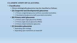
CLASSIFICATION OF GLAUCOMA
• Classification
• Clinico-etiologically glaucoma may be classified as follows:
• (A) Congenital and developmental glaucomas
• 1. Primary congenital glaucoma (without associated anomalies).
• 2. Developmental glaucoma (with associated anomalies).
• (B) Primary adult glaucomas
• 1. Primary open angle glaucomas (POAG)
• 2. Primary angle closure glaucoma (PACG)
• 3. Primary mixed mechanism glaucoma
• (C) Secondary glaucomas
• Depending upon etiology
• Depending upon mechanism of raised IOP
Epidemiology
• Global prevalence of glaucomas
• 2% of those over the age of 40 years , and
• 10% of those over 80 years of age
• POAG versus PACG in different ethnic groups
• Glaucoma blindness
• Global : 8%
• India : 12.8%
Glaucoma – what is happening ?
Either:
the drain blocks
here
Or poor blood
supply here
Damages the optic
nerve..looks ‘caved in’,
called ‘cupped’
Pathogenesis of ocular damage
Death of retinal ganglion cells(RGCs) caused by :
Primary insults
1. Mechanical theory(raised IOP)
Neurotrophins are unable to reach the RGCs due to axonal
deformation and ischaemia caused by mechanical stretch on
lamina cribrosa.
2. Vascular insufficiency theory
Failure of autoregulatory mechanism
Vasospasm
Systemic hypotension
Secondary insults
Toxic factors like glutamate, oxygen free radicals, nitric oxide
released when RGCs undergo apoptosis due to primary
insults
What happens in glaucoma
CONGENITAL / DEVELOPMENTAL
GLAUCOMAS
• TERMINOLOGY
• Types
1. Primary
developmental/congenital
glaucoma
2. Developmental glaucoma
with associated ocular anomalies
3. developmental glaucoma
with associated systemic
anomalies
PRIMARY DEVELOPMENTAL/CONGENITAL GLAUCOMA
1. Newborn glaucoma
2. Infantile glaucoma
3. Juvenile glaucoma
• Prevalence and genetic pattern
• Pathogenesis
• Maldevelopment of trabeculum including the iridotrabecular
junction (trabeculodysgenesis) is responsible for impaired
aqueous outflow resulting in raised IOP
• Flat iris insertion is more common than the concave iris insertion
• Concave iris insertion is less common
Clinical features:-
• 1.Blephrospasm
• 2.Photophobia
• 3.Lacrimation
Triad
Blephrospasm and Photophobia
Clinical features
1. Photophobia, blepharospasm, lacrimation ( classic triad)
2. Corneal signs
1. Corneal oedema
2. Corneal enlargement
3. Tears and breaks in Descemet’s membrane(Haab’s striae)
3. Sclera
4. Anterior chamber becomes deep
5. Iris may show iridodonesis and atrophic patchesin late stage
6. Lens becomes flat due to stretching of zonules and may even
subluxate
7. Optic disc may show variable cupping and atrophy especially
after third year
8. IOP is raised which is neither marked nor acute
9. Axial myopia
Haab Striae
Corneal Haze
Examination (Evaluation)
• A complete examination under general anaesthesia (EUA) should be
performed on each child suspected of having congenital glaucoma
• The examination should include following
1. Measurement of IOP
2. Measurement of corneal diameter
3. SLIT LAMP EXAMINATION
4. Gonioscopic examination of angle of anterior chamber
5. Ophthalmoscopy
Differential diagnosis
• It is to be considered for different presenting signs as
follows
• Cloudy cornea, Large cornea, Lacrimation, Photophobia, Raised
IOP, Optic disc changes
Cloudy Cornea- STUMPED
Sclerocornea
Tears (e.g. tears in Descemets due to forceps delivery)
Ulcers
Mucopolysaccharidoses
Peters anomaly
Endothelial dystrophy
Dermoids
Treatment
• Medical treatment
• Surgical procedures for
congenital glaucoma
1. Incisional angle surgery
1. Goniotomy
2. Trabeculotomy
2. Filteration surgery
1. Trabeculectomy with
antimetabolites
2. Combined trabeculotomy
and trabeculectomy with
antimetabolites
3. Glaucoma drainage
devices(GDD)
Technique of goniotomy : A, showing position
of goniotomy knife in the angle under direct
visualization; B, showing procedure of
sweeping the knife in the angle
Technique of trabeculotomy
Follow-Up
• Patient should be reviewed 1 month after initial surgery.
• IOP & Corneal diameters should be monitored at regular interval.
• Cycloplegic refraction should be done at 6 monthly interval.
• DEVELOPMENTAL GLAUCOMAS WITH ASSOCIATED
OCULAR ANOMALIES
• Glaucoma associated with iridodysgenesis
• ANIRIDIA
• FAMILIAL IRIS HYPOPLASIA
• CONGENITAL ECTROPION UVEA
• WITH CONGENITAL MICROCORNEA
• CONGENITAL NANOPHTHALMOS
• Glaucoma associated with iridocornealdysgenesis
• POSTERIOR EMBRYOTOXON
• AXENFELD-RIEGER SYNDROME
• PETER’S ANOMOLY COMBINED REIGER’S
SYNDROME AND PEYERS ANOMALY
DEVELOPMENTAL GLAUCOMAS WITH ASSOCIATED
SYSTEMIC ANOMALIES
1. Chromosomal disorders
2. Ectopic lentis syndromes which include Marfan’s
syndrome, Weil-Marchesani syndrome and
homocystinuria.
3. Glaucoma associated with phakomatosis is seen in
Sturge-Weber syndrome ( 50% cases) and Von
Recklinghausen’s neurofibromatosis (25% cases)
4. Metabolic syndromes

More Related Content

What's hot

Strabismus
StrabismusStrabismus
Strabismus
gladismathew
 
Management of paediatric cataract DrBP
Management of paediatric cataract DrBPManagement of paediatric cataract DrBP
Management of paediatric cataract DrBPdrbhushan17
 
Management of Cataract
Management of CataractManagement of Cataract
Management of Cataract
Kevin Ambadan
 
Retinal diseases of eye
Retinal diseases of eyeRetinal diseases of eye
Retinal diseases of eye
Sayed Sara
 
Contact lenses in Ophthalmology
Contact lenses in OphthalmologyContact lenses in Ophthalmology
Contact lenses in Ophthalmology
DrArvindMorya
 
DISORDERS OF THE CRYSTALLINE LENS
DISORDERS OF THE CRYSTALLINE LENSDISORDERS OF THE CRYSTALLINE LENS
DISORDERS OF THE CRYSTALLINE LENSHossein Mirzaie
 
Tear film
Tear filmTear film
Refrective surgery ppt
Refrective surgery pptRefrective surgery ppt
Refrective surgery ppt
subhadri manna
 
Intraocular pressure.pdf
Intraocular pressure.pdfIntraocular pressure.pdf
Intraocular pressure.pdf
Mohan
 
Visual acuity testing
Visual acuity testing Visual acuity testing
Visual acuity testing
Aayush Tandon
 
Primary secindary and tertiary eye care
Primary secindary and tertiary eye care Primary secindary and tertiary eye care
Primary secindary and tertiary eye care
Nayab Farhana
 
Aphakia
AphakiaAphakia
Importance of diurnal variation
Importance of diurnal variationImportance of diurnal variation
Importance of diurnal variation
Dr Samarth Mishra
 
Refractive Errors
Refractive ErrorsRefractive Errors
Refractive Errors
Surendran Radjou
 
AqueoUS HUMOUR DYNAMICS
AqueoUS HUMOUR DYNAMICSAqueoUS HUMOUR DYNAMICS
AqueoUS HUMOUR DYNAMICS
SSSIHMS-PG
 
Peadiatric Eye Conditions
Peadiatric Eye ConditionsPeadiatric Eye Conditions
Peadiatric Eye ConditionsVishakh Nair
 
Uveitis
UveitisUveitis
Uveitis
Amr Mounir
 
Contact lenses
Contact lensesContact lenses
Contact lenses
Kevin Ambadan
 

What's hot (20)

Strabismus
StrabismusStrabismus
Strabismus
 
Management of paediatric cataract DrBP
Management of paediatric cataract DrBPManagement of paediatric cataract DrBP
Management of paediatric cataract DrBP
 
Management of Cataract
Management of CataractManagement of Cataract
Management of Cataract
 
Retinal diseases of eye
Retinal diseases of eyeRetinal diseases of eye
Retinal diseases of eye
 
Contact lenses in Ophthalmology
Contact lenses in OphthalmologyContact lenses in Ophthalmology
Contact lenses in Ophthalmology
 
DISORDERS OF THE CRYSTALLINE LENS
DISORDERS OF THE CRYSTALLINE LENSDISORDERS OF THE CRYSTALLINE LENS
DISORDERS OF THE CRYSTALLINE LENS
 
Tear film
Tear filmTear film
Tear film
 
Acquired cataract
Acquired cataractAcquired cataract
Acquired cataract
 
Refrective surgery ppt
Refrective surgery pptRefrective surgery ppt
Refrective surgery ppt
 
Intraocular pressure.pdf
Intraocular pressure.pdfIntraocular pressure.pdf
Intraocular pressure.pdf
 
Visual acuity testing
Visual acuity testing Visual acuity testing
Visual acuity testing
 
Primary secindary and tertiary eye care
Primary secindary and tertiary eye care Primary secindary and tertiary eye care
Primary secindary and tertiary eye care
 
Aphakia
AphakiaAphakia
Aphakia
 
Importance of diurnal variation
Importance of diurnal variationImportance of diurnal variation
Importance of diurnal variation
 
Refractive Errors
Refractive ErrorsRefractive Errors
Refractive Errors
 
AqueoUS HUMOUR DYNAMICS
AqueoUS HUMOUR DYNAMICSAqueoUS HUMOUR DYNAMICS
AqueoUS HUMOUR DYNAMICS
 
Peadiatric Eye Conditions
Peadiatric Eye ConditionsPeadiatric Eye Conditions
Peadiatric Eye Conditions
 
Uveitis
UveitisUveitis
Uveitis
 
Contact lenses
Contact lensesContact lenses
Contact lenses
 
Congenital cataract
Congenital cataractCongenital cataract
Congenital cataract
 

Viewers also liked

Physiology of aqueous humor
Physiology of aqueous humorPhysiology of aqueous humor
Physiology of aqueous humor
Rohit Rao
 
Aqueous humor dynamics
Aqueous humor dynamicsAqueous humor dynamics
Aqueous humor dynamics
Laxmi Eye Institute
 
Pharmacotherapy of glaucoma
Pharmacotherapy of glaucomaPharmacotherapy of glaucoma
Pharmacotherapy of glaucoma
Dr Manju prasad
 
congenital glaucoma part 1
 congenital glaucoma part 1 congenital glaucoma part 1
congenital glaucoma part 1
Nidhi Thaker
 
Aqueous humor dynamics
Aqueous humor dynamics Aqueous humor dynamics
Aqueous humor dynamics
Mohmmad Dmour , MD
 
Chapter 015
Chapter 015Chapter 015
Chapter 015
kevinyocum4
 
Aqueous humor dynamics sivateja
Aqueous humor dynamics sivatejaAqueous humor dynamics sivateja
Aqueous humor dynamics sivatejaSivateja Challa
 
visual pathway
visual pathwayvisual pathway
visual pathway
azizkhan1995
 
Gonioscopy and optic nerve head evaluation
Gonioscopy and optic nerve head evaluationGonioscopy and optic nerve head evaluation
Gonioscopy and optic nerve head evaluation
Ahmedfaik
 
Aqueous humor outflow
Aqueous humor outflowAqueous humor outflow
Aqueous humor outflowJagdish Dukre
 
Primary Glaucoma
Primary GlaucomaPrimary Glaucoma
Primary Glaucoma
John Paul Taday
 
congenital glaucoma and congenital cataract
congenital glaucoma and congenital cataractcongenital glaucoma and congenital cataract
congenital glaucoma and congenital cataract
Regina Lr
 
Pharmacotherapy & recent advances in glaucoma management
Pharmacotherapy & recent advances in glaucoma managementPharmacotherapy & recent advances in glaucoma management
Pharmacotherapy & recent advances in glaucoma management
Jeffrey Pradeep Raj
 
Primary open angle glaucoma (poag)
Primary open angle glaucoma (poag)Primary open angle glaucoma (poag)
Primary open angle glaucoma (poag)
Om Patel
 
The vascular coat of the eye
The vascular coat of the eyeThe vascular coat of the eye
The vascular coat of the eye
Anis Suzanna Mohamad
 
Anatomy of Uvea track
Anatomy of Uvea trackAnatomy of Uvea track
Anatomy of Uvea track
Lhacha
 
Diabetic macular edema-Current treatment Modalities
Diabetic macular edema-Current treatment ModalitiesDiabetic macular edema-Current treatment Modalities
Diabetic macular edema-Current treatment Modalities
Central Park Medical College and WAPDA Teaching Hospital Lahore
 
Trabeculectomy, trabeculotomy, goniotomy and their complications
Trabeculectomy, trabeculotomy, goniotomy and their complicationsTrabeculectomy, trabeculotomy, goniotomy and their complications
Trabeculectomy, trabeculotomy, goniotomy and their complications
Namrata Gupta
 
ASSESSMENT AND MANAGEMENT OF PATIENT WITH EYE AND VISION DISORDER
ASSESSMENT AND MANAGEMENT OF PATIENT WITH EYE AND VISION DISORDERASSESSMENT AND MANAGEMENT OF PATIENT WITH EYE AND VISION DISORDER
ASSESSMENT AND MANAGEMENT OF PATIENT WITH EYE AND VISION DISORDER
Kalinga Institute of Medical Sciences
 
ophthalomolgy.Glaucoma 1 lectures (dr. ali)
ophthalomolgy.Glaucoma 1 lectures (dr. ali)ophthalomolgy.Glaucoma 1 lectures (dr. ali)
ophthalomolgy.Glaucoma 1 lectures (dr. ali)student
 

Viewers also liked (20)

Physiology of aqueous humor
Physiology of aqueous humorPhysiology of aqueous humor
Physiology of aqueous humor
 
Aqueous humor dynamics
Aqueous humor dynamicsAqueous humor dynamics
Aqueous humor dynamics
 
Pharmacotherapy of glaucoma
Pharmacotherapy of glaucomaPharmacotherapy of glaucoma
Pharmacotherapy of glaucoma
 
congenital glaucoma part 1
 congenital glaucoma part 1 congenital glaucoma part 1
congenital glaucoma part 1
 
Aqueous humor dynamics
Aqueous humor dynamics Aqueous humor dynamics
Aqueous humor dynamics
 
Chapter 015
Chapter 015Chapter 015
Chapter 015
 
Aqueous humor dynamics sivateja
Aqueous humor dynamics sivatejaAqueous humor dynamics sivateja
Aqueous humor dynamics sivateja
 
visual pathway
visual pathwayvisual pathway
visual pathway
 
Gonioscopy and optic nerve head evaluation
Gonioscopy and optic nerve head evaluationGonioscopy and optic nerve head evaluation
Gonioscopy and optic nerve head evaluation
 
Aqueous humor outflow
Aqueous humor outflowAqueous humor outflow
Aqueous humor outflow
 
Primary Glaucoma
Primary GlaucomaPrimary Glaucoma
Primary Glaucoma
 
congenital glaucoma and congenital cataract
congenital glaucoma and congenital cataractcongenital glaucoma and congenital cataract
congenital glaucoma and congenital cataract
 
Pharmacotherapy & recent advances in glaucoma management
Pharmacotherapy & recent advances in glaucoma managementPharmacotherapy & recent advances in glaucoma management
Pharmacotherapy & recent advances in glaucoma management
 
Primary open angle glaucoma (poag)
Primary open angle glaucoma (poag)Primary open angle glaucoma (poag)
Primary open angle glaucoma (poag)
 
The vascular coat of the eye
The vascular coat of the eyeThe vascular coat of the eye
The vascular coat of the eye
 
Anatomy of Uvea track
Anatomy of Uvea trackAnatomy of Uvea track
Anatomy of Uvea track
 
Diabetic macular edema-Current treatment Modalities
Diabetic macular edema-Current treatment ModalitiesDiabetic macular edema-Current treatment Modalities
Diabetic macular edema-Current treatment Modalities
 
Trabeculectomy, trabeculotomy, goniotomy and their complications
Trabeculectomy, trabeculotomy, goniotomy and their complicationsTrabeculectomy, trabeculotomy, goniotomy and their complications
Trabeculectomy, trabeculotomy, goniotomy and their complications
 
ASSESSMENT AND MANAGEMENT OF PATIENT WITH EYE AND VISION DISORDER
ASSESSMENT AND MANAGEMENT OF PATIENT WITH EYE AND VISION DISORDERASSESSMENT AND MANAGEMENT OF PATIENT WITH EYE AND VISION DISORDER
ASSESSMENT AND MANAGEMENT OF PATIENT WITH EYE AND VISION DISORDER
 
ophthalomolgy.Glaucoma 1 lectures (dr. ali)
ophthalomolgy.Glaucoma 1 lectures (dr. ali)ophthalomolgy.Glaucoma 1 lectures (dr. ali)
ophthalomolgy.Glaucoma 1 lectures (dr. ali)
 

Similar to Glaucoma 1

Glaucoma-Presentation new.pptx
Glaucoma-Presentation new.pptxGlaucoma-Presentation new.pptx
Glaucoma-Presentation new.pptx
yashabandil155
 
Glaucoma
Glaucoma Glaucoma
Glaucoma
NthembeMwanza
 
Glaucoma
GlaucomaGlaucoma
Glaucoma
Rajeev614043
 
glaucoma classified .pptx
glaucoma classified .pptxglaucoma classified .pptx
glaucoma classified .pptx
manishajha49
 
Glaucoma
GlaucomaGlaucoma
Glaucoma
theerthapk
 
GLAUCOMA.pdf
GLAUCOMA.pdfGLAUCOMA.pdf
GLAUCOMA.pdf
Meghna Verma
 
Glaucoma
GlaucomaGlaucoma
Glaucoma
nooralsoub1
 
Congenital-glaucoma-final.pptx
Congenital-glaucoma-final.pptxCongenital-glaucoma-final.pptx
Congenital-glaucoma-final.pptx
Harshika Malik
 
Glaucoma -Copy.pdf
Glaucoma -Copy.pdfGlaucoma -Copy.pdf
Glaucoma -Copy.pdf
ssuser9127b3
 
Glaucoma and its classifications.pptx
Glaucoma and its classifications.pptxGlaucoma and its classifications.pptx
Glaucoma and its classifications.pptx
AnamSehreen
 
Glaucoma and its classifications.pptx
Glaucoma and its classifications.pptxGlaucoma and its classifications.pptx
Glaucoma and its classifications.pptx
AnamSehreen3
 
Anatomy of the angle structure (glaucoma)
Anatomy of the angle structure (glaucoma)Anatomy of the angle structure (glaucoma)
Anatomy of the angle structure (glaucoma)
Suleman Muhammad
 
Nuovi approcci sperimentali di terapia genica per promuovere la sopravvivenza...
Nuovi approcci sperimentali di terapia genica per promuovere la sopravvivenza...Nuovi approcci sperimentali di terapia genica per promuovere la sopravvivenza...
Nuovi approcci sperimentali di terapia genica per promuovere la sopravvivenza...Merqurio
 
Cscr ( central serous chorioretinopathy )
Cscr ( central serous chorioretinopathy )Cscr ( central serous chorioretinopathy )
Cscr ( central serous chorioretinopathy )
Vinitkumar MJ
 
GLAUCOMA. Introduction to basis of glaucoma
GLAUCOMA. Introduction to basis of glaucomaGLAUCOMA. Introduction to basis of glaucoma
GLAUCOMA. Introduction to basis of glaucoma
RahulGupta39947
 
Anatomy of the angle structure (glaucoma)
Anatomy of the angle structure (glaucoma) Anatomy of the angle structure (glaucoma)
Anatomy of the angle structure (glaucoma)
Suleman Muhammad
 
Glaucoma
GlaucomaGlaucoma
Primary open angle glaucoma presentation
Primary open angle glaucoma presentationPrimary open angle glaucoma presentation
Primary open angle glaucoma presentation
kadayathsandeep
 

Similar to Glaucoma 1 (20)

Glaucoma-Presentation new.pptx
Glaucoma-Presentation new.pptxGlaucoma-Presentation new.pptx
Glaucoma-Presentation new.pptx
 
Glaucoma
Glaucoma Glaucoma
Glaucoma
 
Glaucoma
GlaucomaGlaucoma
Glaucoma
 
glaucoma classified .pptx
glaucoma classified .pptxglaucoma classified .pptx
glaucoma classified .pptx
 
Glaucoma
GlaucomaGlaucoma
Glaucoma
 
GLAUCOMA.pdf
GLAUCOMA.pdfGLAUCOMA.pdf
GLAUCOMA.pdf
 
Glaucoma
GlaucomaGlaucoma
Glaucoma
 
Congenital-glaucoma-final.pptx
Congenital-glaucoma-final.pptxCongenital-glaucoma-final.pptx
Congenital-glaucoma-final.pptx
 
Glaucoma -Copy.pdf
Glaucoma -Copy.pdfGlaucoma -Copy.pdf
Glaucoma -Copy.pdf
 
Glaucoma and its classifications.pptx
Glaucoma and its classifications.pptxGlaucoma and its classifications.pptx
Glaucoma and its classifications.pptx
 
Glaucoma and its classifications.pptx
Glaucoma and its classifications.pptxGlaucoma and its classifications.pptx
Glaucoma and its classifications.pptx
 
Anatomy of the angle structure (glaucoma)
Anatomy of the angle structure (glaucoma)Anatomy of the angle structure (glaucoma)
Anatomy of the angle structure (glaucoma)
 
Nuovi approcci sperimentali di terapia genica per promuovere la sopravvivenza...
Nuovi approcci sperimentali di terapia genica per promuovere la sopravvivenza...Nuovi approcci sperimentali di terapia genica per promuovere la sopravvivenza...
Nuovi approcci sperimentali di terapia genica per promuovere la sopravvivenza...
 
Cscr ( central serous chorioretinopathy )
Cscr ( central serous chorioretinopathy )Cscr ( central serous chorioretinopathy )
Cscr ( central serous chorioretinopathy )
 
GLAUCOMA. Introduction to basis of glaucoma
GLAUCOMA. Introduction to basis of glaucomaGLAUCOMA. Introduction to basis of glaucoma
GLAUCOMA. Introduction to basis of glaucoma
 
Anatomy of the angle structure (glaucoma)
Anatomy of the angle structure (glaucoma) Anatomy of the angle structure (glaucoma)
Anatomy of the angle structure (glaucoma)
 
Glaucoma
GlaucomaGlaucoma
Glaucoma
 
A SEMINAR ON
A SEMINAR ONA SEMINAR ON
A SEMINAR ON
 
Primary open angle glaucoma presentation
Primary open angle glaucoma presentationPrimary open angle glaucoma presentation
Primary open angle glaucoma presentation
 
ANATOMY AND PHYSIOLOGY OF ANTR AND POSTR CHAMBER (1).pdf
ANATOMY AND PHYSIOLOGY OF ANTR AND POSTR CHAMBER (1).pdfANATOMY AND PHYSIOLOGY OF ANTR AND POSTR CHAMBER (1).pdf
ANATOMY AND PHYSIOLOGY OF ANTR AND POSTR CHAMBER (1).pdf
 

Recently uploaded

Integrating Ayurveda into Parkinson’s Management: A Holistic Approach
Integrating Ayurveda into Parkinson’s Management: A Holistic ApproachIntegrating Ayurveda into Parkinson’s Management: A Holistic Approach
Integrating Ayurveda into Parkinson’s Management: A Holistic Approach
Ayurveda ForAll
 
Ophthalmology Clinical Tests for OSCE exam
Ophthalmology Clinical Tests for OSCE examOphthalmology Clinical Tests for OSCE exam
Ophthalmology Clinical Tests for OSCE exam
KafrELShiekh University
 
KDIGO 2024 guidelines for diabetologists
KDIGO 2024 guidelines for diabetologistsKDIGO 2024 guidelines for diabetologists
KDIGO 2024 guidelines for diabetologists
د.محمود نجيب
 
NVBDCP.pptx Nation vector borne disease control program
NVBDCP.pptx Nation vector borne disease control programNVBDCP.pptx Nation vector borne disease control program
NVBDCP.pptx Nation vector borne disease control program
Sapna Thakur
 
How STIs Influence the Development of Pelvic Inflammatory Disease.pptx
How STIs Influence the Development of Pelvic Inflammatory Disease.pptxHow STIs Influence the Development of Pelvic Inflammatory Disease.pptx
How STIs Influence the Development of Pelvic Inflammatory Disease.pptx
FFragrant
 
Superficial & Deep Fascia of the NECK.pptx
Superficial & Deep Fascia of the NECK.pptxSuperficial & Deep Fascia of the NECK.pptx
Superficial & Deep Fascia of the NECK.pptx
Dr. Rabia Inam Gandapore
 
Thyroid Gland- Gross Anatomy by Dr. Rabia Inam Gandapore.pptx
Thyroid Gland- Gross Anatomy by Dr. Rabia Inam Gandapore.pptxThyroid Gland- Gross Anatomy by Dr. Rabia Inam Gandapore.pptx
Thyroid Gland- Gross Anatomy by Dr. Rabia Inam Gandapore.pptx
Dr. Rabia Inam Gandapore
 
Chapter 11 Nutrition and Chronic Diseases.pptx
Chapter 11 Nutrition and Chronic Diseases.pptxChapter 11 Nutrition and Chronic Diseases.pptx
Chapter 11 Nutrition and Chronic Diseases.pptx
Earlene McNair
 
BRACHYTHERAPY OVERVIEW AND APPLICATORS
BRACHYTHERAPY OVERVIEW  AND  APPLICATORSBRACHYTHERAPY OVERVIEW  AND  APPLICATORS
BRACHYTHERAPY OVERVIEW AND APPLICATORS
Krishan Murari
 
Physiology of Special Chemical Sensation of Taste
Physiology of Special Chemical Sensation of TastePhysiology of Special Chemical Sensation of Taste
Physiology of Special Chemical Sensation of Taste
MedicoseAcademics
 
Pharynx and Clinical Correlations BY Dr.Rabia Inam Gandapore.pptx
Pharynx and Clinical Correlations BY Dr.Rabia Inam Gandapore.pptxPharynx and Clinical Correlations BY Dr.Rabia Inam Gandapore.pptx
Pharynx and Clinical Correlations BY Dr.Rabia Inam Gandapore.pptx
Dr. Rabia Inam Gandapore
 
Hemodialysis: Chapter 4, Dialysate Circuit - Dr.Gawad
Hemodialysis: Chapter 4, Dialysate Circuit - Dr.GawadHemodialysis: Chapter 4, Dialysate Circuit - Dr.Gawad
Hemodialysis: Chapter 4, Dialysate Circuit - Dr.Gawad
NephroTube - Dr.Gawad
 
Cervical & Brachial Plexus By Dr. RIG.pptx
Cervical & Brachial Plexus By Dr. RIG.pptxCervical & Brachial Plexus By Dr. RIG.pptx
Cervical & Brachial Plexus By Dr. RIG.pptx
Dr. Rabia Inam Gandapore
 
Adv. biopharm. APPLICATION OF PHARMACOKINETICS : TARGETED DRUG DELIVERY SYSTEMS
Adv. biopharm. APPLICATION OF PHARMACOKINETICS : TARGETED DRUG DELIVERY SYSTEMSAdv. biopharm. APPLICATION OF PHARMACOKINETICS : TARGETED DRUG DELIVERY SYSTEMS
Adv. biopharm. APPLICATION OF PHARMACOKINETICS : TARGETED DRUG DELIVERY SYSTEMS
AkankshaAshtankar
 
Maxilla, Mandible & Hyoid Bone & Clinical Correlations by Dr. RIG.pptx
Maxilla, Mandible & Hyoid Bone & Clinical Correlations by Dr. RIG.pptxMaxilla, Mandible & Hyoid Bone & Clinical Correlations by Dr. RIG.pptx
Maxilla, Mandible & Hyoid Bone & Clinical Correlations by Dr. RIG.pptx
Dr. Rabia Inam Gandapore
 
Journal Article Review on Rasamanikya
Journal Article Review on RasamanikyaJournal Article Review on Rasamanikya
Journal Article Review on Rasamanikya
Dr. Jyothirmai Paindla
 
Ozempic: Preoperative Management of Patients on GLP-1 Receptor Agonists
Ozempic: Preoperative Management of Patients on GLP-1 Receptor Agonists  Ozempic: Preoperative Management of Patients on GLP-1 Receptor Agonists
Ozempic: Preoperative Management of Patients on GLP-1 Receptor Agonists
Saeid Safari
 
Identification and nursing management of congenital malformations .pptx
Identification and nursing management of congenital malformations .pptxIdentification and nursing management of congenital malformations .pptx
Identification and nursing management of congenital malformations .pptx
MGM SCHOOL/COLLEGE OF NURSING
 
Vision-1.pptx, Eye structure, basics of optics
Vision-1.pptx, Eye structure, basics of opticsVision-1.pptx, Eye structure, basics of optics
Vision-1.pptx, Eye structure, basics of optics
Sai Sailesh Kumar Goothy
 
micro teaching on communication m.sc nursing.pdf
micro teaching on communication m.sc nursing.pdfmicro teaching on communication m.sc nursing.pdf
micro teaching on communication m.sc nursing.pdf
Anurag Sharma
 

Recently uploaded (20)

Integrating Ayurveda into Parkinson’s Management: A Holistic Approach
Integrating Ayurveda into Parkinson’s Management: A Holistic ApproachIntegrating Ayurveda into Parkinson’s Management: A Holistic Approach
Integrating Ayurveda into Parkinson’s Management: A Holistic Approach
 
Ophthalmology Clinical Tests for OSCE exam
Ophthalmology Clinical Tests for OSCE examOphthalmology Clinical Tests for OSCE exam
Ophthalmology Clinical Tests for OSCE exam
 
KDIGO 2024 guidelines for diabetologists
KDIGO 2024 guidelines for diabetologistsKDIGO 2024 guidelines for diabetologists
KDIGO 2024 guidelines for diabetologists
 
NVBDCP.pptx Nation vector borne disease control program
NVBDCP.pptx Nation vector borne disease control programNVBDCP.pptx Nation vector borne disease control program
NVBDCP.pptx Nation vector borne disease control program
 
How STIs Influence the Development of Pelvic Inflammatory Disease.pptx
How STIs Influence the Development of Pelvic Inflammatory Disease.pptxHow STIs Influence the Development of Pelvic Inflammatory Disease.pptx
How STIs Influence the Development of Pelvic Inflammatory Disease.pptx
 
Superficial & Deep Fascia of the NECK.pptx
Superficial & Deep Fascia of the NECK.pptxSuperficial & Deep Fascia of the NECK.pptx
Superficial & Deep Fascia of the NECK.pptx
 
Thyroid Gland- Gross Anatomy by Dr. Rabia Inam Gandapore.pptx
Thyroid Gland- Gross Anatomy by Dr. Rabia Inam Gandapore.pptxThyroid Gland- Gross Anatomy by Dr. Rabia Inam Gandapore.pptx
Thyroid Gland- Gross Anatomy by Dr. Rabia Inam Gandapore.pptx
 
Chapter 11 Nutrition and Chronic Diseases.pptx
Chapter 11 Nutrition and Chronic Diseases.pptxChapter 11 Nutrition and Chronic Diseases.pptx
Chapter 11 Nutrition and Chronic Diseases.pptx
 
BRACHYTHERAPY OVERVIEW AND APPLICATORS
BRACHYTHERAPY OVERVIEW  AND  APPLICATORSBRACHYTHERAPY OVERVIEW  AND  APPLICATORS
BRACHYTHERAPY OVERVIEW AND APPLICATORS
 
Physiology of Special Chemical Sensation of Taste
Physiology of Special Chemical Sensation of TastePhysiology of Special Chemical Sensation of Taste
Physiology of Special Chemical Sensation of Taste
 
Pharynx and Clinical Correlations BY Dr.Rabia Inam Gandapore.pptx
Pharynx and Clinical Correlations BY Dr.Rabia Inam Gandapore.pptxPharynx and Clinical Correlations BY Dr.Rabia Inam Gandapore.pptx
Pharynx and Clinical Correlations BY Dr.Rabia Inam Gandapore.pptx
 
Hemodialysis: Chapter 4, Dialysate Circuit - Dr.Gawad
Hemodialysis: Chapter 4, Dialysate Circuit - Dr.GawadHemodialysis: Chapter 4, Dialysate Circuit - Dr.Gawad
Hemodialysis: Chapter 4, Dialysate Circuit - Dr.Gawad
 
Cervical & Brachial Plexus By Dr. RIG.pptx
Cervical & Brachial Plexus By Dr. RIG.pptxCervical & Brachial Plexus By Dr. RIG.pptx
Cervical & Brachial Plexus By Dr. RIG.pptx
 
Adv. biopharm. APPLICATION OF PHARMACOKINETICS : TARGETED DRUG DELIVERY SYSTEMS
Adv. biopharm. APPLICATION OF PHARMACOKINETICS : TARGETED DRUG DELIVERY SYSTEMSAdv. biopharm. APPLICATION OF PHARMACOKINETICS : TARGETED DRUG DELIVERY SYSTEMS
Adv. biopharm. APPLICATION OF PHARMACOKINETICS : TARGETED DRUG DELIVERY SYSTEMS
 
Maxilla, Mandible & Hyoid Bone & Clinical Correlations by Dr. RIG.pptx
Maxilla, Mandible & Hyoid Bone & Clinical Correlations by Dr. RIG.pptxMaxilla, Mandible & Hyoid Bone & Clinical Correlations by Dr. RIG.pptx
Maxilla, Mandible & Hyoid Bone & Clinical Correlations by Dr. RIG.pptx
 
Journal Article Review on Rasamanikya
Journal Article Review on RasamanikyaJournal Article Review on Rasamanikya
Journal Article Review on Rasamanikya
 
Ozempic: Preoperative Management of Patients on GLP-1 Receptor Agonists
Ozempic: Preoperative Management of Patients on GLP-1 Receptor Agonists  Ozempic: Preoperative Management of Patients on GLP-1 Receptor Agonists
Ozempic: Preoperative Management of Patients on GLP-1 Receptor Agonists
 
Identification and nursing management of congenital malformations .pptx
Identification and nursing management of congenital malformations .pptxIdentification and nursing management of congenital malformations .pptx
Identification and nursing management of congenital malformations .pptx
 
Vision-1.pptx, Eye structure, basics of optics
Vision-1.pptx, Eye structure, basics of opticsVision-1.pptx, Eye structure, basics of optics
Vision-1.pptx, Eye structure, basics of optics
 
micro teaching on communication m.sc nursing.pdf
micro teaching on communication m.sc nursing.pdfmicro teaching on communication m.sc nursing.pdf
micro teaching on communication m.sc nursing.pdf
 

Glaucoma 1

  • 2. GLAUCOMA  Definition The term glaucoma refers to the group of diseases that have in common a characteristic optic neuropathy with associated visual function loss. Although IOP is one of the primary risk factors, it’s presence or absence doesn’t have a role in the definition of the disease. Glaucoma leads to a characteristic appearance of the optic nerve head and a specific pattern of irreversible visual field defects.
  • 3. Why glaucoma is important?  Second leading cause of global blindness  Leading cause of irreversible visual loss.  Visual impairment from glaucoma affects adults more than children and women more than men.  By the year 2020 : 80.5 million people in the world with glaucoma
  • 4. ANATOMY AND PHYSIOLOGY APPLIED ANATOMY • Pathophysiology of glaucoma revolves around the aqueous humour dynamics • It is essential to understand the production and drainage of aqueous humour as well as the anatomy of the angle of the anterior chamber • The principal ocular structures concerned with it are • Ciliary body • Angle of anterior chamber • Aqueous outflow system
  • 5. CILIARY BODY • Ciliary body is forward continuation of the choroid at ora serrata • Triangular in shape • The anterior side of the triangle forms the part of the angle of anterior and posterior chambers • The anterior part (about 2 mm) having finger-like ciliary processes is called pars plicata and the posterior smooth part (about 4 mm) is called pars plana
  • 6. Angle of anterior chamber • Angle of anterior chamber plays an important role in the process of aqueous drainage • Formed by root of iris, anterior-most part of ciliary body, scleral spur, trabecular meshwork and Schwalbe’s line (prominent end of Descemet’s membrane of cornea) • Fig. Section of the anterior ocular structures showing region of the anterior chamber
  • 7.  Clinically the angle structures can be visualised by gonioscopic examination • Gonioscopic grading of the angle width • The most commonly used is Shaffer’s system • Diagrammatic depiction of various angle structures (SL, Schwalbe's line; TM, trabecular meshwork; SS, scleral spur; CBB, ciliary body band; ROI, root of iris) as seen in different grades of angle width (Schaffer's gradingsystem): A, Gonioscopic view; B, Configuration of the angle in cross section of the anterior chamber
  • 8. Aqueous outflow system • IT includes • Trabecular meshwork • sieve-like structure through which aqueous humour leaves the eye • consists of three layers,which from inside out are I. Uveal meshwork II. Corneoscleral meshwork III. Juxtacanalicular (endothelial) meshwork Schlemm’s canal Collector channels  Direct( aqueous veins)  Indirect Episcleral veins The aqueous outflow system
  • 9. APPLIED PHYSIOLOGY • Concerned with the dynamics of aqueous humour are its production, drainage and maintenance of intraocular pressure • Aqueous humour and its production • Functions and compostion of aqueous humour • Volume : Anterior chamber (0.25 ml) and posterior chamber (0.06 ml) • Functions of aqueous humour are • Maintains a proper intraocular pressure • Metabolic and nutritional role • Optical function • Clearing function
  • 10. • Functions and compostion of aqueous humour cont. • Refractive index of aqueous humour is 1.336 • Composition • Water 99.9% , solids 0.1% • Proteins (colloid content)(5-6 mg%) • Amino acid 5 mg/kg water • Non-colloid constituents • Oxygen • Aqueous humour: anterior chamber versus posterior chamber • main differences are : HCO3 and Ascorbate in posterior chamber Cl in posterior chamber
  • 11. Aqueous humour formation • Ultrafiltration • Secretion • Diffusion Control of aqueous formation Drainage of aqueous humour Trabecular (conventional) outflow Uveoscleral (unconventional) outlow Flow chart depicting drainage of aqueous humour
  • 12. Mechanism of aqueous transport across inner wall of Schlemm’s canal Passive filter mechanism Leaky pores in endothelial cells Contraction microfilaments Sondermann’channels Vacuolation theory-it was one of the most accepted view till recent past Aqueous outflow active pump mechanism
  • 13. Vacuolation theory of aqueous transport across the inner wall of the Schlemm's canal: 1. Non-vacuolated stage; 2. Stage of early infolding of basal surface of the endothelial cell; 3. Stage of macrovacuolar structure formation; 4. Stage of vacuolar transcellular channel formation;5.Stage of occlusion of the basal infolding.
  • 14. Aqueous outflow pump mechanism, a part of muscular circulatory loop: (A) during diastole; (B) during systole
  • 15. Intraocular pressure  The intraocular pressure (IOP) refers to the pressure exerted by the intraocular fluids on the coats of the eyeball.  Normal IOP = 10-21 mmHg  It is determined by the rate of aqueous secretion and rate of outflow.
  • 16. Factors influencing intraocular pressure  Local factors 1. Rate of aqueous formation which in turn is influenced by permeability of ciliary capillaries and osmotic pressure of the blood. 2. Resistance to aqueous outflow 3. Increased episcleral venous pressure may result in rise of IOP 4. Dilatation of pupil in patients with narrow angle can cause rise in IOP 5. Refractive errors
  • 17.  General factors 1. Hereditary 2. Age - increases after 40 years 3. Sex- mean IOP is greater in females in the older age group 4. Diurnal variation- usually the IOP is greater in the morning than evening ( normal <5mmHg) 5. Postural variation- increases from sitting to supine 6. Blood pressure- glaucoma is more prevalent in hypertensives 7. Osmotic pressure of blood- increase in plasma osmolarity is associated with decrease in IOP and vice versa 8. General anaesthetics and drugs
  • 18. CLASSIFICATION OF GLAUCOMA • Classification • Clinico-etiologically glaucoma may be classified as follows: • (A) Congenital and developmental glaucomas • 1. Primary congenital glaucoma (without associated anomalies). • 2. Developmental glaucoma (with associated anomalies). • (B) Primary adult glaucomas • 1. Primary open angle glaucomas (POAG) • 2. Primary angle closure glaucoma (PACG) • 3. Primary mixed mechanism glaucoma • (C) Secondary glaucomas • Depending upon etiology • Depending upon mechanism of raised IOP
  • 19. Epidemiology • Global prevalence of glaucomas • 2% of those over the age of 40 years , and • 10% of those over 80 years of age • POAG versus PACG in different ethnic groups • Glaucoma blindness • Global : 8% • India : 12.8%
  • 20. Glaucoma – what is happening ? Either: the drain blocks here Or poor blood supply here Damages the optic nerve..looks ‘caved in’, called ‘cupped’
  • 21. Pathogenesis of ocular damage Death of retinal ganglion cells(RGCs) caused by : Primary insults 1. Mechanical theory(raised IOP) Neurotrophins are unable to reach the RGCs due to axonal deformation and ischaemia caused by mechanical stretch on lamina cribrosa. 2. Vascular insufficiency theory Failure of autoregulatory mechanism Vasospasm Systemic hypotension Secondary insults Toxic factors like glutamate, oxygen free radicals, nitric oxide released when RGCs undergo apoptosis due to primary insults
  • 22. What happens in glaucoma
  • 23. CONGENITAL / DEVELOPMENTAL GLAUCOMAS • TERMINOLOGY • Types 1. Primary developmental/congenital glaucoma 2. Developmental glaucoma with associated ocular anomalies 3. developmental glaucoma with associated systemic anomalies
  • 24. PRIMARY DEVELOPMENTAL/CONGENITAL GLAUCOMA 1. Newborn glaucoma 2. Infantile glaucoma 3. Juvenile glaucoma • Prevalence and genetic pattern • Pathogenesis • Maldevelopment of trabeculum including the iridotrabecular junction (trabeculodysgenesis) is responsible for impaired aqueous outflow resulting in raised IOP • Flat iris insertion is more common than the concave iris insertion • Concave iris insertion is less common
  • 25. Clinical features:- • 1.Blephrospasm • 2.Photophobia • 3.Lacrimation Triad Blephrospasm and Photophobia
  • 26. Clinical features 1. Photophobia, blepharospasm, lacrimation ( classic triad) 2. Corneal signs 1. Corneal oedema 2. Corneal enlargement 3. Tears and breaks in Descemet’s membrane(Haab’s striae) 3. Sclera 4. Anterior chamber becomes deep 5. Iris may show iridodonesis and atrophic patchesin late stage 6. Lens becomes flat due to stretching of zonules and may even subluxate 7. Optic disc may show variable cupping and atrophy especially after third year 8. IOP is raised which is neither marked nor acute 9. Axial myopia
  • 29. Examination (Evaluation) • A complete examination under general anaesthesia (EUA) should be performed on each child suspected of having congenital glaucoma • The examination should include following 1. Measurement of IOP 2. Measurement of corneal diameter 3. SLIT LAMP EXAMINATION 4. Gonioscopic examination of angle of anterior chamber 5. Ophthalmoscopy Differential diagnosis • It is to be considered for different presenting signs as follows • Cloudy cornea, Large cornea, Lacrimation, Photophobia, Raised IOP, Optic disc changes
  • 30. Cloudy Cornea- STUMPED Sclerocornea Tears (e.g. tears in Descemets due to forceps delivery) Ulcers Mucopolysaccharidoses Peters anomaly Endothelial dystrophy Dermoids
  • 31. Treatment • Medical treatment • Surgical procedures for congenital glaucoma 1. Incisional angle surgery 1. Goniotomy 2. Trabeculotomy 2. Filteration surgery 1. Trabeculectomy with antimetabolites 2. Combined trabeculotomy and trabeculectomy with antimetabolites 3. Glaucoma drainage devices(GDD) Technique of goniotomy : A, showing position of goniotomy knife in the angle under direct visualization; B, showing procedure of sweeping the knife in the angle
  • 33. Follow-Up • Patient should be reviewed 1 month after initial surgery. • IOP & Corneal diameters should be monitored at regular interval. • Cycloplegic refraction should be done at 6 monthly interval.
  • 34. • DEVELOPMENTAL GLAUCOMAS WITH ASSOCIATED OCULAR ANOMALIES • Glaucoma associated with iridodysgenesis • ANIRIDIA • FAMILIAL IRIS HYPOPLASIA • CONGENITAL ECTROPION UVEA • WITH CONGENITAL MICROCORNEA • CONGENITAL NANOPHTHALMOS • Glaucoma associated with iridocornealdysgenesis • POSTERIOR EMBRYOTOXON • AXENFELD-RIEGER SYNDROME • PETER’S ANOMOLY COMBINED REIGER’S SYNDROME AND PEYERS ANOMALY
  • 35. DEVELOPMENTAL GLAUCOMAS WITH ASSOCIATED SYSTEMIC ANOMALIES 1. Chromosomal disorders 2. Ectopic lentis syndromes which include Marfan’s syndrome, Weil-Marchesani syndrome and homocystinuria. 3. Glaucoma associated with phakomatosis is seen in Sturge-Weber syndrome ( 50% cases) and Von Recklinghausen’s neurofibromatosis (25% cases) 4. Metabolic syndromes