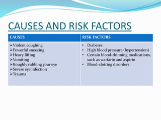
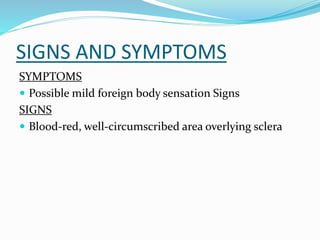
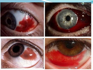
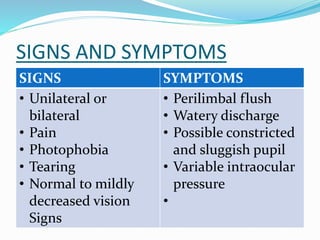
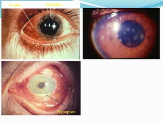
Subconjuctival haemorrhage
uveitis
• A subconjunctival haemorrhage occurs
when a tiny blood vessel breaks just
underneath the clear surface of your eye
(conjunctiva).
CAUSES AND RISK FACTORS
CAUSES RISK FACTORS
Violent coughing
Powerful sneezing
Heavy lifting
Vomiting
Roughly rubbing your eye
Severe eye infection
Trauma
• Diabetes
• High blood pressure (hypertension)
• Certain blood-thinning medications,
such as warfarin and aspirin
• Blood-clotting disorders
SIGNS AND SYMPTOMS
SYMPTOMS
 Possible mild foreign body sensation Signs
SIGNS
 Blood-red, well-circumscribed area overlying sclera
TREATMENT
 Reassurance
 Cold compresses
 Artificial tears
UVEITIS
 Uveitis is inflammation of the uvea, the middle layer
of the eye. The uvea consists of the iris, choroid and
ciliary body.
 The most common type of uveitis is an inflammation
of the iris called iritis (anterior uveitis).
CAUSES AND RISK FACTORS
The specific cause of uveitis isn't clear. However, in some
people, uveitis is associated with:
 Autoimmune disorders, such as Behcet's disease,
sarcoidosis or ankylosing spondylitis
 Inflammatory disorders, such as Crohn's disease or
ulcerative colitis
 Infections such as cat-scratch disease, herpes, syphilis,
toxoplasmosis, tuberculosis or West Nile virus
 Eye injury
 Certain cancers, such as lymphoma, that can directly or
indirectly affect the eye
SIGNS AND SYMPTOMS
SIGNS SYMPTOMS
• Unilateral or
bilateral
• Pain
• Photophobia
• Tearing
• Normal to mildly
decreased vision
Signs
• Perilimbal flush
• Watery discharge
• Possible constricted
and sluggish pupil
• Variable intraocular
pressure
•
WORK UP
 Complete ocular history and exam
 Systemic history and exam for various associated
conditions
Slit lamp exam
Deposits on posterior surface of cornea (keratic
precipitates)
Inflammatory cells and protein (flare) in AC
Adhesions of iris to surface of lens (posterior
synechiae)
Hypopyon
TREATMENT
1. Anti-inflammatory medication. E.g corticosteroids
2. Antibiotic or antiviral medication. If uveitis is caused by an
infection
3. Immunosuppressive or cell-destroying (cytotoxic) medication.
Immunosuppressive or cytotoxic agents may be necessary if your
uveitis doesn't respond well to corticosteroids or becomes severe
enough to threaten your vision.
4. Surgery. Vitrectomy-may be necessary both for diagnosis and
management of your uveitis..

Subconjuctival haemorrhage

  • 1.
  • 2.
    • A subconjunctivalhaemorrhage occurs when a tiny blood vessel breaks just underneath the clear surface of your eye (conjunctiva).
  • 3.
    CAUSES AND RISKFACTORS CAUSES RISK FACTORS Violent coughing Powerful sneezing Heavy lifting Vomiting Roughly rubbing your eye Severe eye infection Trauma • Diabetes • High blood pressure (hypertension) • Certain blood-thinning medications, such as warfarin and aspirin • Blood-clotting disorders
  • 4.
    SIGNS AND SYMPTOMS SYMPTOMS Possible mild foreign body sensation Signs SIGNS  Blood-red, well-circumscribed area overlying sclera
  • 6.
    TREATMENT  Reassurance  Coldcompresses  Artificial tears
  • 7.
    UVEITIS  Uveitis isinflammation of the uvea, the middle layer of the eye. The uvea consists of the iris, choroid and ciliary body.  The most common type of uveitis is an inflammation of the iris called iritis (anterior uveitis).
  • 8.
    CAUSES AND RISKFACTORS The specific cause of uveitis isn't clear. However, in some people, uveitis is associated with:  Autoimmune disorders, such as Behcet's disease, sarcoidosis or ankylosing spondylitis  Inflammatory disorders, such as Crohn's disease or ulcerative colitis  Infections such as cat-scratch disease, herpes, syphilis, toxoplasmosis, tuberculosis or West Nile virus  Eye injury  Certain cancers, such as lymphoma, that can directly or indirectly affect the eye
  • 9.
    SIGNS AND SYMPTOMS SIGNSSYMPTOMS • Unilateral or bilateral • Pain • Photophobia • Tearing • Normal to mildly decreased vision Signs • Perilimbal flush • Watery discharge • Possible constricted and sluggish pupil • Variable intraocular pressure •
  • 10.
    WORK UP  Completeocular history and exam  Systemic history and exam for various associated conditions Slit lamp exam Deposits on posterior surface of cornea (keratic precipitates) Inflammatory cells and protein (flare) in AC Adhesions of iris to surface of lens (posterior synechiae)
  • 11.
  • 12.
    TREATMENT 1. Anti-inflammatory medication.E.g corticosteroids 2. Antibiotic or antiviral medication. If uveitis is caused by an infection 3. Immunosuppressive or cell-destroying (cytotoxic) medication. Immunosuppressive or cytotoxic agents may be necessary if your uveitis doesn't respond well to corticosteroids or becomes severe enough to threaten your vision. 4. Surgery. Vitrectomy-may be necessary both for diagnosis and management of your uveitis..